Multiple Wounds and Comorbidities Drive the Real-World Natural History of Venous Leg Ulcers: A Retrospective Analysis
© 2026 HMP Global. All Rights Reserved.
Any views and opinions expressed are those of the author(s) and/or participants and do not necessarily reflect the views, policy, or position of Wounds or HMP Global, their employees, and affiliates.
Abstract
Background. The natural history of real-world patients with venous leg ulcers (VLUs) is poorly understood, because randomized controlled trials (RCTs) generally enroll a single VLU in a highly selected cohort of patients. Objective. To evaluate the real-world natural history of VLUs. Methods. Retrospective analysis was performed of all VLU and patient data from a wound care–specific electronic health record transmitted to the U.S. Wound Registry from January 1, 2021, through December 31, 2022. Wound outcomes were analyzed through 52 weeks and for eventual closure. Results. Out of a total of 160 341 wounds and 51 708 patients, 30 790 VLUs (19.2%) affecting 11 197 patients (21.7%) were analyzed. Obesity, hypertension, diabetes, and impaired ambulation were common comorbid conditions. Patients had a mean 2.7 VLUs during the study, as well as 2.5 concomitant wounds of any etiology with a combined wound area that was double that of index VLUs. The eventual index VLU closure rate was 74.1%, but closure of all index wounds in a given patient occurred in only 50.7% of patients. Conclusion. Analysis of a large VLU cohort demonstrates that in the real world, patients frequently have multiple wounds that are exacerbated by serious comorbidities and impaired ambulation, take longer to close, and have lower healing rates than typically reported in RCTs.
Chronic venous disease is one of the most common chronic diseases, with a recent report from The Sage Group estimating that more than 190 million Americans had chronic venous disease in 2025.1 Chronic venous disease results in chronic venous insufficiency, chronic edema, and, in some patients, the development of venous leg ulcers (VLUs), which are the most common leg ulceration in the United States. From 2014 to 2019, the prevalence of venous ulcers among Medicare beneficiaries increased by nearly 50%, from 1.0% to 1.4%, and more than doubled in patients younger than 65 years (from 0.9% to 1.9%).2 Although overall costs per Medicare beneficiary for chronic wounds and other specific wound types decreased during this period, the cost of venous ulcer care increased by approximately 50%, from $1206 to $1803 per Medicare beneficiary.
VLUs usually appear in the gaiter region and are frequently associated with venous hypertension, postthrombotic syndrome, and varicose veins, although their pathophysiology is poorly understood.3 When wounds are diagnosed early and are managed with compression therapy (the standard for VLU care) and good wound care, wound closure can be expected within 6 months.3,4 Unfortunately, it is frequently reported that only a minority of VLUs receive adequate compression therapy, mainly due to patient and clinician nonadherence resulting from behavioral barriers and limited time, financial, and human resources.5-14 Consequently, average closure times can extend to 1 year.3,4 A majority of the wounds that do close recur, which can be due to nonadherence to prophylactic compression therapy, worsening chronic venous disease, incorrect diagnoses, and/or the presence of mixed venous-arterial disease. Obesity, malnutrition, and underlying chronic venous disease are known to delay VLU healing,3 while significant factors of failure to heal include the number of concomitant wounds on the body (regardless of etiology), increased wound duration and size, presence of bioburden or infection, and impaired ambulation.15
The regulatory approval process and clinical treatment decisions are influenced by the safety and efficacy data produced by randomized controlled trials (RCTs), which are the standard for clinical evaluation.16-20 However, the complexity of VLUs and their natural history has not been comprehensively captured in RCTs, which are designed to enroll highly selected patients with few comorbidities and small ulcers, so that wound closure can be assessed within 12 weeks to 16 weeks.20 Consequently, RCTs evaluating VLU have limited external validity and certainty for real-world healing trajectories.16-20 The US Food and Drug Administration (FDA) promotes the use of real-world evidence (RWE) analysis of real-world data (RWD) to address these knowledge gaps. As of December 2025, the FDA now allows for deidentified RWD to be submitted with medical device marketing applications, removing the longstanding requirement that RWE submissions include identifiable individual‑level patient data.21 This change lifts a major barrier to the use of RWE in regulatory decision-making.
Given the gap in knowledge concerning the effectiveness of wound treatments in real-world patients compared with carefully controlled trial populations, a timely investigation has been undertaken by the RWE Group of the Wound Care Collaborative Community (WCCC), a 501(c)3 nonprofit recognized by the FDA, to understand the natural history of real-world diabetic foot ulcers (DFUs) and VLUs, defined as the progression of ulcer disease from onset to final outcomes, using real-world clinical treatment data from the U.S. Wound Registry (USWR), a national registry populated by direct transmission from a structured, wound care–specific electronic health record (EHR). The findings of this 3-part retrospective analysis of the WCCC natural history project (NHP) are intended to inform health care policies and improve trial design to facilitate access to effective wound treatments. The first article in the NHP series examines shared comorbidities, concomitant medications, and wound characteristics of 2 large separate cohorts of VLUs and DFUs, finding that DFUs and VLUs may develop from a shared underlying patient phenotype predisposed to chronic and overlapping ulceration and disease.22 Whereas RCTs traditionally evaluate the outcome of a single wound, the second NHP article comprehensively examined the natural history of DFUs in the DFU cohort, finding that the predominance of multiple wounds combined with comorbidities in patients with DFUs prolongs time in service (ie, number of days the patient or wound received care at the USWR facility), increases morbidity (especially amputations), and inhibits overall healing status, which cannot be represented by the healing trajectory of a single DFU alone.23
In this third article, the real-world natural history of VLUs is analyzed by examining long-term closure rates (by maximum wound area), amputation and mortality rates, and time in service to capture the full picture of disease severity, progression, and both wound and patient outcomes, and to further compare with the natural history of DFUs.
Methods
Study Design and Data Collection
The first 2 articles of the WCCC NHP series comprehensively describe the study design and data collection methods.22,23 In summary, clinicians used a highly structured, wound care–specific EHR (Intellicure, LLC, The Woodlands, TX]) to collect patient and wound data at point of care. The authors of the current study retrospectively analyzed USWR data that were prospectively collected from the entire EHR from January 1, 2021, through December 31, 2022, via automatic, secure, and direct transmission at the end of each clinical encounter at 149 outpatient clinics, physician offices, and mobile practices in 29 US states. Upon transmission, research-grade data were aggregated, deidentified, and stored in a relational USWR database. WCCC experts formed a Clinical Advisory Panel (CAP) to oversee the study. The Woodlands IRB (The Woodlands, TX) reviewed the study protocol and determined that secondary analysis of deidentified data was considered exempt research.
When the study database was captured on April 5, 2024, the entire USWR dataset included data on 191 654 patients with 740 167 wounds. To identify the potential study database, filters were simultaneously applied to the overall USWR dataset to include only the following data registered from January 1, 2021, through December 31, 2022: patients with 3 or more total visits, visits involving an advanced practitioner, in-person visits at the registered USWR facility, and clinical problems related to a wound. This identified 160 341 wounds in 51 708 patients in the USWR that could be retrospectively screened for study eligibility.
Documentation of VLUs
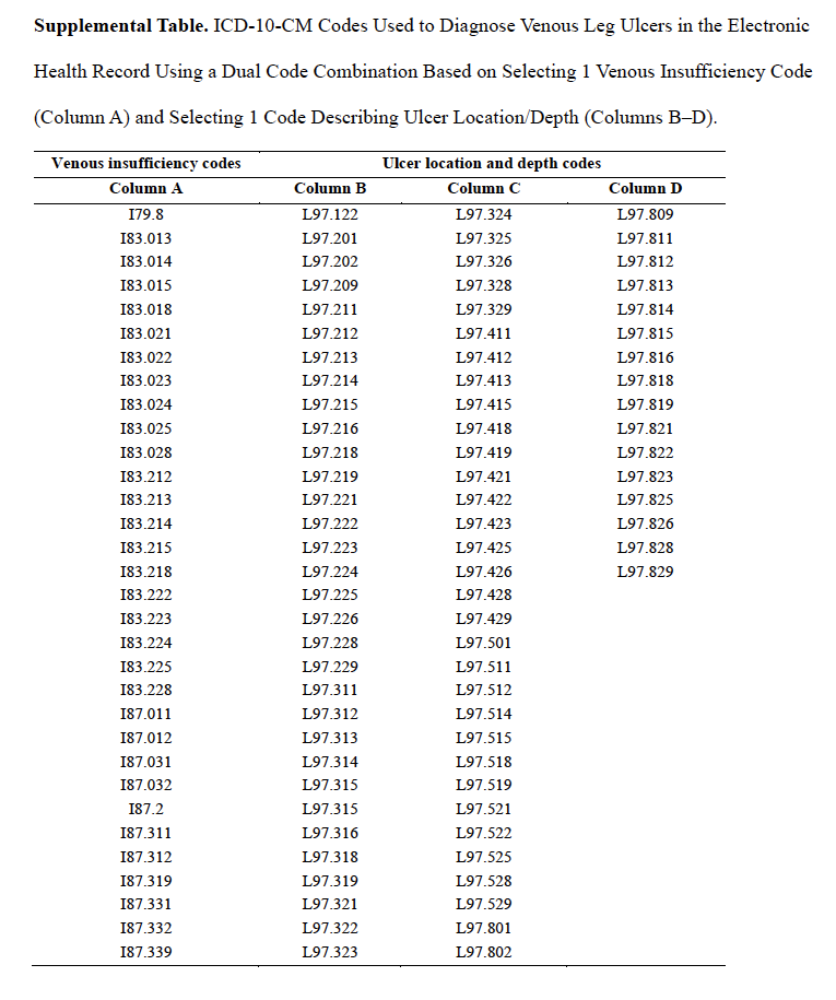
The study cohort comprised data from all study-eligible patients with VLUs (index wounds), their wounds (of any type), and their related visits documented during the study period. The structured EHR uses International Classification of Diseases, Tenth Revision, Clinical Modification (ICD-10-CM) to standardize diagnosis coding, but it lacks specific codes to diagnose a VLU. Therefore, the USWR identified VLUs by combining venous insufficiency codes with the appropriate code for ulcer location and depth. The Supplemental Table lists all the ICD-10-CM codes used to diagnose VLUs based on venous insufficiency (Column A) and ulcer location and depth (Columns B–D). To document a VLU in the EHR, the clinician selected a dual code combination based on 1 code listed in Column A plus 1 code listed in Column B, C, or D. To mitigate differential diagnoses, the CAP excluded any VLUs with exposed deep structures or with eschar coverage, because these manifestations likely indicated arterial ulcers. If the thigh was documented as the ulcer location, then it was also excluded from this analysis, because this location is clinically unlikely.
All VLUs were documented. When multiple VLUs were present in the same limb, they were counted and measured separately, provided that their wound edges were not touching each other. Wound area (length × width) was measured using a ruler. Individual index wound area and concomitant wound area were calculated.
Variables Related to Clinical Complexity
Medication data were linked to an electronic prescribing module (DrFirst, version 4.41.3.2) in the EHR and were analyzed for the VLU cohort in the first article in this NHP series.22
A patient was considered to have home health care if their EHR documented a home health agency for some part of their time in service. Dressing orders from a durable medical equipment company implied that dressings were changed at home in between study visits.
At each VLU visit, the clinician documented the wound area and reported wound infection (when there was a definitive diagnosis of osteomyelitis and/or cellulitis). Any other possible infection was counted based on the documentation of at least 1 of the following: periwound erythema; periwound induration; systemic fever; wound culture ordered at visit; antibiotics prescribed at visit; and/or green, purulent, or malodorous exudate.
A previous USWR publication reported that impaired ambulation based on the patient’s arrival method (using a cane, in a wheelchair or a scooter, or in a bed or stretcher) was a significant predictive factor of nonhealing in real-world VLUs.15 Because ambulation status could change over the course of care and because many patients developed additional wounds during the study period, ambulatory status was determined on the first visit date documented for each VLU.
Outcome Documentation
Clinician documentation at the time the study database was captured (April 5, 2024) determined both wound and patient outcomes. If a VLU lacked a documented outcome at this time, then the database was refreshed through August 31, 2025, to determine final outcomes. The designation wound closure documented described VLUs based on the preferred wording of the clinician to describe a healed or closed wound from the following options documented: healed, resolved, goal achieved, or surgical closure. The designation wound closure not documented was based on any of the following outcomes documented: major or minor amputation (involving the index ulcer), merged with another wound (when wound area grew to merge with another nearby wound, forming a single, larger wound), patient deceased, wound still in service, or lost to follow-up (which included a subset of outcomes: transferred care, diagnosis change, VLU and patient both lost to follow-up, and patient still in service but VLU lost to follow-up). It is important to note that when merging occurred between 2 VLUs, the resulting VLU was counted as a new wound. VLUs were considered to be still in service if visits for their management continued between July 1 and August 31, 2025, approximately 4.5 years after the retrospective study start date and 2.5 years after the study end date.
Data Analysis
The primary end point was percentage of VLUs with wound closure documented at 12 weeks, 16 weeks, 26 weeks, and 52 weeks, which was grouped by maximum wound area documented at any time during the study period: 0 cm2–5 cm2, >5 cm2–10 cm2, >10 cm2–25 cm2, >25 cm2–50 cm2, >50 cm2–100 cm2, and >100 cm2. The percentage of VLUs that eventually or never went on to close (beyond the study timeframe) was also analyzed.
Secondary end points were the number of visits per VLU, wound time in service (in weeks), patient time in service (in weeks), the number or percentage of VLU visits with infection, the number of patients who presented with a new VLU during the study timeframe, and the mean number of wounds of any type a patient had during a 52-week rolling period. As noted previously, time in service was the number of days the patient or VLU received wound care at the USWR facility. If a patient temporarily discontinued wound care or was hospitalized for less than 60 days (8.6 weeks), that period of interruption was omitted from the time-in-service calculation. If the interruption in wound care lasted 60 days or more, then a new time-in-service period began when the patient resumed treatment. The percentages of patients whose time spanned at least 52 weeks (≥1 year) and at least 104 weeks (≥2 years) were also analyzed. Deceased patients during the study timeframe were counted to determine the 1-year mortality rate.
Excel (Version 2608, Microsoft 365) was used to summarize categorical variables by counts and percentages and continuous variables by descriptive statistics. The CAP recommended that potentially undocumented comorbidities should be imputed based on the documentation of specific medications. Atrial fibrillation was counted if apixaban was documented in the patient’s record. Documented amlodipine, carvedilol, lisinopril, and/or losartan implied that the patient had hypertension. Clopidogrel was imputed as peripheral arterial disease. Atorvastatin indicated that the patient had hyperlipidemia.
Results
Cohort Wound and Patient Population
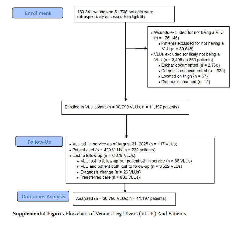
A total of 160 341 wounds or ulcers affecting 51 708 patients were registered in the USWR during the study period and screened for study eligibility. The Supplemental Figure shows the VLU cohort flowchart. There were 126 145 non-VLUs excluded from analysis (78.7%) and 39 648 patients without VLUs excluded from analysis (76.7%). Additionally, 3 406/34 196 VLUs (10%) in 863 patients were excluded from analysis because these wounds were likely not true VLUs for the following reasons: 2 768/34 196 (8.1%) were excluded for documented eschar, 935/34 196 (2.7%) for documented deep tissue (muscle, tendon, joint capsule, bone, or other deep tissue), and 67/34 196 (0.2%) for being located on the thigh. Finally, 2 VLUs in a pediatric patient were removed from the dataset because clinicians’ notes indicated they were, in fact, insect bites. There were 30 790 (19.2%) VLUs affecting 11 197 (21.7%) patients eligible for retrospective analysis; all these VLUs and patients were analyzed in this study (Supplemental Figure).
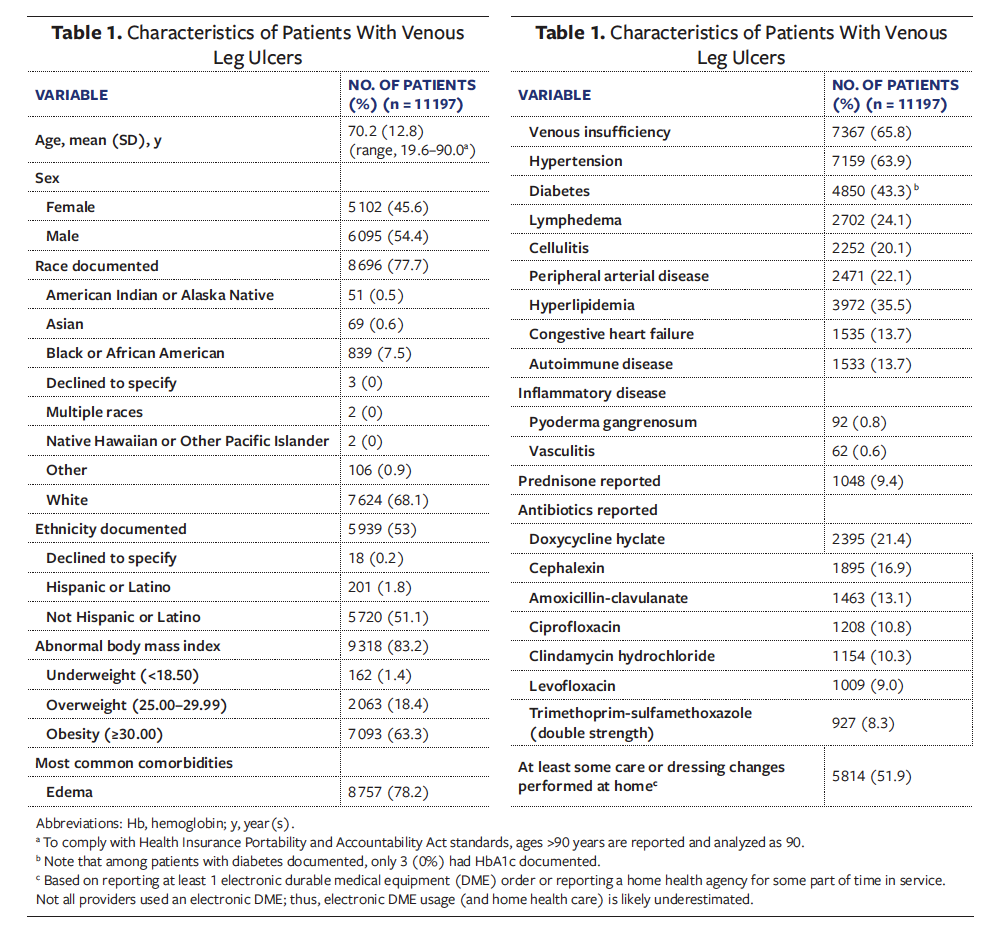
Patient characteristics. Table 1 summarizes the patient characteristics in the current study. The mean (SD) age was 70.2 (12.8) years (range, 19.6 years–90.0 years). The slight majority of patients in this study were male, 6 085 (54.4%), compared with 5102 female patients (45.6%). Patients were predominantly White (n = 7 624 [68.1%]); 839 (7.5%) were Black or African American, and 201 (1.8%) identified as Hispanic or Latino. Abnormal body mass index was predominant in this cohort (n = 9 318 [78.2%]), and the majority of patients had obesity (n = 7 093 [63.3%]), based on body mass index greater than or equal to 30. In addition to venous insufficiency and edema or lymphedema, other common comorbidities documented were hypertension, diabetes, peripheral arterial disease, hyperlipidemia, congestive heart failure (CHF), and autoimmune disease.
VLU characteristics. The VLU characteristics are described in Table 2. The mean (SD) wound age of the index VLUs reported by patients at their first visit was 14.1 (39) weeks (range, 0.1 weeks–52.1 weeks), but 2 775 VLUs (9%) had a wound duration greater than 26 weeks and 1 466 (4.8%) reported a duration greater than 52 weeks. The largest mean wound area documented was 74.1 (219.7) cm2, which indicates that areas varied widely.
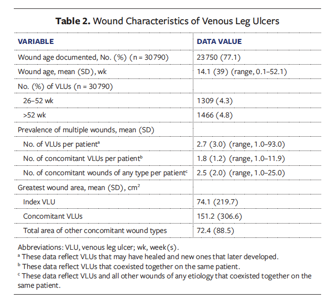
Burden of multiple wounds. There were 2 rolling periods of 52 weeks each during the study timeframe, with a mean (SD) 5.1 (3.6) wounds of any type documented during each rolling period. A patient might have had only 1 VLU at a time if a VLU healed and another subsequent ulcer later formed. During the study timeframe, 40.6% of patients (n = 4 551) developed at least 1 new VLU in a different location than the index ulcer. Patients also had multiple, coexisting wounds, which are analyzed as the concomitant wound count. On average, patients had nearly 3 VLUs at any given time during the study period, as well as an average of nearly 2 concomitant VLUs coexisting with their index ulcer. Furthermore, they had an average of 2.5 concomitant wounds (including concomitant VLUs) of any type coexisting with the index VLU (Table 2). Importantly, the total wound area of concomitant VLUs was 151.2 cm2, which was more than double the area of index ulcers, suggesting that these patients have a serious burden of chronic venous disease.
Additional clinical complexity to consider. Of the 210 302 visits that occurred in this cohort during the study period, signs of infection in index VLUs were documented in 67 173 (31.9%), and 20.1% of patients had documented cellulitis. Further contributing to clinical complexity, 9.4% of patients received long-term prednisone therapy, 0.8% had a diagnosis of pyoderma gangrenosum, and 0.6% had vasculitis, 2 conditions that often are mistaken for VLUs but do not respond to routine wound care. Home health care was documented in more than half of patients (52.2%).
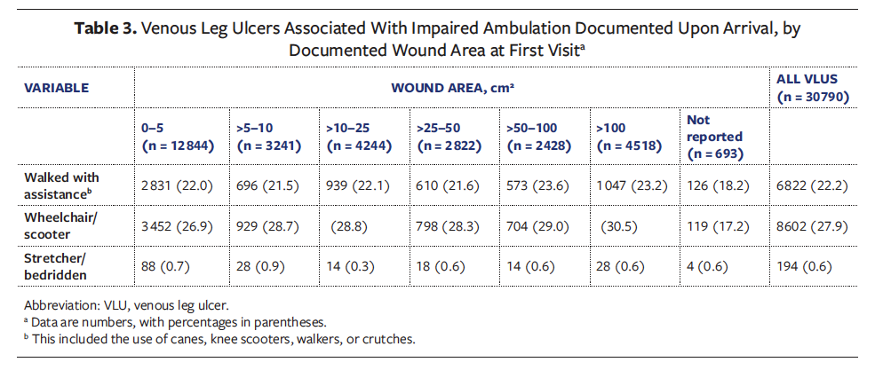
For approximately half of VLUs (50.7%), impaired ambulation was documented at the first VLU visit (Table 3). Although only 0.6% of VLUs occurred in bedridden patients, more than a quarter of VLUs developed in patients arriving in a wheelchair or scooter. Impaired ambulation status was nearly consistent across ulcer sizes, with only very slight increases in rates observed among larger wound sizes, with 49.6% of patients arriving with impaired ambulation among VLUs 0 cm2–5 cm2 in size versus 54.3% among VLUs >100 cm2 in size. Patients with smaller ulcers had frailty comparable to patients with larger ulcers.
Wound and Patient Outcomes Based on Index VLUs
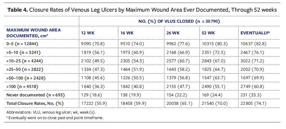
Documented closure rates for index VLUs. Table 4 summarizes the VLU closure rates documented at various time points, by maximum wound area. At 12 weeks, 55.9% of all VLUs closed, and 29.2% of the smallest VLUs remained open. At 52 weeks, closure was reported for 80.3% of the smallest VLUs (0 cm2–5 cm2). Closure rates decreased as wound area increased, and only a minority of the largest ulcers were closed at 12 weeks. Closure rates reached a majority by 52 weeks for all sizes, when 70.0% of all VLUs had documented closure in the EHR (n = 21 540). Furthermore, 74.1% (n = 22 805) eventually closed after the study period. Most closure rates were approximately 20% higher for VLUs that eventually went on to close compared with those that closed after only 12 weeks. Interestingly, the smallest change in closure rates was observed among the smallest VLUs, with only 9.5% more of these closing by 52 weeks and 12% more eventually closing, while closure rates noticeably improved over time among the larger VLUs. At 12 weeks, only 36.3% of VLUs larger than 100 cm2 closed; by 52 weeks the majority had closed (55.1%), with 60.8% eventually closing.
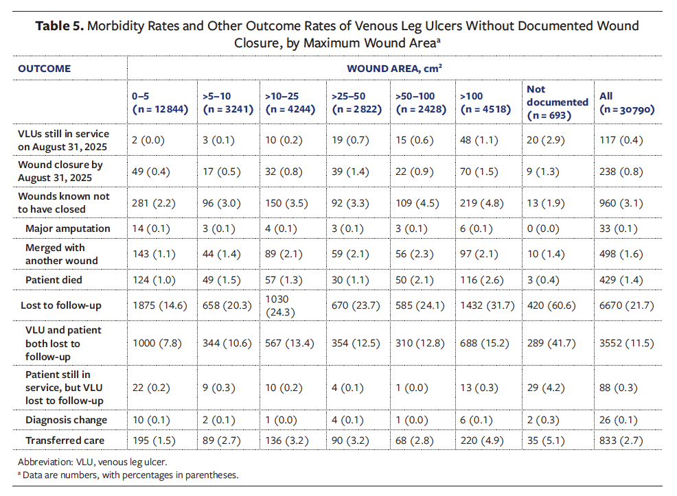
Wound closure not documented. The documented 1-year mortality rate among patients in this cohort was 2% (n = 222 patients) (Supplemental Figure). However, since patients generally had multiple wounds, there were 429 VLUs without outcomes reported because the patient died, but for the particular index VLU, death was not documented. For example, a patient could have had 2 VLUs and died, but only 1 VLU had death documented as final outcome. Lost to follow-up rates increased with increasing wound area and approached one-third among the largest ulcers (>100 cm2) (Table 5). This trend was likely due to some patient deaths not reported or documented. Among the 693 VLUs for which wound area was not documented, only 33.3% had documented closure (Table 4), and the lost to follow-up rate was high, at 60.6% (Table 5). The major amputation rate was only 0.1% overall and was consistent across wound areas.
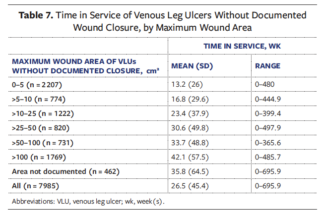
Time in service. A majority of patients (57.3% [n = 6 414]) had time in service of 52 weeks or longer, and 41.0% (n = 4 592) were in service for 104 weeks or longer. Among VLUs with documented closure, the mean time in service was 13 (26.6) weeks (range, 0 weeks–508.1 weeks) (Table 6). Mean time in service was shortest for the smallest VLUs at 8.3 (19.1) weeks (range, 0 weeks–477.4 weeks) and longest for the largest VLUs at 21 (33.7) weeks (range, 0 weeks–354.3 weeks). Among VLUs without documented wound closure (Table 7), time in service was unsurprisingly longer, with similar trends of time increasing as wound sizes increased.
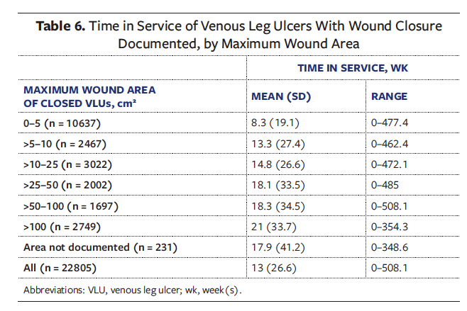
Outcomes when multiple wounds present. The closure rate among patients in the VLU cohort who eventually achieved documented closure of all their wounds was 50.7% (Table 4). This rate was 23.4% less than the overall VLU closure rate of 74.1% in the current study, demonstrating that even if their VLUs healed, patients still had other wounds of different etiologies that remained open. Consequently, a longer time in service could have been documented for patients with multiple wounds compared to the time in service documented for a single index VLU on the same patients. That is to say, the time in service of a single wound does not reflect the total time in service of a patient. Among patients for whom wound closure was documented in all their wounds, the mean time in service was 36.8 (45.9) weeks (range, 0 weeks–516.9 weeks), which was more than double the mean time in service for an index VLU (Table 6), indicating that patients with multiple wounds had to stay in service for care of their remaining wounds.
An increased morbidity burden in the presence of multiple wounds is also demonstrated by the major amputation rate at the patient level of 0.2% (n = 25 patients), which was double the major amputation rate of index VLUs (Table 5). This difference could be explained by the presence of multiple wounds in an amputated limb; for example, a VLU may have healed and been documented as such, but another VLU in the same limb may have required amputation.
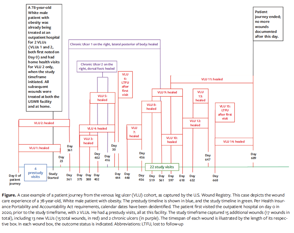
Case example of a patient journey in wound care. The Figure describes the wound care experience of a randomly selected 78-year-old, White male patient with obesity. The patient first visited the outpatient hospital on day 0 in 2020, prior to the study timeframe, with 2 VLUs, thus serving as an example of VLUs and patients already in service at study onset. All 4 prestudy visits occurred at that same facility. The study timeframe captured 15 additional wounds (17 in total), including 13 new VLUs (15 total) and 2 documented as chronic ulcers (which were of another etiology). The timespan of each wound is captured by the length of their respective boxes. For example, VLU 4 was first noted on day 375 and healed on day 430. During the study timeframe, all wounds also received home health care, except for VLU 1, which healed quickly, on day 23. The patient journey lasted 689 days and involved 26 visits (22 of which occurred during the study timeframe), during which 13 of 15 VLUs healed (86.7%), 2 VLUs did not have their outcomes documented and were lost to follow-up, and both chronic ulcers healed. After day 689 (when VLUs 11 and 14 were noted as healed), the patient did not have any more new wounds documented.
Discussion
This retrospective analysis of real-world outcomes of patients with VLUs supports the findings previously reported in the second article of the NHP series for the DFU cohort—that the patient’s experience of care is not represented by the clinical course of 1 ulcer.23 As with patients with DFUs,22,23 the majority of real-world patients with VLUs have obesity and serious comorbid conditions, including diabetes and autoimmune disease (Table 1). Nearly 1 in 10 patients in the present cohort took immunosuppressants (prednisone), which interact with wound healing. The poor health state of this cohort is further emphasized by the fact that impaired ambulation was documented at the first visit for approximately half of index VLUs, with similar rates of impaired ambulation observed across all wound areas (Table 3).
In a systematic review of 144 RCTs that evaluated VLUs, the calculated weighted mean wound area was only 13.2 cm2.19 The VLUs in the present cohort were on average 5.6 times larger. Although it is true that higher closure rates were associated with smaller, less severe VLUs at all time points throughout the current study (Table 4), the least improvement in closure rates over time occurred, paradoxically, among the smallest VLUs. Nearly 20% of the smallest VLUs never achieved documented closure. There are many reasons why very small wounds diagnosed as VLUs are slow to improve or heal, including the difficulty of accurately measuring very small changes in a small wound using a ruler, and misdiagnosis (eg, skin cancers diagnosed as VLUs). However, the WCCC CAP attempted to mitigate differential diagnoses of VLUs in the current study by requesting that wounds with exposed bone, muscle, and tendon be removed from the VLU cohort, because they were likely other types of wounds. It is important to note that never-healed VLUs are often associated with impaired ambulation, which may reflect the presence of serious comorbidities.4,15 It is interesting to note that among patients with VLU, those with small ulcers were just as likely to have impaired ambulation as those with large ulcers (Table 3). This implies that even patients with very small and presumably less severe VLUs are still frail, with serious comorbidities that could impede healing.
Given time, regardless of size the majority of VLUs eventually close, but the time to closure extends well beyond the 3- to 4-month windows of evaluation used in trials and can require years (Table 4). This finding brings into question the real-world relevance of 12-week healing rates. In a retrospective analysis of USWR data from 2001 through 2016, the 12-week healing rate of VLUs was 44.1%, of which 56.9% eventually closed after 12 weeks passed.20 These historic RWD contrasted sharply with what wound clinics were publicly reporting on their websites at the time, which were 12-week healing rates of 90% or higher. The current analysis found that 55.9% of VLUs healed at 12 weeks, a somewhat higher rate than in the previous USWR analysis; however, that first analysis included data from 25 years ago, and it is possible that newer treatments or more consistent evidence-based use of compression have improved outcomes. Nevertheless, clinicians must be cognizant that drastically high closure rates after only 12 weeks are highly unlikely in the real world.
The effect of chronic venous disease on wound closure should not be underestimated. Patients had multiple VLUs develop during this study, both concomitantly and independently. That the size of the combined wound area of concomitant VLUs was more than twice the area of a single VLU speaks to the extent of chronic venous disease affecting the patients in this cohort (Table 2). Although high recurrence of VLU is well known, the development of entirely new VLUs is not well studied. In the current study cohort, 40.6% of patients developed at least 1 new VLU, distinguished from the initial index ulcer by forming in a different anatomic location. The patient case example in the Figure exemplifies the realities of wound multiplicity and new VLU development in patients with VLU. Similar to the majority of the patients analyzed in the present VLU cohort, this patient was in service for longer than 1 year (689 days); in fact, he was in service for nearly 2 years. On average, 5.1 wounds per patient were documented in the cohort per year. In his first year in service, the patient described in the Figure had 3 documented wounds (2 VLUs and 1 chronic ulcer). Within the first few months of his second year in service (beginning on day 375), his condition seemed to deteriorate, as evidenced by the development of many new wounds. During his second year in service 14 wounds formed, including 13 new VLUs and 1 other chronic ulcer. His first chronic ulcer, which appeared on day 361, coexisted over the course of 200 days with 8 different wounds, including 7 VLUs, before it closed on day 561. It is not known why 2 VLUs were lost to follow-up during the second year of service, but a possible factor is documentation fatigue, which can occur when clinicians have to report and follow-up on many wounds in a short time at a single visit.
The development of new VLUs while another VLU is still in treatment suggests that patients receiving wound care are still at risk of new ulceration; wound therapies do not correct underlying conditions, such as venous hypertension, obesity, poor nutrition, and CHF. The majority of patients in the present cohort had venous insufficiency (65.8%) and edema (78.2%), the hallmark conditions associated with venous ulceration, but the importance of CHF (13.7%) and lymphedema (24.1%) in this patient population also supports volume overload as a contributor to the development of edema-related ulceration24,25 (Table 1). Venous ablation is a minimally invasive treatment that blocks refluxing veins, facilitating the reduction of pressure in affected limbs and demonstrating superior VLU outcomes when used in combination with compression compared with compression alone.26 It is increasingly understood that controlling venous hypertension by removing or blocking superficial incompetent veins that drive venous reflux and lead to ulceration is essential in combination with compression therapy for VLU management and healing.26-28
In the present VLU cohort, 63.9% of patients had hypertension and 43.3% had diabetes. The overlap between chronic venous insufficiency and diabetes among patients with DFUs and VLUs was reported in the first article in the NHP series, in which it was also reported that 15.5% of patients with VLUs had either a concomitant DFU or a nonfoot ulcer attributed to diabetes.22 Among patients with DFUs, 19.4% had both diabetes and venous insufficiency documented. A recent, albeit small, systematic review and meta-analysis of 4 studies estimated that the prevalence of chronic venous insufficiency in patients with type 2 diabetes was 55%.29 A larger systematic review of 20 studies analyzed comorbidities in 3 395 patients with VLUs, noting that hypertension and diabetes were important, prevalent conditions in this patient population that can inhibit treatments, particularly compression therapy.30 Other authors have also advised that wound care should approach the patient as a whole,31 recognizing that VLUs coexist with serious comorbidities that interfere with treatment and inhibit healing. The control of some comorbidities (eg, diabetes, heart failure, obesity, edema) can vary over time, affecting the likelihood of ulcer recurrence over time.32 In the current study, only 3 patients with diabetes in the VLU cohort had hemoglobin A1c documented in laboratory results, and nearly half of all patients underwent dressing changes by either skilled home health nurses or at home by the patients themselves or their caregivers, which has implications for the implementation of needed compression bandaging. The findings of the NHP promote the vital importance of both providing and documenting the basic elements of DFU and VLU care (eg, off-loading, compression, and arterial assessment) to provide optimal clinical care and increase the value of RWD. As concluded with our DFU findings,23 future VLU RWD analysis should also examine compression adherence and other practice trends, including venous ablation, to understand their influence on outcomes.
Comparing RWD vs recent RCT data. The NHP revelation that patients with VLU are in service typically for at least 1 year and have overlapping comorbidities and ulcerations with patients with DFUs has very important implications for RCT design that will be examined in detail by the WCCC in a future study. In the case of DFU trials, recent publications demonstrated that trial design is gradually becoming more inclusive of more severe DFUs that have deep tissue exposure, osteomyelitis, cellulitis, and/or gangrene.23 While not exhaustive or systematic, Table 8 provides some insight into the comorbidities, medications, and wound severity evaluated in 10 VLU RCTs published since 2019 to compare prospective trial generalizability to the VLU natural history findings in the present cohort. For the most part, VLU trials continued to evaluate a single, noninfected index ulcer in relatively small cohorts (<100 VLUs to <200 VLUs) over a short 12- to 24-week period. The size of the wounds included in VLU trials varied widely, but it was generally much smaller (<25 cm2) than the RWD in the current study. Aside from underlying chronic venous disease, comorbidities included in the trial populations were few; hypertension, CHF, and diabetes were rarely reported among the trial populations, and autoimmune diseases were completely excluded. Nearly 1 in 4 patients in the present real-world cohort had lymphedema, but none of the trials included in Table 8 reported any comorbidity data for lymphedema. However, none of them excluded this condition; thus, there could be a gap in reporting. Nevertheless, it would appear that lymphedema, which causes fluid buildup and damage to surrounding tissue that leads to ulceration, is not being evaluated in VLU RCTs. Thus, the generalizability of VLU RCTs remains limited, and great potential exists to use the present RWD analysis to guide trial reform to include multiple wounds and more severe cases, thus strengthening the evidence base for regulatory decision-making processes.
Limitations
The limitations of this retrospective analysis have been described in detail in the first 2 articles of this NHP series.22,23 As with DFUs,23 it remains necessary to investigate the potential influence and correlation of comorbidities and medications on VLU outcomes among patients with single wounds vs multiple wounds. Important gaps in documentation were observed in the VLU cohort, such as the 693 VLUs for which wound area was never documented (the majority of which were lost to follow-up and the minority of which had documented closure). Wound measurement is the most basic documentation element for any clinician purporting to specialize in wound management.43 If this basic aspect of documentation is ignored, then it is not surprising that other documentation elements are missing, such as final wound outcome. To strengthen future RWD analysis, it would be informative to analyze documentation trends and standards and their associations with outcomes, especially when a patient has multiple wounds. In general, wound care research remains in need of standardized, universal metrics for data entry to facilitate the calibration of RWE findings with RCT results.44
Conclusion
In this third article in the NHP series, impaired ambulation and wound multiplicity were predominant in patients with VLUs, findings that were also observed in the DFU cohort in the second article. Less severe and smaller ulcerations may remain unhealed after 1 year in the presence of coexisting comorbidities, multiple wounds of all types, and impaired ambulation. Among patients with VLUs who are seen by wound care experts, a single VLU does not reflect the patient’s full experience of care, nor does the outcome of a single VLU reflect the outcome of all concomitant wounds. These findings are applicable to both DFUs and VLUs and support the hypothesis introduced in the first article in the NHP series that there may be a potential shared phenotype driving chronic ulceration in patients with VLUs and DFUs, calling into question the paradigm of distinct and easily classified wound types. These findings have significant implications for the generalizability of effective treatments in wound care; advanced therapies for which RCTs have proved efficacy in targeted index wounds may have limited benefit among real-world patients with multiple ulcerations and serious underlying diseases. Future research should analyze RWD to understand whether healing outcomes are clearly different based on diagnosed wound “type” or are primarily determined by patient comorbidities, wound severity, and/or total wound area affected by multiple wounds. The overlap of comorbid conditions and wound types in real-world patients revealed by the WCCC NHP suggests the need for a less siloed, “wound-type specific” approach to clinical care standards. Instead, wound care clinicians should shift focus to the management of specific barriers to healing (eg, management of diabetes, edema, ischemia, nutritional status, obesity), which is necessary regardless of the wound diagnosis.
Overall, patients with VLU were found to be much sicker than subjects typically enrolled in RCTs; their VLUs were larger, more numerous, and took longer to heal. Additionally, it was found that new VLUs or other wounds commonly developed while the patient was still under care for the index VLU. The 3- to 4-month evaluation period of RCTs is a mere snapshot of the real-world prolonged time in service that spans months, if not years.
It is urgently necessary to look past the wound as a single “hole” in the patient and to consider the patient as a whole. Wound care practitioners need to clearly define their field of medicine and recognize and accept that chronic ulceration is more than a symptom of underlying disease, with lifelong prophylactic management required to mitigate new and/or recurring frequent ulceration.
This third article also is meant to jumpstart dialogue between the WCCC and the FDA to incorporate the insightful findings of this series into the FDA’s RWE evidence and decision-making process. The WCCC is currently drafting new recommendations based on the NHP findings to improve RCT design in wound care to better reflect the entire patient journey of care.
Author and Public Information
Authors: Vickie R. Driver, DPM, MS1; Lucian G. Vlad, MD2; Marissa J. Carter, PhD, MA3; Joseph Rolley, BS, MSIA4; David J. Margolis, MD, PhD5;Lisa Gould, MD, PhD6; and Caroline E. Fife, MD7,8
Affiliations: 1Elson S. Floyd College of Medicine, Washington State University, Spokane, WA, USA; 2Wake Forest University School of Medicine, Winston-Salem, NC, USA; 3Strategic Solutions, Bozeman, MT, USA; 4JTR Business Consulting, LLC, Doylestown, PA, USA; 5Department of Dermatology, University of Pennsylvania Perelman School of Medicine, Philadelphia, PA, USA; 6Warren Alpert Medical School, Brown University, Providence, RI, USA; 7Baylor College of Medicine, Houston, TX, USA; 8Intellicure, LLC, The Woodlands, TX, USA
Contributions: All authors contributed equally to this work.
Funding: The Wound Care Collaborative Community funded this study.
Disclosure: V.R.D. is president, chair, and a volunteer member of the Wound Care Collaborative Community (WCCC). M.J.C is a volunteer member of and consultant for WCCC. C.E.F. is the chief medical officer of Intellicure, LLC, which received funding for this project. All remaining authors are volunteer members of the WCCC and disclose no financial or other conflicts of interest.
Acknowledgements: The authors would like to thank Kristen Eckert (independent consultant) for her assistance in data analysis, drafting, and editing the manuscripts and Ben LeBoutillier (Intellicure, LLC) for his assistance with data collection and extraction.
Ethics Statement: The Woodlands IRB (The Woodlands, TX) reviewed the study protocol and determined that secondary analysis of deidentified data was considered exempt research.
Correspondence: Lucian G. Vlad, MD; Wake Forest University School of Medicine, 1 Medical Center Blvd, Winston-Salem, NC 27157: lucian.vlad@wfusm.edu
Manuscript Accepted: June 10, 2026
References
1. The Sage Group. Chronic Venous Disease (CVD) Epidemiology, Prevalence, Incidence and Progression. The Sage Group; 2026. Accessed January 22, 2026. https://www.thesagegroup.us/reports/chronic-venous-disease-cvd-epidemiology-prevalence-incidence-and-progression
2. Carter MJ, DaVanzo J, Haught R, Nusgart M, Cartwright D, Fife CE. Chronic wound prevalence and the associated cost of treatment in Medicare beneficiaries: changes between 2014 and 2019. J Med Econ. 2023;26(1):894-901. doi:10.1080/13696998.2023.2232256
3. Raffetto JD, Ligi D, Maniscalco R, Khalil RA, Mannello F. Why venous leg ulcers have difficulty healing: overview on pathophysiology, clinical consequences, and treatment. J Clin Med. 2020;10(1):29. doi:10.3390/jcm10010029
4. Smith J, Carville K, Smith K, Alan J. The impact of comorbidities on wound healing in the community: the value of using linked hospital data. Wound Practice and Research. 2024;32(3):136-144. doi: 10.33235/wpr.32.3.136-144
5. Harding K. Challenging passivity in venous leg ulcer care - the ABC model of management. Int Wound J. 2016;13(6):1378-1384. doi:10.1111/iwj.12608
6. Harding K, Dowsett C, Fias L, et al. Simplifying venous leg ulcer management. consensus recommendations. Wounds International. 2015. Accessed January 26, 2026. https://woundsinternational.com/consensus-documents/simplifying-venous-leg-ulcer-management-consensus-recommendations/
7. Weller CD, Bouguettaya A, Britt H, Harrison C. Management of people with venous leg ulcers by Australian general practitioners: an analysis of the national patient-encounter data. Wound Repair Regen. 2020;28(4):553-560. doi:10.1111/wrr.12820
8. Perry C, Atkinson RA, Griffiths J, et al. What promotes or prevents greater use of appropriate compression in people with venous leg ulcers? A qualitative interview study with nurses in the north of England using the theoretical domains framework. BMJ Open. 2022;12(8):e061834. doi:10.1136/bmjopen-2022-061834
9. Bar L, Brandis S, Marks D. Improving adherence to wearing compression stockings for chronic venous insufficiency and venous leg ulcers: a scoping review. Patient Prefer Adherence. 2021;15:2085-2102. doi:10.2147/PPA.S323766
10. Fife CE, Carter MJ, Walker D. Why is it so hard to do the right thing in wound care? Wound Repair Regen. 2010;18(2):154-158. doi:10.1111/j.1524-475X.2010.00571.x
11. Lian Y, Birt L, Poland F, Naughton F, Moffatt C, Wright D. A qualitative study examining the application of compression therapy for inpatients with venous leg ulcers—perspectives of hospital staff where it is routinely applied. Int Wound J. 2026;23(2):e70810. doi:10.1111/iwj.70810
12. Heyer K, Protz K, Glaeske G, Augustin M. Epidemiology and use of compression treatment in venous leg ulcers: nationwide claims data analysis in Germany. Int Wound J. 2017;14(2):338-343. doi:10.1111/iwj.12605
13. Mahoney K. More wounds, less time to treat them: 1717 nurses discuss the challenges in wound care in a series of study days. Br J Community Nurs. 2017;22(Sup6):S33-S38. doi:10.12968/bjcn.2017.22.Sup6.S33
14. Gray TA, Rhodes S, Atkinson RA, et al. Opportunities for better value wound care: a multiservice, cross-sectional survey of complex wounds and their care in a UK community population. BMJ Open. 2018;8(3):e019440. doi:10.1136/bmjopen-2017-019440
15. Fife CE, Horn SD. The wound healing index for predicting venous leg ulcer outcome. Adv Wound Care (New Rochelle). 2020;9(2):68-77. doi:10.1089/wound.2019.1038
16. Eckert KA, Fife CE, Carter MJ. The impact of underlying conditions on quality-of-life measurement among patients with chronic wounds, as measured by utility values: a review with an additional study. Adv Wound Care (New Rochelle). 2023;12(12):680-695. doi:10.1089/wound.2023.0098
17. Carter MJ, Fife CE, Walker D, Thomson B. Estimating the applicability of wound care randomized controlled trials to general wound-care populations by estimating the percentage of individuals excluded from a typical wound-care population in such trials. Adv Skin Wound Care. 2009;22(7):316-324. doi:10.1097/01.ASW.0000305486.06358.e0
18. Serena TE, Fife CE, Eckert KA, Yaakov RA, Carter MJ. A new approach to clinical research: integrating clinical care, quality reporting, and research using a wound care network-based learning healthcare system. Wound Repair Regen. 2017;25(3):354-365. doi:10.1111/wrr.12538
19. Gethin G, Ivory JD, Connell L, McIntosh C, Weller CD. External validity of randomized controlled trials of interventions in venous leg ulceration: a systematic review. Wound Repair Regen. 2019;27(6):702-710. doi:10.1111/wrr.12756
20. Fife CE, Eckert KA, Carter MJ. Publicly reported wound healing rates: the fantasy and the reality. Adv Wound Care (New Rochelle). 2018;7(3):77-94. doi:10.1089/wound.2017.0743
21. U.S. Food and Drug Administration. Use of Real-World Evidence to Support Regulatory Decision-Making for Medical Devices. Guidance for Industry and Food and Drug Administration Staff. Published December 18, 2025. Accessed January 16, 2026. https://www.fda.gov/media/190201/download
22. Driver VR, Vlad LG, Carter MJ, et al. Is there a chronic wound-patient phenotype? A retrospective analysis of real-world overlap of diabetic foot and venous leg ulcers. Wounds. 2026;38(10):251-261. doi:10.25270/wnds/26031.
23. Driver VR, Vlad LG, Carter MJ, et al. Re-examining the real-world natural history of diabetic foot ulcers in a large, retrospective analysis reveals patients with multiple wounds and comorbidities. Wounds. 2026;38(10):262-277. doi:10.25270/wnds/26032.
24. Bräutigam P, Földi E, Schaiper I, Krause T, Vanscheidt W, Moser E. Analysis of lymphatic drainage in various forms of leg edema using two compartment lymphoscintigraphy. Lymphology. 1998;31(2):43-55.
25. Schellong SM, Wollina U, Unger L, Machetanz J, Stelzner C. Leg swelling. Article in German. Internist (Berl). 2013;54(11):1294-1303. doi:10.1007/s00108-013-3339-z
26. Cai PL, Hitchman LH, Mohamed AH, Smith GE, Chetter I, Carradice D. Endovenous ablation for venous leg ulcers. Cochrane Database Syst Rev. 2023;7(7):CD009494. doi:10.1002/14651858.CD009494.pub3
27. Gohel MS, Heatley F, Liu X, et al. A randomized trial of early endovenous ablation in venous ulceration. N Engl J Med. 2018;378(22):2105-2114. doi:10.1056/NEJMoa1801214
28. Puggina J, Sincos IR, Campos W Jr, et al. A randomized clinical trial of the effects of saphenous and perforating veins radiofrequency ablation on venous ulcer healing (VUERT trial). Phlebology. 2021;36(3):194-202. doi:10.1177/0268355520951697
29. de Castro ACF, Barbosa AV, Lima DF, Vidal JVR, Reis JMC, Carneiro SR. Systematic review and meta-analysis of the impact of diabetes mellitus on chronic venous insufficiency. J Vasc Bras. 2025;24:e20250006. doi:10.1590/1677-5449.202500062
30. Gethin G, Vellinga A, Tawfick W, et al. The profile of patients with venous leg ulcers: a systematic review and global perspective. J Tissue Viability. 2021;30(1):78-88. doi:10.1016/j.jtv.2020.08.003
31. Garavello A, Fransvea P, Gilardi S, Fiamma P. Venous ulcers: look at the patient, not at the ulcer! Risk factors and comorbid conditions in venous ulcers of lower limbs. Minerva Cardiol Angiol. 2023;71(5):575-581. doi:10.23736/S2724-5683.23.06130-6
32. Tiwary SK, Choubey KK, Nath G, Kumar P, Khanna AK. Effect of four-layer dressing on the microbiological profile of venous leg ulcer. J Wound Care. 2023;32(Sup3):S22-S30. doi:10.12968/jowc.2023.32.Sup3.S22
33. Beraldo S, Ljungqvist J, Rodger R, Hanson B, Saavedra C. Effectiveness of an enhanced silver-containing dressing in hard-to-heal venous leg ulcers: a randomised controlled trial. J Wound Care. 2025;34(3):170-178. doi:10.12968/jowc.2025.0023
34. Bakker O, Smits P, van Weersch C, et al. Improved wound healing by direct cold atmospheric plasma once or twice a week: a randomized controlled trial on chronic venous leg ulcers. Adv Wound Care (New Rochelle). 2025;14(1):1-13. doi:10.1089/wound.2023.0196
35. Onida S, Tan M, Balan V, et al. Decellularized dermis allograft for the treatment of venous leg ulceration: the DAVE RCT. Br J Surg. 2025;112(2):znae330. doi:10.1093/bjs/znae330
36. Zhang L, Yang J, Wang H, et al. A non-inferiority study to compare the effect of silica gel fiber dressing with alginate dressing on healing of venous leg ulcers. Wound Manag Prev. 2023;69(4). doi:10.25270/wmp.22091
37. Serena TE, Orgill DP, Armstrong DG, et al. A multicenter, randomized, controlled, clinical trial evaluating dehydrated human amniotic membrane in the treatment of venous leg ulcers. Plast Reconstr Surg. 2022;150(5):1128-1136. doi:10.1097/PRS.0000000000009650
38. Mahlapuu M, Sidorowicz A, Mikosinski J, et al. Evaluation of LL-37 in healing of hard-to-heal venous leg ulcers: a multicentric prospective randomized placebo-controlled clinical trial. Wound Repair Regen. 2021;29(6):938-950. doi:10.1111/wrr.12977
39. Mościcka P, Cwajda-Białasik J, Szewczyk MT, Jawień A. Healing process, pain, and health-related quality of life in patients with venous leg ulcers treated with fish collagen gel: a 12-week randomized single-center study. Int J Environ Res Public Health. 2022;19(12):7108. doi:10.3390/ijerph19127108
40. Jull A, Wadham A, Bullen C, Parag V, Weller C, Waters J. Wool-derived keratin dressings versus usual care dressings for treatment of slow healing venous leg ulceration: a randomised controlled trial (Keratin4VLU). BMJ Open. 2020;10(7):e036476. doi:10.1136/bmjopen-2019-036476
41. Gohel MS, Mora J, Szigeti M, et al. Long-term clinical and cost-effectiveness of early endovenous ablation in venous ulceration: a randomized clinical trial. JAMA Surg. 2020;155(12):1113-1121. doi:10.1001/jamasurg.2020.3845
42. Nunes CAB, Melo PG, Malaquias SG, et al. Effectiveness of two bundles in venous leg ulcer healing: a randomized controlled trial. J Vasc Nurs. 2019;37(4):232-245. doi:10.1016/j.jvn.2019.09.004
43. Fife CE, Eckert KA. Harnessing electronic healthcare data for wound care research: standards for reporting observational registry data obtained directly from electronic health records. Wound Repair Regen. 2017;25(2):192-209. doi:10.1111/wrr.12523
44. Driver VR, Walthall H, Oropallo A, et al. Collaboration encourages innovation: setting new standards in wound care with the Wound Care Collaborative Community expert panel consensus recommendations. Wounds. 2024;36(12):410-418. doi:10.25270/wnds/24186