Reexamining the Real-World Natural History of Diabetic Foot Ulcers in a Large, Retrospective Analysis Reveals Patients with Multiple Wounds and Comorbidities
© 2026 HMP Global. All Rights Reserved.
Any views and opinions expressed are those of the author(s) and/or participants and do not necessarily reflect the views, policy, or position of Wounds or HMP Global, their employees, and affiliates.
Abstract
Background. The natural history of diabetic foot ulcer (DFU) is not well defined, because most prospective studies enroll small cohorts and/or highly selected populations. Objective. To evaluate the real-world natural history of DFUs. Methods. Aggregated patient and DFU data from a wound care–specific electronic health record used by 149 registered facilities in 29 states transmitted to the U.S. Wound Registry from January 1, 2021, to December 31, 2022, were retrospectively analyzed. Wound outcome was analyzed by Wagner grade, tissue type exposed, and maximum wound area. Results. Among 160 341 wounds, there were 26 042 DFUs in 10 966 patients. Most patients had obesity and hypertension; 44.4% had impaired ambulation at the first visit for their index DFU. On average, patients had more than 2 DFUs develop at any time during the study period, including approximately 3 concomitant wounds of any type (and 2 concomitant DFUs) coexisting with their index DFU. The index DFU closure rate was 57.4% at 52 weeks, but only 38.8% of patients (n = 4 248) had eventual closure of all of their wounds. Conclusion. This analysis of one of the largest real-world DFU datasets revealed that the outcome of a single DFU is not representative of patient outcome, that time to closure is long, and that serious comorbidities and multiple wounds are common.
The United States (US) has the third highest burden of diabetes worldwide, and the prevalence of this life- and limb-threatening disease has been increasing drastically during the first quarter of the 21st century, now affecting 13.7% of the population.1 Consequently, the burden of diabetic foot ulcers (DFUs) among Medicare beneficiaries is growing, with a reported 25% increase from 406 000 ulcers in 2014 to 507 000 in 2019.2 DFUs are a debilitating complication of diabetes that commonly result from neuropathy-induced loss of protective sensation and ischemia secondary to peripheral arterial disease (PAD).3 In the United States, nearly 1 in 3 patients with DFU does not survive 5 years, a mortality rate equivalent to that of cancer.4 Importantly, just having had a previous healed DFU is significantly associated with increased mortality and morbidity, including increased risk of lower extremity amputation (LEA).5,6 To better understand these mortality and LEA trends, a patient with a healed DFU or multiple healed DFUs should be considered to be in ulcer remission, given that they are likely to have new DFUs develop, with DFU recurrence reported to occur in 42% of patients after 1 year and the majority of patients (65%) after 5 years.4,6,7
A meta-analysis of 16 cohort studies evaluating major LEA among 5 689 patients with DFUs estimated that the incidence of major LEA is approximately 31%,8,9 nearly the same as the 5-year mortality rate reported previously. Patients often view LEA as an outcome worse than death.4,10 An alarming 92% of DFUs involving infected bone result in amputation,11-13 and approximately 25% of patients with DFUs die from infection.14 Patients with DFUs and PAD are more than 3 times as likely to develop recurring DFUs than patients with DFUs and without PAD.15 Similarly, patients with recurrent DFUs are at more than twice the risk of LEA compared to patients without recurring DFUs.14
To reduce the confounding influence of comorbid conditions on DFU outcomes, randomized controlled trials (RCTs) frequently exclude patients with PAD, cardiovascular disease, autoimmune or connective tissue disorders, chronic kidney disease, and other life- and limb-threatening conditions common in patients with DFUs.16-23 Medications used to manage these comorbidities, such as antiplatelet agents, immunosuppressants, vasodilators, or cancer therapies, are typically excluded due to their potential effect on wound healing. Most prospective trials do not enroll DFUs larger than 25 cm², because such ulcers may be 11 times less likely to heal than smaller ulcers.11 Another group rarely enrolled in prospective trials is patients with Wagner grade 3 and 4 ulcers, which are deep ulcers complicated by osteomyelitis or partial gangrene, which are predictive factors of nonhealing.11,24 By narrowly defining the cohort of patients with DFUs, treatment efficacy can be prospectively demonstrated with the primary end point of DFU healing over a 12- to 16-week period. In contrast, real-world data (RWD) analyses show that less than one-third of DFUs achieve closure in 12 weeks, and perhaps half of DFUs never heal.18
Although RCTs remain indispensable for establishing safety and efficacy, and form the foundation of the regulatory approval process, the primary reliance of health care coverage decisions on data from RCTs has led to benchmarks for treatment duration, intensity, and wound response that may not accurately represent real-world clinical trajectories.16,18-20 To address this gap, the Real-World Evidence (RWE) Group of the Wound Care Collaborative Community (WCCC), a 501(c)3 nonprofit recognized by the U.S. Food and Drug Administration (FDA), carried out a natural history project (NHP) of real-world DFUs and venous leg ulcers (VLUs) using RWD from the U.S. Wound Registry (USWR), a national registry populated by clinical treatment data directly transmitted from a structured, wound care–specific electronic health record (EHR). The goal of the WCCC is to use RWD to guide innovation, by identifying the patients who need more effective treatments, highlighting shortcomings in care delivery, and informing health care policies that shape access to care. The current RWE analysis is reported on the heels of a December 2025 FDA RWE guidance update, which now allows for the submission of deidentified RWD in marketing applications for medical devices25; this update will greatly increase the application of RWD from deidentified patient databases to regulatory processes, which previously required identifiable data. The first article in the 3-part retrospective analysis of the natural history of chronic DFUs and VLUs analyzed 2 cohorts of DFUs and VLUs derived from a large, prospectively collected USWR dataset, examining patients’ overlapping comorbidities, concomitant medications, and wound characteristics to better understand the severity of disease and wound burden.26 Initial analysis suggests that DFUs and VLUs may not represent distinct clinical entities, but rather may be manifestations of a shared underlying chronic wound-patient phenotype that predisposes patients to chronic and overlapping ulceration. This second article examines the natural history of the DFU cohort in greater detail through a retrospective analysis of ulcer progression (Wagner grades 1–4), closure rate, amputation frequency, and mortality, providing a comprehensive view of the patient care journey and the effect of multiple wounds and comorbidities on real-world outcomes and time in service.
Methods
Study Design and Data Collection
This retrospective analysis evaluated data that were directly transmitted from the EHR completed by 527 providers at 149 registered outpatient clinics, physician offices, and mobile practices, who reported data from 164 diverse sites of care (home, inpatient, residential, and outpatient) in 29 US states between January 1, 2021, and December 31, 2022.26 A WCCC Clinical Advisory Panel (CAP) of wound care experts was consulted for study oversight.
The USWR data collection methods were described in detail in the first article in the WCCC NHP series.26 Clinicians prospectively captured research-grade data at point of care using a highly structured, wound care–specific EHR (Intellicure, LLC), which directly, automatically, and securely transmitted data to the USWR at the end of each signed clinical encounter. All patient data were aggregated, deidentified, and stored in a relational database within the USWR. At the time the project database was captured on April 5 2024, the USWR contained data on 191 654 patients with 740 167 wounds.
Documentation of DFUs
The study cohort comprised data from all patients with DFUs, their wounds (of any type), and their related visits recorded during the study period. To determine the study dataset that could be derived from the USWR, filters were first simultaneously applied to the overall USWR dataset by excluding patients with fewer than 3 total visits, nurse-only visits that did not involve an advanced practitioner, telehealth visits, and clinical problems that were not related to a wound (eg, lymphedema). This process identified 160 341 potential wounds in 51 708 potential patients in the USWR that could be retrospectively screened for study inclusion.
Next, all non-DFUs documented by clinicians were excluded. Wound type was documented within the EHR using standardized diagnosis coding from the International Classification of Diseases, Tenth Revision, Clinical Modification (ICD-10-CM).27 It is critically important to note that, because the ICD-10-CM does not have specific codes for DFU, the EHR interface guides practitioners to achieve DFU diagnosis in the source data to combine a diabetes code, such as E10.621, E11.621, or E13.621 (diabetes with foot ulcer), with a practitioner-
selected nonpressure ulcer code describing location and severity, such as L97.4 or L97.5 (chronic ulcer of the heel or foot). The combinations of ICD-10-CM codes representing DFUs that were used in the current study were listed in detail in a previous publication.2
Variables Related to Clinical Complexity
Medication data from an electronic prescribing module (DrFirst) are integrated into the EHR. (Medication data were reported and analyzed for the DFU cohort in the first article of this NHP series).26
Patients were considered to have skilled home health services when the involvement of a home health agency was documented for some part of their time in service. Dressing orders placed to a durable medical equipment (DME) company indicated that interval dressing changes were performed at home by an individual other than a home health nurse between outpatient clinic visits.
Previous analysis of RWD derived from the USWR determined that impaired ambulation, based on the patient arriving using a cane, in a wheelchair or a scooter, or in a bed or stretcher, was a significant predictive factor of nonhealing in DFUs.24 Because many patients had multiple wounds develop over the 2-year study period and because ambulation status could change over the course of care, in the current study ambulatory status was determined on the date each DFU was first documented.
Wound infection was documented for patients with a definitive diagnosis of osteomyelitis and/or cellulitis. For other patients, the presence of possible infection was documented if at least 1 of the following was documented during a wound visit: periwound erythema, periwound induration, systemic fever, wound culture ordered at visit, antibiotics prescribed at visit, and/or green, purulent, or malodorous exudate.
Outcome Documentation
Wound and patient outcomes were based on clinician documentation at the time the study database was captured on April 5, 2024. For DFUs with no outcome captured at this date, final outcomes were determined by refreshing the database through August 31, 2025. This approach allowed capture of the frequent occurrence of patients whose wound care extended over multiple years. The primary end point of outcome status was summarized as wound closure documented versus wound closure not documented, and percentage of DFUs with wound closure documented at 12 weeks, 16 weeks, 26 weeks, and 52 weeks. Wound closure documented described DFUs with the following clinician-selected outcomes, based on their preferences for describing a “healed wound” from the following options: healed, resolved, goal achieved, or surgical closure. Wound closure not documented described DFUs with the following outcomes: major or minor amputation (involving the index ulcer), merged with another wound (when a wound increased in size to the point it merged with another nearby wound, forming 1 larger wound), patient deceased, wound still in service, and lost to follow-up (which included a subset of outcomes: transferred care, diagnosis change, DFU and patient both lost to follow-up, and patient still in service but DFU lost to follow-up). Wounds were considered still in service when the index DFU continued to generate visits between July 1, 2025, and August 31, 2025, approximately 4.5 years after the start of the retrospective study and 2.5 years after the end of the original study time frame. Outcomes were summarized by wounds and by patients.
Data Analysis
The primary end point was percentage of DFUs closed through 52 weeks, which included DFUs that had wound closure documented and were grouped by Wagner grade, by deepest exposed tissue level (partial thickness, subcutaneous, muscle, tendon or joint capsule, or bone), and by maximum wound area documented at any time during the study period (0 cm2 to 5 cm2, >5 cm2 to 10 cm2, >10 cm2 to 25 cm2, >25 cm2 to 50 cm2, >50 cm2 to 100 cm2, and >100 cm2). Although the limitations of the Wagner grading system are well known, Medicare and other payers incorporate Wagner grade into coverage criteria for certain DFU treatments.26 During treatment, wounds may increase in surface area due to debridement or because they simply worsen; thus, the largest wound surface area at any visit was used to calculate the mean area for each Wagner grade. Additionally, separate from Wagner grade, at each wound visit the deepest tissue type exposed was documented (eg, subcutaneous tissue, muscle, tendon, or bone).
Secondary end points were the number of visits per DFU by Wagner grade, wound time in service (in weeks), patient time in service (in weeks), number/percentage of DFU visits with infection, and the number of patients who presented with a new DFU during the study. The mean number of wounds of any type a patient had during a 52-week rolling period was also calculated. Time in service included the number of days the patient or the DFU specifically received wound care at the study facility or practice. Time in service is not equivalent to time to closure, nor does it reflect wound duration, because day 0 for some patients is not day 0 (day of onset) for some wounds. When patients were hospitalized or temporarily discontinued wound care for less than 60 days (8.6 weeks), those interruption days were omitted from the time in service calculation. Although patients typically returned to the outpatient wound center after hospitalization, the authors of the current study cannot be sure if any wounds closed and/or reopened during interruptions in wound care due to hospitalization or any other reason for temporarily interrupting care. Consequently, interruptions in wound care of 60 days or more were treated as separate episodes, with a new time in service period beginning when the patient resumed treatment. Time in service was also analyzed by calculating the percentage of patients whose time in service spanned at least 52 weeks (≥1 year) and at least 104 weeks (≥2 years). Patients who died during the study were counted to determine the 1-year mortality rate.
Additional statistical analysis was performed using Excel (Version 2608, Microsoft 365). Categorical variables were summarized by counts and percentages. Descriptive statistics summarized continuous variables. For potentially missing comorbidities not recorded by clinicians, the CAP requested that common comorbid conditions be imputed from these medications as follows: apixaban was imputed as atrial fibrillation; amlodipine, carvedilol, lisinopril, and/or losartan were imputed as hypertension; clopidogrel was imputed as PAD; and atorvastatin was imputed as hyperlipidemia. Obesity was imputed for body mass index greater than or equal to 30.
Results
Cohort Population Characteristics of DFUs and Patients
A total of 160 341 wounds or ulcers were present in 51 708 patients during the study period; 134 299 wounds (83.8%) were excluded for not being a DFU, and 40 742 patients (78.8%) were excluded for not having a DFU. Figure 1 presents the flow diagram of DFUs in the current study. The outcomes of 26 042 DFUs in 10 966 patients included in the current study were analyzed.
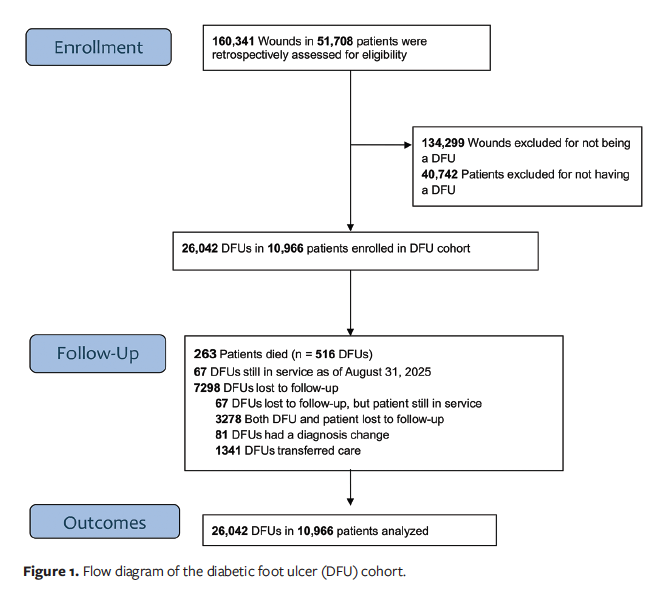
Table 1 summarizes patient demographic and clinical characteristics. The mean (SD) patient age was 63.2 (12.8) years (range, 18.8 years–90.0 years); 7 649 patients were male (69.8%) and 3 317 were female (30.2%). The majority of patients were White (n = 6 964 [63.5%]), 8.5% (n = 937) were Black or African American, and 4.7% (n = 512) identified as Hispanic or Latino. A substantial majority of patients had an abnormal body mass index (84.6% [n = 9 278]), of which 59.2% had obesity (n = 6 492). Most patients had type 2 diabetes (n = 10 267 [93.6%]). Hemoglobin A1c (HbA1c) level was documented for only 16.6% of patients (n = 1 824); for these patients, the mean HbA1c level was 8.0 (1.9) (range, 3.8–16.6). There were 10 264 patients with documented comorbidities (93.6%), excluding diabetes. Hypertension and venous, arterial, and autoimmune disease were among the most common comorbidities other than diabetes (Table 1). Of the 200 768 visits that occurred in this cohort during the study period, signs of infection were documented in the index DFU in 29.1% (n = 58 372). Additionally, osteomyelitis was diagnosed in 25.9% of DFUs (n = 2 837) and cellulitis was diagnosed in 16.8% of patients (n = 1 839) (Table 1). In addition to the wound care documented in the clinical setting, at some point in their course of care 50% of the patients received additional wound care at home.
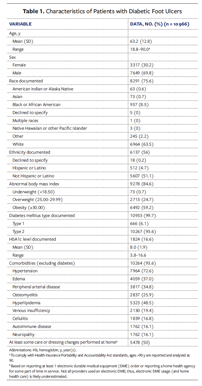
Table 2 summarizes the wound characteristics of the DFUs. The mean (SD) wound age reported by patients at their first visit was 12.4 (29.7) weeks (range, 0.1 week–521.7 weeks), although nearly 10% of DFUs had a historical duration greater than 26 weeks (n = 2 479), and 1 111 patients (4.3%) reported wound duration longer than 52 weeks. The mean greatest wound area documented was 9.0 (29.1) cm2. Importantly, 25.8% of DFUs (n = 6 703) had a Wagner grade of 3 or higher, and 2118 DFUs (8.1%) were documented as extending to bone.
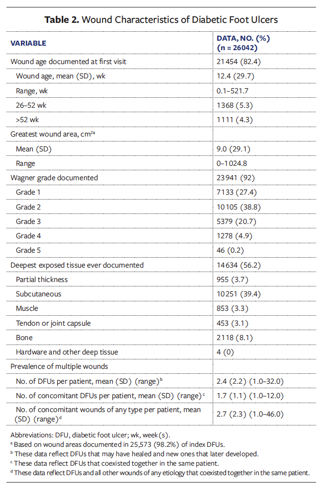
The study time frame included 2 rolling periods of 52 weeks each; during each period, patients had a mean (SD) of 4.2 (3.4) wounds of any type documented. In some cases, 1 DFU may have healed and another may have developed, although the patient may have had only 1 DFU at a time. While in service, 4 520 patients (41.2%) developed DFUs in a location different from that of the index ulcer. Patients also had multiple wounds present simultaneously, which was dubbed the “concomitant” wound count. Concomitant wounds were most often another DFU, but patients typically had wounds of several different types. On average, patients had more than 2 DFUs develop at any time during the study period, as well as an average of 2 concomitant DFUs coexisting with their index DFU. However, they also had on average nearly 3 concomitant wounds (including concomitant DFUs) of any type coexisting with the index DFU (Table 2).
Table 3 summarizes wound characteristics by Wagner grade. Whereas the mean (SD) wound age of Wagner grade 1 DFUs was 9.7 (25.6) weeks (range, 0.1 week–521.7 weeks), it increased to 15.1 (31.4) weeks (range, 0.1 week–425.7 weeks) for Wagner grade 3 DFUs. Mean DFU area increased as Wagner grade increased. Wagner grade 1 ulcers had the smallest mean area (5.0 cm²), while Wagner grade 3 ulcers averaged 12.2 cm², more than twice that of Wagner grade 1 ulcers, and Wagner grade 4 ulcers averaged 23.8 cm², nearly fivefold larger. It is notable that aggregate wound area of all concomitant wounds among these patients remained relatively consistent across Wagner grades 1 through 3 (26 cm²–27.3 cm²). Although individual Wagner grade 1 and 2 DFUs were substantially smaller than higher-grade ulcers, their combined mean area of all concomitant wounds was comparable to that of Wagner grade 3 DFUs. In contrast, patients with Wagner grade 4 ulcers demonstrated a marked increase in concomitant wound area, with a mean of 50.0 (58.8) cm² (range, 0.5 cm²–451.5 cm²).
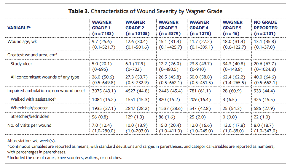
For 11 787 DFUs (45.3%), patients had impaired ambulation documented upon wound onset (Table 3). While only 318 (1.2%) of DFUs occurred in patients who were bedridden, a substantial proportion of DFUs developed in patients using a wheelchair or scooter. For Wagner grade 1 through 3 ulcers, approximately 28% of DFUs presented in patients in a wheelchair or scooter; this burden increased with increasing Wagner severity to nearly double that percentage for Wagner grade 5 DFUs (54.3%). The number of visits per wound also more than doubled, from a mean (SD) of 7.0 (12.4) visits (range, 1.0 visit–280.0 visits) for Wagner grade 1 DFUs to 15.0 (20.4) visits (range, 1.0 visit–411.0 visits) for Wagner grade 3 DFUs.
Wound and Patient Outcomes Based on Index DFUs
Table 4 summarizes the outcomes of index DFUs by Wagner grade. Most index DFUs, had wound closure documented by the clinician (62.5% [n = 16 283]), with closure reported in 71.9% of Wagner grade 1 DFUs and with that rate decreasing across Wagner grades. Wagner grade 5 involves necrosis of the foot; thus, it is surprising that 30.4% of Wagner grade 5 DFUs were documented as healed. More likely, in those cases the Wagner grade was misclassified upon documentation.
The documented 1-year mortality rate among patients in this cohort was 2.4% (n = 263 patients). However, as shown in Table 4, patient death at any time during the study occurred in 2.1% of DFUs (n = 535). This difference could be explained by the presence of multiple wounds in a single patient and by death not being documented for the particular index DFU. Furthermore, among DFUs with Wagner grade 3 or higher, the lost to follow-up rate was 32.3% to 43.5%, which was likely due to lack of reporting or documentation of some patient deaths (Table 4). The wound-related amputation rate for all DFUs was 4.9%, but this rate approached 9% for Wagner grade 3 DFUs and was highest for Wagner grade 4 DFUs (17.4%), with 16.0% of those patients undergoing major amputation (Table 4).
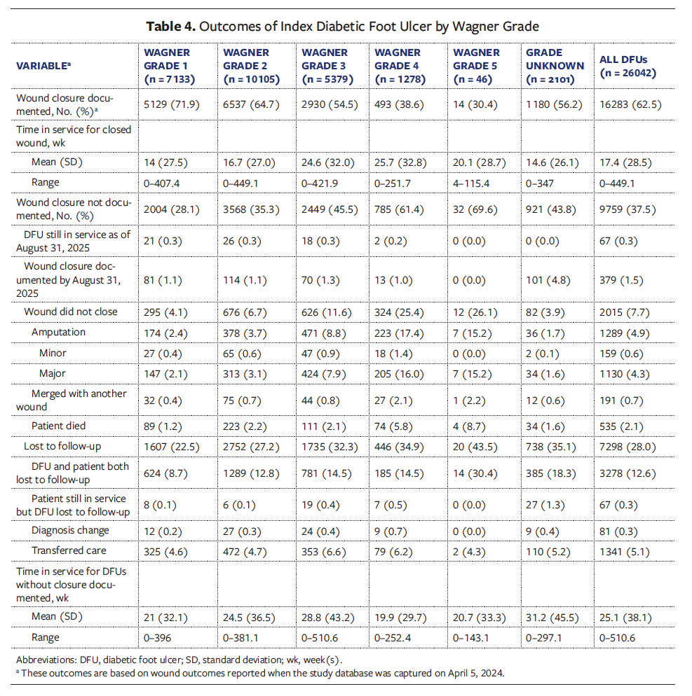
For a majority of patients (n = 6 593 [60.1%]) time in service was 52 weeks or longer, and a substantial proportion (n = 4 644 [42.3%]) were in service for 104 weeks or longer. Among wounds with documented closure, the mean (SD) time in service for all DFUs was 17.4 (28.5) weeks (range, 0 weeks–449.1 weeks) (Table 4). Mean time in service was shortest for Wagner grade 1 DFUs, at 14 (27.5) weeks (range, 0 weeks–407.4 weeks), and longest for Wagner grade 4 DFUs, at 25.7 (32.8) weeks (range, 0 weeks–251.7 weeks).
Figure 2 depicts the percentage of DFUs with wound closure documented at various time points, by Wagner grade, through 52 weeks. At 12 weeks, 39.8% of all DFUs were closed, and surprisingly, only 52.3% of Wagner grade 1 ulcers were closed at that same time point. For all grades, DFU closure rates improved with time.
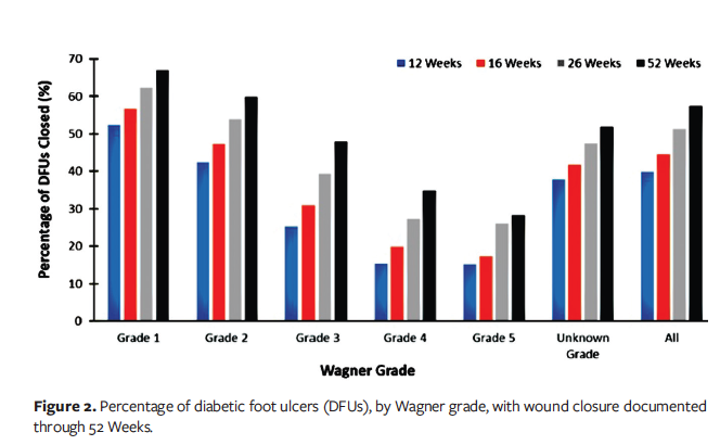
Figure 3 shows the percentage of DFUs with closure documented at various time points, by deepest exposed tissue level, through 52 weeks. At 12 weeks, the majority of partial-thickness DFUs closed (62.6%), with closure rates decreasing with deepening level of exposed tissue. Only 14% of DFUs with exposed tendon, joint capsule, or bone were closed at 12 weeks. By 52 weeks, 72.5% of partial-thickness wounds had closed, while only 35.6% of bone-exposed DFUs ever closed.
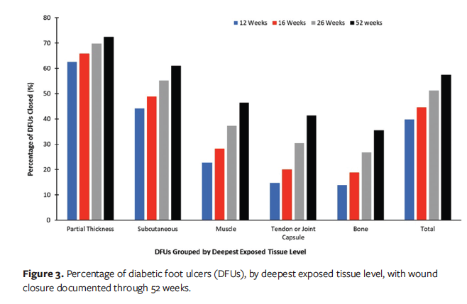
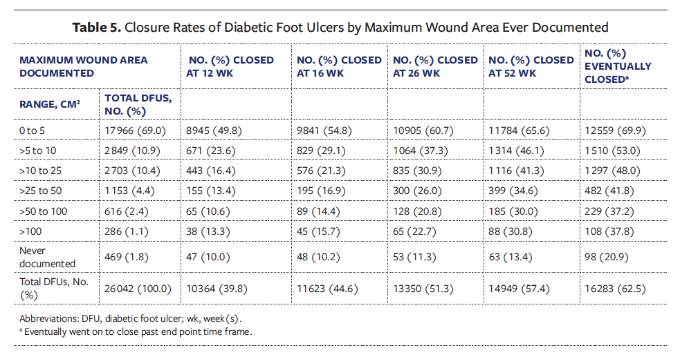
Table 5 reports the closure rates of DFUs with documented closure by maximum wound area. Interestingly, the largest DFUs (>100 cm2) consistently had higher closure rates than those between 50 cm2 and 100 cm2 in area. Closure rates were lower for larger DFUs than for smaller DFUs at all time points; interestingly, the differences in closure rates between wounds grouped by size did not vary much over time. For example, the difference in closure rates between the smallest and largest DFUs was 36.5% at 12 weeks and 34.8% at 52 weeks. A majority of the smallest DFUs closed by 16 weeks (54.8%), and 65.6% closed by 52 weeks. However, 30.1% of the smallest ulcers never healed.
Closure Rates for Multiple Wounds
Patients in the DFU cohort had multiple wounds (both other DFUs and other wound types) during the study period (Table 2), and only 38.7% of patients in the DFU cohort (n = 4 248 patients) had closure documented in all their wounds; that is, even if their DFU or DFUs healed, these patients had other wounds of different etiologies that remained unhealed. Similarly, 34.3% of patients with DFUs had wound closure documented in at least 1 wound, but not all of their wounds (n = 3 760 patients).
Patients with multiple wounds stayed in service considerably longer than the time required to close a given DFU. Among patients for whom wound closure was documented in all wounds, their time in service was more than twice that of a given index DFU (36.8 weeks; 45.9; 0–516.9). That is, even if a DFU healed, patients with multiple wounds remained in service for treatment of their remaining wounds.
The amputation rate among patients further reveals the increased morbidity in the presence of multiple wounds. The minor amputation rate among patients was 1.2% (n = 134 patients), but the major amputation rate was relatively high at 6.8% (n = 750 patients) and was much higher than the 4.9% overall amputation rate reported among index DFUs. The higher amputation rate among patients compared with index DFUs may be explained by the presence of multiple DFUs in the same limb that underwent amputation.
Case Example of a Patient Journey in Wound Care
Figure 4 shows the wound care experience of a randomly selected 46-year-old male patient with autoimmune disease, congestive heart failure, depression, diabetes, hypertension, and PAD. Per Health Insurance Portability and Accountability Act (HIPAA) requirements, calendar dates were deidentified.
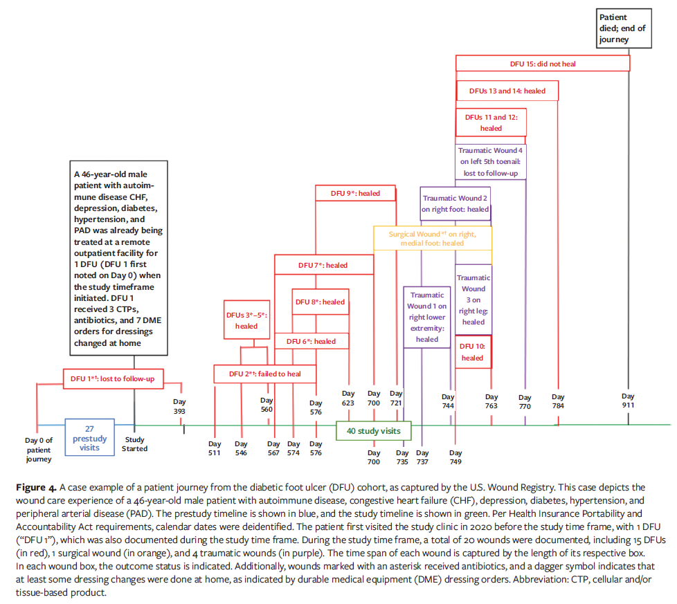
The patient first visited the study clinic in 2020 before the study time frame, at which point he had 1 DFU (labeled “DFU 1”) documented at 27 visits. After the study time frame began, he had 4 more visits for this wound, which illustrates that some DFUs in the current study cohort were already in service at study onset. DFU 1 was associated with 7 DME orders for home dressing changes and was treated with systemic antibiotics and with 3 applications of cellular and/or tissue-based products (CTPs). The outcome of DFU 1 was unknown at the patient’s last visit, but presumably, it remained open when the patient was lost to follow-up after a total of 31 visits spanning 393 days. The patient returned on day 511 when a DFU (dubbed “DFU 2” in the current study) did not close. The lesions identified as DFU 1 and DFU 2 were both located in the right plantar foot and both had the same date of onset, suggesting that they were the same lesion. The authors of the current study do not know whether, during the interval, DFU 1 healed and recurred, or whether it remained open the entire time the patient was absent from treatment. The ambiguity of determining whether a wound is new or recurring and characterizing outcome status is a common challenge in real-world clinical practice, particularly when patients’ visit schedules are erratic.
During the study time frame, 18 additional wounds were documented. The first documented DFU was the only wound that did not coexist with another wound or wounds during the study. There were 13 unambiguously new DFUs (Figure 4), none of which was treated with CTPs. There was 1 surgical wound in the right medial foot. Of the 4 traumatic wounds, the first and third were located at the right lower extremity, the second was located at the right foot, and the fourth was located at the left fifth toenail.
Figure 4 also shows the time span of each wound, whether antibiotics were received, and whether at least some dressing changes occurred at home, as indicated by DME orders. For example, surgical wound 1 was first noted on day 700, antibiotics were administered, and the wound was managed with some dressing changes at home and healed on day 763.
During the study time frame, only 3 wounds were reported to have DME orders, including DFU 1 and DFU 2 (possibly the same wound), which were the only DFUs for which some dressings were changed at home. Ten wounds (50%), including 9 separate DFUs (60%) were treated with antibiotics. Traumatic wounds 1 and 4 were the only wounds that did not undergo debridement. The patient had 40 additional visits during the study time frame (67 visits in total, spanning 721 days) before he died on day 911. Sixteen wounds healed before the patient’s death (80%), including 12 DFUs (80%). All 15 DFUs were analyzed in the DFU cohort based on data collected across 210 days at 18 separate visits.
Discussion
In this real-world, retrospective analysis of prospectively collected data, the more severe the DFU, the less likely it was to heal at any time, and the longer it took to achieve closure. As Wagner grade increased, there was a trend toward increasing wound size, wound time in service, and number of visits (Tables 3 and 4). For the entire cohort of DFUs, 6 months (26 weeks) of service was required for closure of greater than 50% of the wounds (Figure 2). Surprisingly, 38% of Wagner grade 1 DFUs remained open at 6 months (Figure 2). Similar closure rates were also observed among the smallest DFUs (0 cm2–5 cm2), with 39.3% of these open at 6 months and 30.1% never healing (Table 5). These findings align with previous RWD studies evaluating large cohorts of DFUs, which reported that approximately 25% to 50% of DFUs do not heal.18,24,28
The potentially long time frames needed to reach a DFU outcome, even for small Wagner grade 1 DFUs, has important implications in the development of DFU registries and prospective clinical trials. These data confirm that in the real world, DFU healing takes longer than the length of the typical prospective clinical trial, even among DFUs that should be the easiest to heal. However, these data suggest some explanations for the aforementioned findings. Given that ambulation status has been significantly associated with healing likelihood,11,24 the longer healing times for less severe DFUs could partially be explained by the associated ambulatory impairment, suggesting that some patients with low-grade DFUs were quite debilitated. The data in Table 3 suggest that ambulatory status seems to be largely independent of Wagner grade, being similarly distributed among Wagner grade 1 through 3 DFUs. The proportion of DFUs that presented on patients with impaired ambulation approached half.
A previous USWR RWD analysis reported that only 30.5% of DFUs healed at 12 weeks.18 In the current study, which has a larger sample size and spans a longer time frame, 39.8% of DFUs had closure documented at 12 weeks. Thus, real-world healing rates greater than 40% are not likely during the time frame of the typical RCT, even though wound clinics are known to frequently report healing rates of 95% or higher.18 In the current study, wound closure was determined by the provider-selected outcomes of “healed,” based on their preferred terms among the following choices that represented “wound closures”: “healed,” “resolved,” “goal achieved,” and “surgical closure.” This choice of terms to describe a healed wound reflects the discomfort some clinicians have in documenting a DFU as “healed,” given the rate of recurrence and new ulceration. Increasingly, researchers and clinicians prefer to use the term “remission” to describe DFU closure, recognizing that patients with closed DFUs still face the constant threat of ulcers from the underlying chronic disease of diabetes as well as their comorbidities.29-31 There is also a perverse incentive not to label a wound as healed, because dressings are covered only for open wounds under Medicare rules. If a newly closed ulceration still requires protection via dressings, then clinicians may continue to document the wound as open until the epithelium matures.
The case study in Figure 4 illustrates how difficult it may be in the real world to identify new vs recurrent ulceration. Considerable effort has been directed at understanding whether a specific DFU recurs, but little attention has been paid to the frequency with which entirely new DFUs develop. In the current study, 41.2% of patients developed a new DFU while in service for another DFU. These data emphasize that the patient’s experience of care is not represented by the course of a single DFU, but rather, as described by authors in Singapore, as a “cyclical lifelong illness threat alongside a vicious foot cycle.”30 The development of new DFUs, even while the patient is under active treatment, demonstrates that the current suite of wound treatments, nearly all of which are focused on the wound itself, do not address the underlying causal factors for ulcer development, such as diabetes, neuropathy and the loss of protective sensation, and vascular disease.29-46
The case example in the current study had 20 wounds of 3 different etiologies during a 2.5-year period. Although this may seem at first to be an extreme or atypical case, this patient is, in fact, representative of the study’s real-world dataset. Approximately 8 new wounds were documented each year in this patient, and the present dataset revealed that the included patients had on average 4 or more different wounds documented each year, and nearly 3 different concomitant wounds of any type (Table 2). The reality of multiple concomitant wounds, the development of new wounds, and the prolonged time frames needed for healing complicate the creation and implementation of payer coverage policies, especially if the documentation does not allow the reporting of the breadth of possible concomitant wounds. Within the same calendar year, when new DFUs develop or a previously closed ulcer recurs in the same anatomical location, a new treatment episode may be initiated, which has important implications to coverage limitations on utilization. However, without a way to designate on Medicare claims that these are new and different DFUs, the clinician will appear to be exceeding treatment limitations for the same ulceration.
The current study shows a stark contrast between patient-level and wound-level healing outcomes. The overall closure rate was 1.6 times higher for DFUs than for patients, reflecting that not all wounds in a patient achieved closure. Patient time in service was substantially longer than wound time in service. Previous research has shown that the outcome of any given wound is influenced by the total number of concomitant wounds.24 On average, patients in this study whose wounds all achieved closure required more than 6 months of care, approximately twice the duration observed for the closing of individual DFUs. These findings indicate that among patients with DFUs, the “wound care experience” is not adequately reflected by the trajectory of a single DFU but rather comprises the cumulative effect of multiple concomitant wounds, a conclusion with significant implications for longer closure rates and the design of clinical trials and prospective registries. This has been previously reported in other real-world studies. In a 4-year retrospective analysis, 90% of patients with DFUs had multiple wounds of various types documented; approximately half received treatment for more than 6 wounds.33
Additionally, the effect of comorbidities other than diabetes on patient and/or wound outcomes and new ulcer formation cannot be understated. The first article in this NHP series reported that in the DFU cohort, 41.2% of patients received metformin and 11.7% received insulin to manage their diabetes; 37.1% were on statins to manage cholesterol and heart disease; 33.3% received gabapentin for neuropathic pain relief; and at least 24.4% were on antibiotics.26 PAD is among the most significant comorbidities influencing DFU development, progression, and healing. PAD (along with a history of previous DFUs) significantly increases chronicity, major amputations, and mortality in patients with DFUs.34,35 In the current study, PAD and neuropathy were important comorbidities (Table 1), similar to a real-world study performed in Singapore.36 The rate of major amputation was quite similar between studies, with 6.8% of patients in the present cohort experiencing major amputation compared with 6.5% of patients in Singapore.36
The first article of this 3-part NHP series revealed that 52% of patients in the DFU cohort were younger than 65 years, the age at which most patients qualify for Medicare (unless they are classified as disabled).26 A recent analysis of Medicare claims data showed an increase in DFUs among beneficiaries younger than 65 years.2 Figure 4 illustrates the patient journey of a relatively young man (aged 46 years) with 15 predominantly overlapping DFUs (20 wounds total) and 6 comorbidities contributing to DFU development, progression, and outcome. He had both PAD and congestive heart failure, 2 comorbidities associated with increased mortality.33,34 His wound care patient journey alone lasted 2.5 years and ended with his premature death at age 48 years. The NHP real-world findings underscore the serious consequences of poor health, particularly among patients with a DFU who are younger than 65 years.
Table 2 shows that Wagner grade 1 DFUs represented only about 27% of the study cohort, with the largest percentage being Wagner grade 2 (nearly 39%) and approximately 26% being Wagner grade 3 or higher. The preponderance of the DFUs seen by wound care experts are more severe than Wagner grade 1 ulcers; this finding has important implications for clinical trial design and payer coverage policy. There is a significant need for trials to enroll DFUs more severe than Wagner grade 1 ulcers. The results in the current study indicate that DFUs often need substantially more time to heal than is typically captured in RCTs, where follow-up duration is limited by study resources.
Compared with a 2009 analysis of prospective trial generalizability,16 recent RCTs have included larger, more severe, and deeper DFUs. Table 6 summarizes the patient and wound inclusion and exclusion criteria of 10 DFU RCTs, including 9 trials published in 2024 or 2025. The trial published in 2017 evaluated Wagner grade 3 and 4 DFUs as well as multiple DFUs in the same foot,45 whereas the other 9 trials evaluated a single DFU and excluded patients with multiple wounds. Three trials included a majority of patients with Wagner grade 3 DFUs or osteomyelitis (55.4%–100%), 2 of which also included Wagner 4 DFUs and localized gangrene.37,43,44 The majority of DFUs enrolled in a 2024 trial were Wagner grade 3 (55.4%), and patients with osteomyelitis and cellulitis were included, 23.6% of whom had been treated for infection prior to randomization.38 Interestingly, a 2024 trial included Wagner grade 3 and 4 DFUs but still reported an 80% screen failure rate, likely because that trial tightly controlled for comorbidities.44 Two trials included postamputation open wounds,32,37 with these wounds comprising 51% of the sample in 1 study.32 Four trials did not impose a limit on wound area,32,41,42,45 while 1 allowed larger wounds up to 40 cm2.44 Autoimmune disease and cancer, as well as liver, heart, and renal failure, were excluded comorbidities from trials. However, some of the recent RCTs reported in Table 6 did include patients with varying degrees of PAD, neuropathy, hypertension, and venous disease, in addition to the aforementioned osteomyelitis and cellulitis, which are among the most common real-world comorbidities listed in Table 1. It is encouraging to note that DFU trials are becoming more inclusive, enrolling larger ulcers, higher Wagner grades, potentially infected DFUs, and those with a history of osteomyelitis, cellulitis, or even gangrene (Table 6). However, with the exception of various levels of PAD, comorbidities continue to be tightly controlled.
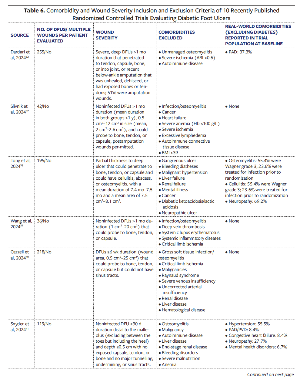
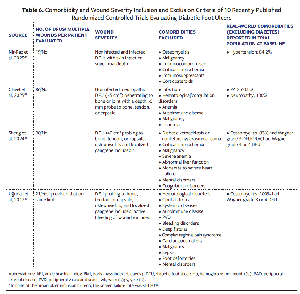
One RCT included Wagner grade 3 through 5 ulcers and open amputation wounds, with an extended evaluation period of up to 24 weeks.32 Interestingly, closure rates among DFUs treated with standard of care in that trial were nearly identical to the rates observed in Wagner grade 3 to 5 DFUs in the present cohort. At 24 weeks, approximately 37.8% of the trial DFUs treated with standard of care closed,32 while 37% of the Wagner grade 3 to 5 DFUs in the current study closed at 26 weeks. In an RCT comparing the effectiveness of 2 different applications of negative pressure wound therapy on Wagner grade 3 or 4 ulcers, long follow-up times of approximately 9 months were reported in both groups, with all of these severe ulcers healing.45
The authors of the current study applaud these trials and encourage future investigators to embrace trial designs that reflect real-world DFU severity and healing timelines and that will yield outcomes more representative of actual patient experience. Not surprisingly, trials enrolling larger and more severe wounds generally report lower overall healing rates compared with trials of superficial DFUs. Developing a mechanism to identify prospective trials with higher generalizability could help ensure that such studies are appropriately valued and that their findings are recognized as being more clinically relevant.
Limitations
This retrospective analysis has the same limitations that were reported in the first NHP article.26 At the initial wound assessment, wound age was documented according to the patient’s reported duration, which could not be independently confirmed and may be subject to recall bias. Further statistical investigation is warranted to compare DFU outcomes among patients with single vs multiple wounds of varying types, to assess the effect of comorbidities on healing at both the patient and the wound level, and to clarify whether real-world outcomes are driven primarily by wound diagnosis or by a shared underlying chronic wound-
patient phenotype.26 Previous studies have suggested that the likelihood of any 1 wound healing is inversely proportional to the total number of wounds present, a factor that is almost never taken into account when wound outcome is analyzed in isolation.24
The authors of the current study also recognize the challenges inherent to understanding RWD. Clinical outcome concepts such as “healed” are inconsistently applied and variously reported. These clinical data represent only a portion of a much larger clinical picture that may have included months or even years of prior care, subsequent care elsewhere, and hospitalizations. It is possible to link USWR clinical data to payer claims in a HIPAA-compliant fashion through tokenization to obtain a more complete understanding of the patient journey.
The data related to level of tissue exposed in Table 2 do not necessarily align with Wagner classifications, because Wagner grade 2 and 3 ulcers include the same level of tissue exposure. Wagner grade 3 criteria are more complicated than level of tissue exposure, also including osteomyelitis and pyarthrosis. In wound care research and clinical practice, not everyone agrees that the Wagner classification is most appropriate for DFU grading. Although the Wagner system is the classification system most frequently used in the literature and is widespread in practice, the International Working Group on the Diabetic Foot does not recommend the Wagner system to classify DFUs in its clinical practice guidelines.45 The major limitations of the Wagner system are that it does not address wound size, neuropathy, or PAD, and it indirectly addresses infection in deep ulcers only.46-48
In an outpatient setting, patient death is recorded only if a family member informs the clinician of the patient’s death and the clinician then records it in the EHR, or when the clinical staff contact a family member to determine the reason for a missed visit. Thus, the recorded mortality rate of patients with DFUs almost certainly underestimates the actual death rate while in active DFU treatment, and the higher rates lost to follow-up reported for higher Wagner grades are likely due at least in part to patient mortality.
Outcomes analysis in the current study considers neither the effect of ulcer location (plantar vs dorsal) nor clinical practice patterns or quality of care. For example, the current study does not analyze off-loading rates and whether they could influence DFU outcomes. Other limitations related to diabetes management in the current study are that for most patients (83.4%), HbA1c level was not documented (Table 1), and only 37% of the cohort reported statin therapy, even though it is standard of care for all individuals with diabetes older than age 40 years.48 A possible explanation may be that these data were not captured by the structured language interface of the EHR; additionally, most wound care clinicians do not directly manage diabetes and blood sugar control. This limitation harkens back to a greater challenge of coordinated care faced by real-world patients, who often see multiple providers across multiple sites of care, which is explained in the first article of this NHP series.26 Without HbA1c data, it is not possible to know whether lack of diabetes control accounts in part for poor outcomes in less severe ulcers. Future USWR research will analyze HbA1c data extracted from free text fields.
Conclusion
This second article in the NHP series highlights the relationship between ambulatory status and wound multiplicity on DFU outcome in a very large DFU cohort. Even smaller, less severe DFUs frequently do not close within 1 year in the presence of coexisting comorbidities, multiple ulcers of other types, and impaired ambulation. When multiple DFUs are present, a patient’s experience of care cannot be represented by a single wound, and closure of 1 ulcer does not equate to complete patient healing. Although highly sophisticated local treatments may facilitate healing of the index wound, there is limited effect on the overall wound care journey of a debilitated patient with multiple ulcerations. The prolonged time often required for complete DFU healing, which extends well beyond the length of RCTs, and the frequent development of new DFUs during treatment for an existing ulcer, underscore the need for more effective, perhaps systemic therapies.
It is hoped that these observations will help identify and encourage more generalizable prospective trials and serve as a framework for further discussions with the FDA and payers about the use of RWD in the treatment of patients with DFU. In addition to identifying the need for reform of clinical trial design, which will be explained further in a future article, findings from this RWD analysis represent a renewed call to action to consider a change in longitudinal clinical care and the patient journey in wound care.
Author and Public Information
Authors: Vickie R. Driver, DPM, MS1; Lucian G. Vlad, MD2; Marissa J. Carter, PhD, MA3; Joseph Rolley, BS, MSIA4; David J. Margolis, MD, PhD5;Teresa L. Z. Jones, MD6; Lisa Gould, MD, PhD7; and Caroline E. Fife, MD8,9
Affiliations: 1Elson S. Floyd College of Medicine, Washington State University, Spokane, WA, USA; 2Wake Forest University School of Medicine, Winston-Salem, NC, USA; 3Strategic Solutions, Bozeman, MT, USA; 4JTR Business Consulting, LLC, Doylestown, PA, USA; 5Department of Dermatology, University of Pennsylvania Perelman School of Medicine, Philadelphia, PA, USA; 6National Institute of Diabetes and Digestive and Kidney Diseases, National Institutes of Health, Bethesda, MD, USA; 7Warren Alpert Medical School, Brown University, Providence, RI, USA; 8Baylor College of Medicine, Houston, TX, USA; 9Intellicure, LLC, The Woodlands, TX, USA
Acknowledgments: The authors would like to thank Kristen Eckert (independent consultant) for her assistance in data analysis, drafting, and editing the manuscript, and Ben LeBoutillier (Intellicure, LLC) for his assistance with data collection and extraction.
Author Contributions: All authors contributed equally to this work.
Funding: The Wound Care Collaborative Community funded this study.
Disclosure: V.R.D. is president, chair, and a volunteer member of the Wound Care Collaborative Community (WCCC). M.J.C is a volunteer member of and consultant for the WCCC. C.E.F. is the chief medical officer of Intellicure, LLC, which received funding for this project. All remaining authors are volunteer members of the WCCC and disclose no financial or other conflicts of interest.
Ethics Statement: The Woodlands IRB (The Woodlands, TX) reviewed the study protocol and determined that secondary analysis of deidentified data was considered exempt research.
Manuscript Accepted: August 14, 2026
References
1. International Diabetes Federation (IDF). United States of America: Diabetes country report 2000–2050. IDF Atlas. 2025. Accessed September 29, 2026. https://diabetesatlas.org/data-by-location/country/united-states-of-america/
2. Carter MJ, DaVanzo J, Haught R, Nusgart M, Cartwright D, Fife CE. Chronic wound prevalence and the associated cost of treatment in Medicare beneficiaries: changes between 2014 and 2019. J Med Econ. 2023;26(1):894-901. doi:10.1080/13696998.2023.2232256
3. McDermott K, Fang M, Boulton AJM, Selvin E, Hicks CW. Etiology, epidemiology, and disparities in the burden of diabetic foot ulcers. Diabetes Care. 2023;46(1):209-221. doi:10.2337/dci22-0043
4. Armstrong DG, Swerdlow MA, Armstrong AA, Conte MS, Padula WV, Bus SA. Five year mortality and direct costs of care for people with diabetic foot complications are comparable to cancer. J Foot Ankle Res. 2020;13(1):16. doi:10.1186/s13047-020-00383-2
5. Martins-Mendes D, Monteiro-Soares M, Boyko EJ, et al. The independent contribution of diabetic foot ulcer on lower extremity amputation and mortality risk. J Diabetes Complications. 2014;28(5):632-638. doi:10.1016/j.jdiacomp.2014.04.011
6. Armstrong DG, Tan TW, Boulton AJM, Bus SA. Diabetic foot ulcers: a review. JAMA. 2023;330(1):62-75. doi:10.1001/jama.2023.10578
7. Armstrong DG, Boulton AJM, Bus SA. Diabetic foot ulcers and their recurrence. N Engl J Med. 2017;376(24):2367-2375. doi:10.1056/NEJMra1615439
8. Luo Y, Liu C, Li C, Jin M, Pi L, Jin Z. The incidence of lower extremity amputation and its associated risk factors in patients with diabetic foot ulcers: a meta-analysis. Int Wound J. 2024;21(7):e14931. doi:10.1111/iwj.14931
9. Lin C, Liu J, Sun H. Risk factors for lower extremity amputation in patients with diabetic foot ulcers: a meta-analysis. PLoS One. 2020;15(9):e0239236. doi:10.1371/journal.pone.0239236
10. Wukich DK, Raspovic KM, Suder NC. Patients with diabetic foot disease fear major lower-extremity amputation more than death. Foot Ankle Spec. 2018;11(1):17-21. doi:10.1177/1938640017694722
11. Cazzell S, Moyer PM, Samsell B, Dorsch K, McLean J, Moore MA. A prospective, multicenter, single-arm clinical trial for treatment of complex diabetic foot ulcers with deep exposure using acellular dermal matrix. Adv Skin Wound Care. 2019;32(9):409-415. doi:10.1097/01.ASW.0000569132.38449.c0
12. Armstrong DG, Lavery LA, Harkless LB. Validation of a diabetic wound classification system. The contribution of depth, infection, and ischemia to risk of amputation. Diabetes Care. 1998;21(5):855-859. doi:10.2337/diacare.21.5.855
13. Oyibo SO, Jude EB, Tarawneh I, Nguyen HC, Harkless LB, Boulton AJ. A comparison of two diabetic foot ulcer classification systems: the Wagner and the University of Texas wound classification systems. Diabetes Care. 2001;24(1):84-88. doi:10.2337/diacare.24.1.84
14. Chen L, Sun S, Gao Y, Ran X. Global mortality of diabetic foot ulcer: a systematic review and meta-analysis of observational studies. Diabetes Obes Metab. 2023;25(1):36-45. doi:10.1111/dom.14840
15. Lin C, Tian J, Zhang Z, Zheng C, Liu J. Risk factors associated with the recurrence of diabetic foot ulcers: a meta-analysis. PLoS One. 2025;20(2):e0318216. doi:10.1371/journal.pone.0318216
16. Carter MJ, Fife CE, Walker D, Thomson B. Estimating the applicability of wound care randomized controlled trials to general wound-care populations by estimating the percentage of individuals excluded from a typical wound-care population in such trials. Adv Skin Wound Care. 2009;22(7):316-324. doi:10.1097/01.ASW.0000305486.06358.e0
17. Fortin M, Dionne J, Pinho G, Gignac J, Almirall J, Lapointe L. Randomized controlled trials: do they have external validity for patients with multiple comorbidities?. Ann Fam Med. 2006;4(2):104-108. doi:10.1370/afm.516
18. Fife CE, Eckert KA, Carter MJ. Publicly reported wound healing rates: the fantasy and the reality. Adv Wound Care (New Rochelle). 2018;7(3):77-94. doi:10.1089/wound.2017.0743
19. Eckert KA, Fife CE, Carter MJ. The impact of underlying conditions on quality-of-life measurement among patients with chronic wounds, as measured by utility values: a review with an additional study. Adv Wound Care (New Rochelle). 2023;12(12):680-695. doi:10.1089/wound.2023.0098
20. Serena TE, Fife CE, Eckert KA, Yaakov RA, Carter MJ. A new approach to clinical research: integrating clinical care, quality reporting, and research using a wound care network-based learning healthcare system. Wound Repair Regen. 2017;25(3):354-365. doi:10.1111/wrr.12538
21. Wigston C, Hassan S, Turvey S, et al. Impact of medications and lifestyle factors on wound healing: a pilot study. Wounds UK. 2013;9(1):22-28.
22. Beitz JM. Pharmacologic impact (aka “breaking bad”) of medications on wound healing and wound development: a literature-based overview. Ostomy Wound Manage. 2017;63(3):18-35.
23. Bennett G, Abbott J, Sussman G. The negative impact of medications on wound healing. Wound Practice and Research: Journal of the Australian Wound Management Association. 2024;32(1):17-24. doi:10.33235/wpr.32.1.17-24
24. Fife CE, Horn SD, Smout RJ, Barrett RS, Thomson B. A predictive model for diabetic foot ulcer outcome: the wound healing index. Adv Wound Care (New Rochelle). 2016;5(7):279-287. doi:10.1089/wound.2015.0668
25. U.S. Food and Drug Administration. Use of real-world evidence to support regulatory decision-making for medical devices. Guidance for industry and Food and Drug Administration staff. December 18, 2025. Accessed January 16, 2026. https://www.fda.gov/media/190201/download
26. Driver VR, Vlad LG, Carter MJ, et al. Is there a chronic wound-patient phenotype? A retrospective analysis of real-world overlap of diabetic foot and venous leg ulcers. Wounds. 2026;38(10):251-261. doi:10.25270/wnds/26031.
27. World Health Organization. International Statistical Classification of Diseases and Related Health Problems. 10th rev. World Health Organization;2016. Accessed September 14, 2026. who.int
28. Prompers L, Schaper N, Apelqvist J, et al. Prediction of outcome in individuals with diabetic foot ulcers: focus on the differences between individuals with and without peripheral arterial disease. The EURODIALE Study. Diabetologia. 2008;51(5):747-755. doi:10.1007/s00125-008-0940-0
29. Fernando ME, Blanchette V, Woelfel SL, et al. Prescribing home digital thermometry coupled with activity dosing and optimized offloading to prolong diabetic foot remission: a case report. Int J Low Extrem Wounds. 2026;25(2):629-635. doi:10.1177/15347346231184008
30. Zhu X, Lee ES, Lim PXH, Chan FHF, Chen YC, Griva K. “It is a constant battle”: a qualitative study of the lived experience of individuals with diabetic foot ulcer and post-healing challenges in primary care. Diabet Med. 2025;42(9):e70096. doi:10.1111/dme.70096
31. Bouly M, Laborne FX, Tourte C, Henry E, Penfornis A, Dardari D. Post-healing follow-up study of patients in remission for diabetic foot ulcers Pied-REM study. PLoS One. 2022;17(5):e0268242. doi:10.1371/journal.pone.0268242
32. Dardari D, Piaggesi A, Potier L, et al. Intact fish skin graft to treat deep diabetic foot ulcers [published correction appears in NEJM Evid. 2025;4(3):EVIDx2500032.]. NEJM Evid. 2024;3(12):EVIDoa2400171. doi:10.1056/EVIDoa2400171
33. Raspovic KM, Wukich DK, Naiman DQ, et al. Effectiveness of viable cryopreserved placental membranes for management of diabetic foot ulcers in a real world setting. Wound Repair Regen. 2018;26(2):213-220. doi:10.1111/wrr.12635
34. Meloni M, Izzo V, Giurato L, Lázaro-Martínez JL, Uccioli L. Prevalence, clinical aspects and outcomes in a large cohort of persons with diabetic foot disease: comparison between neuropathic and ischemic ulcers. J Clin Med. 2020;9(6):1780. doi:10.3390/jcm9061780
35. Sayiner ZA, Can FI, Akarsu E. Patients’ clinical characteristics and predictors for diabetic foot amputation. Prim Care Diabetes. 2019;13(3):247-251. doi:10.1016/j.pcd.2018.12.002
36. Lo ZJ, Surendra NK, Saxena A, Car J. Clinical and economic burden of diabetic foot ulcers: a 5-year longitudinal multi-ethnic cohort study from the tropics. Int Wound J. 2021;18(3):375-386. doi:10.1111/iwj.13540
37. Slivnik M, Navodnik Preložnik M, Fir M, et al. A randomized, placebo-controlled study of chitosan gel for the treatment of chronic diabetic foot ulcers (the CHITOWOUND study). BMJ Open Diabetes Res Care. 2024;12(3):e004195. doi:10.1136/bmjdrc-2024-004195
38. Tong J, Zhang J, Xiang L, et al. Continuous intrafemoral artery infusion of urokinase improves diabetic foot ulcers healing and decreases cardiovascular events in a long-term follow-up study. BMJ Open Diabetes Res Care. 2024;12(1):e003414. doi:10.1136/bmjdrc-2023-003414
39. Wang G, Li X, Ju S, et al. Effect of electrospun poly (L-lactide-co-caprolactone) and formulated porcine fibrinogen for diabetic foot ulcers. Eur J Pharm Sci. 2024;198:106800. doi:10.1016/j.ejps.2024.106800
40. Cazzell SM, Caporusso J, Vayser D, Davis RD, Alvarez OM, Sabolinski ML. Dehydrated amnion chorion membrane versus standard of care for diabetic foot ulcers: a randomised controlled trial. J Wound Care. 2024;33(Sup7):S4-S14. doi:10.12968/jowc.2024.0139
41. Snyder R, Nouvong A, Ulloa J, et al. Efficacy and safety of autologous whole blood clot in diabetic foot ulcers: a randomised controlled trial. J Wound Care. 2024;33(9):688-700. doi:10.12968/jowc.2024.0195
42. Nir-Paz R, Onallah H, Dekel M, et al. Randomized double-blind study on safety and tolerability of TP-102 phage cocktail in patients with infected and non-infected diabetic foot ulcers. Med. 2025;6(5):100565. doi:10.1016/j.medj.2024.11.018
43. Clavel S, Denizot C, Boëzennec B, Turzi A, Albache N. A randomised controlled clinical study comparing the efficacy and safety of an autologous standardised leukocyte-poor platelet gel with standard care for the treatment of chronic neuropathic diabetic foot ulcers. Int Wound J. 2025;22(4):e70495. doi:10.1111/iwj.70495
44. Sheng X, Hu L, Li T, et al. Clinical efficacy and mechanism of the combination of autologous platelet-rich gel and recombinant human acidic fibroblast growth factor in the management of refractory diabetic foot. Front Endocrinol (Lausanne). 2024;15:1374507. doi:10.3389/fendo.2024.1374507
45. Uğurlar M, Sönmez MM, Armağan R, Eren OT. Comparison of two different vacuum-assisted closure (VAC) treatments of multiple chronic diabetic foot wounds in the same extremity. Foot Ankle Surg. 2017;23(3):173-178. doi:10.1016/j.fas.2016.05.314
46. Monteiro-Soares M, Hamilton EJ, Russell DA, et al. Guidelines on the classification of foot ulcers in people with diabetes (IWGDF 2023 update). Diabetes Metab Res Rev. 2024;40(3):e3648. doi:10.1002/dmrr.3648
47. Stang D, Young M. Selection and classification of a diabetic foot ulcer classification in Scotland: part 2. The Diabetic Foot Journal. 2018;21(2):100-106.
48. American Diabetes Association Professional Practice Committee for Diabetes*. 10. Cardiovascular Disease and Risk Management: Standards of Care in Diabetes-2026. Diabetes Care. 2026;49(Suppl 1):S216-S245. doi:10.2337/