Efficacy of Autologous Blood-Derived Therapies in Diabetic Foot Ulcers: A Systematic Review and Meta-Analysis of Randomized Controlled Trials
© 2026 HMP Global. All Rights Reserved.
Any views and opinions expressed are those of the author(s) and/or participants and do not necessarily reflect the views, policy, or position of Wounds or HMP Global, their employees, and affiliates.
Abstract
Background. Diabetic foot ulcers (DFUs) are a significant complication of diabetes, leading to high morbidity rate, amputation, and health care costs. Autologous blood-derived products, including autologous whole blood clot (AWBC), platelet-rich plasma, and platelet-rich fibrin, have emerged as promising treatments that leverage the body’s own healing mechanisms. Objective. To evaluate the efficacy of autologous blood-derived products compared with standard-of-care (SOC) treatment in achieving complete healing of DFUs by 12 weeks. Methods. A systematic review and meta-analysis of sources published through April 2025 was performed following PRISMA (Preferred Reporting Items for Systematic Reviews and Meta-Analyses) guidelines. PubMed, Embase, and the Cochrane Library were searched. Randomized controlled trials (RCTs) reporting complete healing at 12 weeks were included. Data were pooled using a random effects model to calculate odds ratio (OR) with 95% CI. Results. Six RCTs with a total of 629 patients met the inclusion criteria. Autologous blood-derived products significantly improved the odds of complete healing at 12 weeks compared with SOC (pooled OR, 2.24; 95% CI, 1.47-3.41; P = .0045). Among these products, AWBC demonstrated the highest efficacy, and there was minimal indication of publication bias. Conclusion. Autologous blood-derived products significantly improve healing outcomes in DFUs compared with SOC, supporting their clinical use. Further research should refine product preparation methods and treatment durations to maximize efficacy.
Diabetic foot ulcers (DFUs) are a growing health concern, affecting approximately 18.6 million people worldwide and 1.6 million in the United States each year.1 DFUs are associated with major complications, including amputations and increased mortality.2,3 Patients with DFU have a 2.5-fold higher mortality rate compared with those without DFU,4 with approximately 10% of patients with DFU dying within 1 year of diagnosis.3,5 Approximately 20% of patients with DFU will require amputation, ranging from minor below-ankle procedures to major above-ankle amputations, or both,2 which contributes significantly to the financial burden on the health care system. The direct cost for treating DFUs in the United States is estimated to be in 2018 between $9 and $13 billion annually,6 making DFUs the second most expensive wound type covered by Medicare.7
A meta-analysis estimated a worldwide DFU prevalence of 6.3%, while 19–34% of individuals with diabetes are expected to develop a DFU during their lifetime. As the global diabetes population is projected to increase from 463 million in 2019 to 700 million by 2045, the burden of DFUs is expected to rise substantially in the coming decades.1,8-9 With the increased incidence worldwide of diabetes and, subsequently, of DFUs, these wounds have become an urgent health problem.8 US Wound Registry real-world data indicate that 70% of patients with DFUs do not achieve healing within 12 weeks of receiving standard wound care.10 Current first-line treatments focus on moisture and infection control, regular debridement, glucose monitoring, and appropriate wound dressings.11 These poor outcomes and the need for more effective therapies has led to the emergence of autologous blood-derived products, including autologous whole blood clot (AWBC), platelet-rich plasma (PRP), and platelet-rich fibrin (PRF). These technologies utilize the patient’s own blood to create a therapeutic dressing designed to restore the wound’s natural healing cascade.12,13 The efficacy of autologous blood-derived products in managing DFUs and enhancing healing rates by week 12 has been established in clinical trials.13-18 To further assess the effect of these therapies, the authors of the present review conducted a meta-analysis of all relevant randomized controlled trials (RCTs) with the aim of providing robust evidence supporting their clinical effectiveness.
Methods
Literature Search and Study Selection
A systematic literature search was conducted in accordance with the PRISMA (Preferred Reporting Items for Systematic Reviews and Meta-Analyses) guidelines using 3 databases: PubMed, Embase, and the Cochrane Library. The search for literature published through April 2025 used the terms “diabetic foot ulcer,” “autologous,” and “randomized controlled trial.” Two reviewers (RJS, SC) independently screened the titles and abstracts of all identified articles. Any disagreements regarding a manuscript’s eligibility were resolved through consultation with a third reviewer (SS).
Eligibility Criteria
The inclusion criteria were as follows: (1) studies involving only DFU, (2) RCT design, (3) treatment groups receiving autologous blood-derived products, (4) control groups receiving conventional treatment only, (5) outcomes data reporting complete healing at 12 weeks, (6) full text available, and (7) articles published in English. Exclusion criteria included the following: (1) studies involving combined treatments, (2) retrospective studies, (3) retracted articles, and (4) studies lacking outcomes data.
Data Extraction
Data extracted from the search were entered into an electronic spreadsheet, capturing the full article title, first author’s name, year of publication, and reason for inclusion or exclusion. For studies included in the analysis, additional data were collected, including the number of patients in the intention-to-treat population, duration of active treatment, standard of care (SOC) used in the control group (ie, off-loading, debridement, wound cleansing, moisture and infection control, and the application of wound dressings [eg, petrolatum-based, alginate, hydrocolloid, semiocclusive]), outcome measures, wound duration, and ulcer size. Treatments involving advanced technologies, including hyperbaric oxygen therapy, negative pressure wound therapy, and advanced dressings such as cellular and/or tissue-based products were not considered part of SOC and were excluded from the final analysis.
Statistical Analysis
The meta-analysis used a random effects model because of the initial assumption of heterogeneity in results between the 6 studies that ultimately were selected. The odds ratio (OR) (blood-derived product vs control) in favor of healing was calculated for each of the studies and then pooled using the Mantel-Haenszel method. CIs for each individual study and overall based on the normal distribution approximation were computed. Assessment of the homogeneity of the studies was performed using the Cochran Q test. In addition, the I2 index for the quantification of heterogeneity was determined by the method of Higgins and Thompson.19 A CI for I2 was calculated using the method of Higgins et al.20
Analysis was performed using data on complete healing, which was defined as 100% reepithelialization of the wound surface in the absence of drainage or discharge and without the need for wound dressings. For all analyses, a P value of less than .05 was considered to be significant.
Results
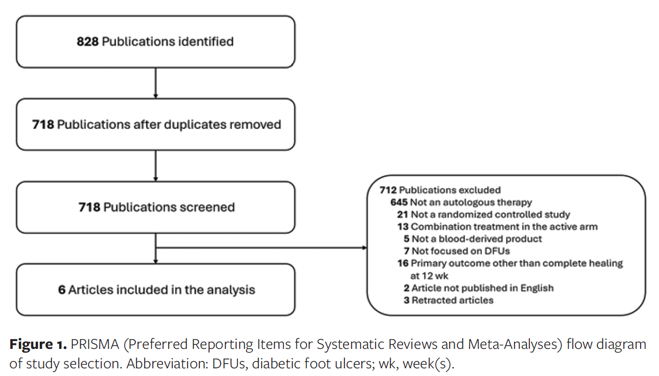
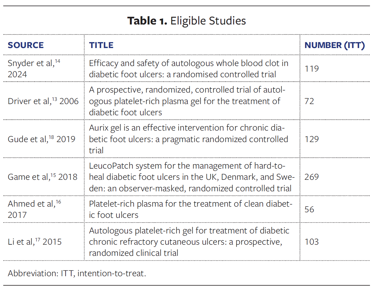
The initial search identified 828 studies, of which 110 were duplicates. Of the remaining studies, 645 were excluded because they investigated treatments that were not autologous. An additional 67 articles were excluded based on the study inclusion and exclusion criteria: 21 were not RCTs, 13 involved combination treatments in the treatment groups, 5 did not use blood-derived products, 7 did not focus on DFUs, 16 assessed outcomes other than complete healing at 12 weeks, 2 were not published in English, and 3 were retracted (Figure 1). Ultimately, 6 articles met the inclusion criteria and were included in the final analysis. These articles are detailed in Table 1.
A meta-analysis was conducted to evaluate wound healing outcomes at 12 weeks after the initiation of treatment with autologous blood-derived products. Only RCTs comparing these products with SOC treatment and reporting complete healing rates at 12 weeks were considered. The 6 studies included in the analysis were Snyder et al,14 Ahmed et al,16 Driver et al,13 Li et al,17 Gude et al18 (limited to patients whose control group received SOC), and Game et al,15 with analysis focused on the 12-week data. The products used in these studies included AWBC, PRP, autologous platelet-rich gel, and PRF.
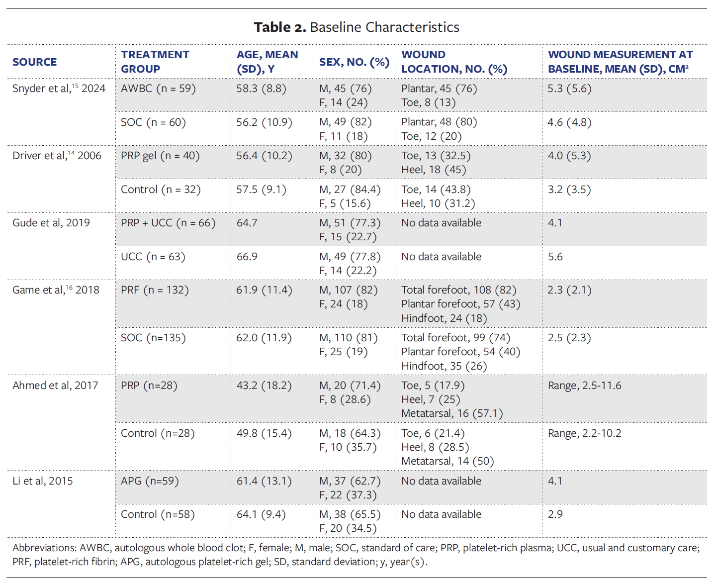
In the analyzed sources, a total of 629 patients were enrolled to receive either an autologous blood product or a control treatment. Most patients were male (92%), with mean age ranging from 43.2 years to 66.9 years across the studies. All wounds were chronic (>28 days), with mean wound size at baseline ranging from 2.2 cm² to 5.6 cm² (Table 2). Wound size assessment varied across the included studies. Snyder et al14 and Li et al17 used validated planimetric digital measurement systems, Gude et al18 and Driver et al13 estimated wound area using linear measurements (length × width), and Game et al15 and Ahmed et al16 did not report the methods used for wound measurement. The wounds included in the current study were predominantly classified as Wagner grade 1.13-16 Wagner grades 2 and 3 were reported in 2 studies.17-18 However, these publications did not provide exact distributions of wound counts across Wagner grade categories.
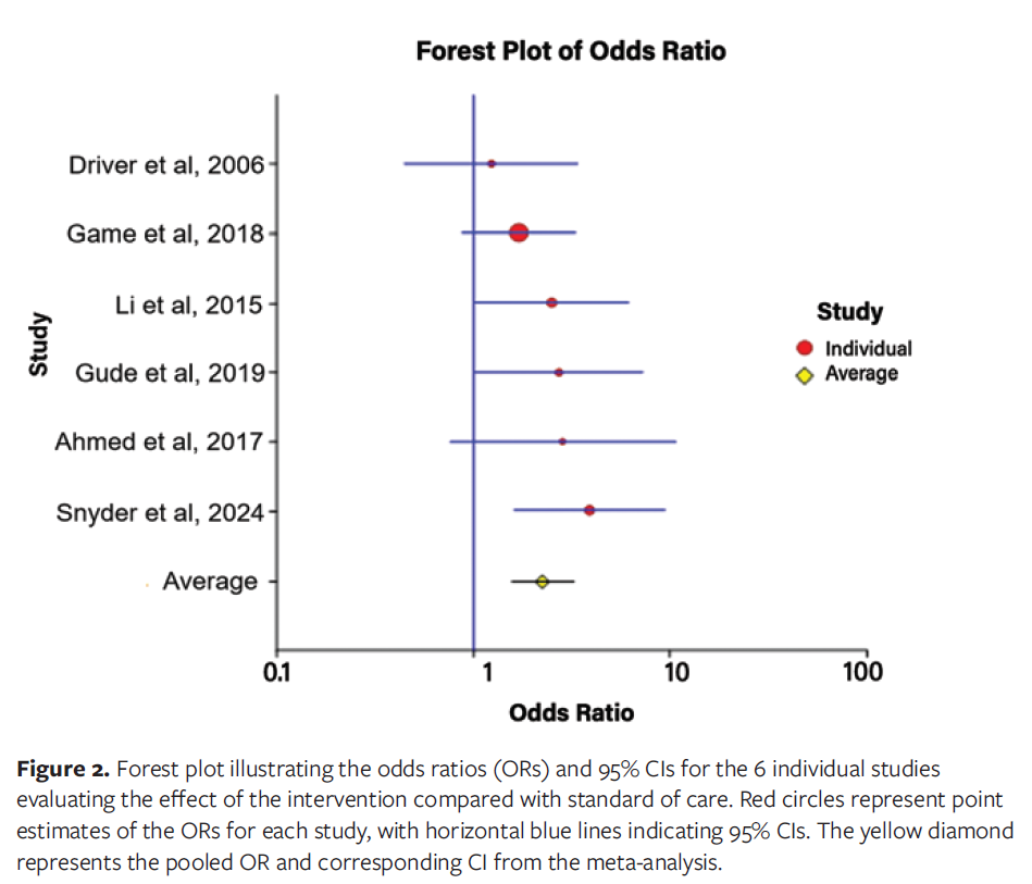
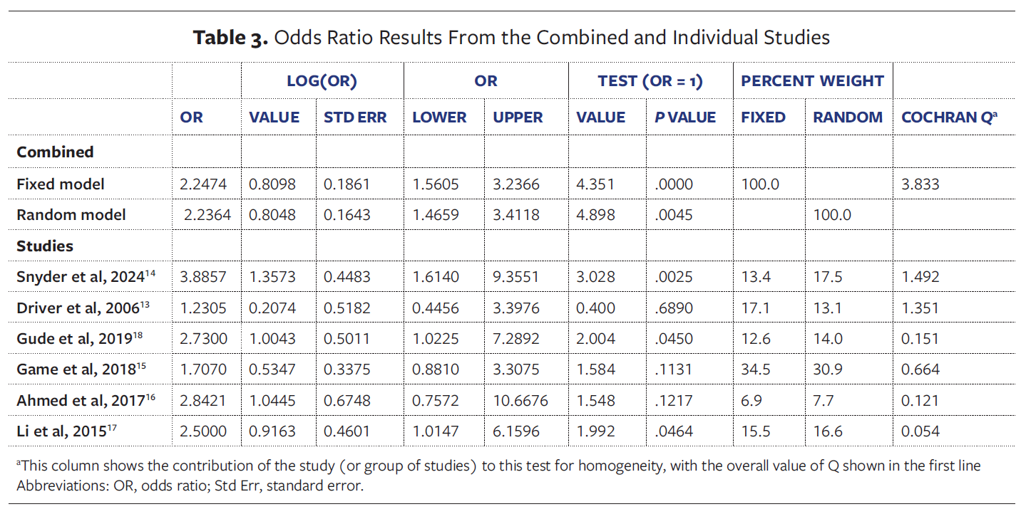
The results of the current study demonstrate that autologous blood-derived products show significantly greater efficacy compared with SOC treatment in DFUs. The ORs across the included studies range from 1.23 in Driver et al13 to 3.89 in Snyder et al,14 with a pooled OR of 2.24 (95% CI, 1.47-3.41; P = .0045) (Figure 2). It is important to note that the authors of the current study used only the reported 12-week efficacy results from Game et al,15 which were substantially lower than the 20-week outcomes (the primary end point of that study). Additionally, concerning the study by Gude et al, the authors of the current study only used data from the control group that received SOC treatment (excluding advanced therapies), based on the reported information.
Cochran Q test of homogeneity yielded a value of 3.833, which was not statistically significant (P = .57). In addition, the I2 index was 0.000 (95% CI, 0.000-66.895), further supporting the lack of heterogeneity in results between the studies; however, it is important to note that with only 6 studies, there is limited power to detect heterogeneity. Nonetheless, a fixed model could have been used; however, as shown in Table 3, it would not have resulted in a different estimate of the combined OR (OR, 2.24; 95% CI, 1.56-3.24; P < .0001).
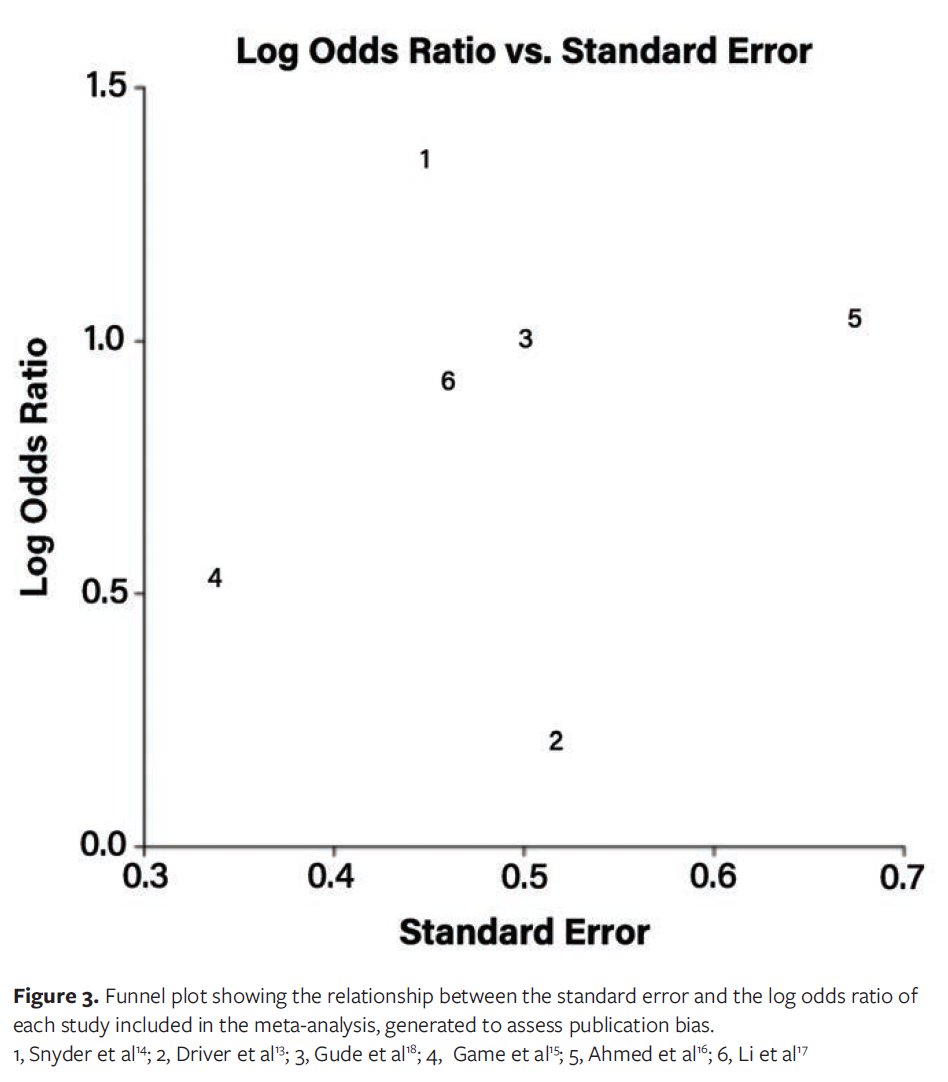
Although only 6 studies were included in the analysis, the distribution of points in the funnel plot appears roughly symmetrical, suggesting a low likelihood of publication bias (Figure 3). However, with so few studies available, a single unpublished negative study could adversely affect this plot.
Discussion
Autologous blood-derived products offer a potential unique advantage by delivering the body’s physiological healing cascade directly into the wound bed. By providing a rich supply of growth factors and cytokines, the autologous blood-derived products form a scaffold, and support cell migration, tissue regeneration, and extracellular matrix (ECM) reconstruction.21-23 This balanced microenvironment actively promotes and accelerates healing. Additionally, the autologous nature of these products carries significantly reduced risk of disease transmission and immune rejection, making them both safe and highly biocompatible.
The present study demonstrates that autologous blood-derived products are highly effective in treating DFUs compared with SOC. The 3 main products in this category—PRP, PRF, and AWBC—have been used as treatments for DFUs. Although all 3 products are derived from the patient’s own blood, they differ in their preparation methods and composition.
PRP is prepared by drawing whole blood into a tube containing anticoagulants, followed by centrifugation steps to concentrate the platelets. However, the procedure lacks standardization, leading to variability in product composition.24 In contrast, PRF preparation does not require anticoagulants or external activators. The blood is drawn and subjected to a single centrifugation step, resulting in 3 layers, 1 of which is the PRF matrix used for treatment.25
Unlike PRP and PRF, the preparation of AWBC does not involve centrifugation. Instead, whole blood is placed into an activation mold that contains clotting factors, which accelerates the clotting process and produces a blood clot within a few minutes.14 Whereas PRP and PRF focus on concentrating activated platelets, which are widely recognized as major contributors to the wound healing process, AWBC incorporates all the blood components, including all activated platelets, in its final product.
Platelets, which are a common component in all the blood-derived products, play a central role in regulating hemostasis and thrombosis.26 When activated, platelets secrete growth factors such as platelet-derived growth factor (PDGF) and epidermal growth factor (EGF), which are vital to wound healing because they promote the production of collagen, glycosaminoglycans, and proteoglycans by fibroblasts.27-29 Together with fibrin, the scaffold formed by platelets delivers a wide array of growth factors, such as vascular endothelial growth factor, PDGF, basic fibroblast growth factor, and EGF, into the wound bed. These factors play crucial roles in the wound healing cascade, supporting reepithelialization, dermal maturation, and cellular proliferation.28 Researchers have found that in blood-derived products, the factors stored within platelets play a key role in influencing the molecular mechanisms and biological processes essential for wound healing.23,30,31
Although there is a focus on platelets when looking at PRP products, the optimal platelet concentration remains a topic of debate. Some studies suggest that a minimum concentration of 1 000 000 platelets/µL is required to achieve therapeutic effects,32 but others caution that excessively high levels of secreted growth factors and proteins may lead to abnormal cell proliferation or dysregulated signaling, potentially disrupting normal tissue repair.33 The optimal platelet concentration for PRP has not been clearly defined. The concentration of growth factors can vary depending on the preparation method and platelet levels, and these variations may significantly affect the efficacy of PRP in healing DFUs.31
In contrast to PRP, which contains high concentrations of platelets, PRF also includes elevated levels of host immune cells.22 During PRF preparation a dense fibrin network forms, allowing for the slow and sustained release of growth factors into the surrounding tissue.34 Unlike PRP, PRF is produced using a standardized protocol.21
Different from both PRP and PRF, the AWBC preparation technique involves no separation of blood components; the final product retains all elements of whole blood, including activated platelets, leukocytes, red blood cells, fibrin, and plasma. Each of these components has been shown to contribute to the healing process by promoting the secretion of growth factors essential for wound repair.23 When examining the mechanism of action of AWBC, the combination of various blood cells and the fibrin mesh, which mimics the properties of the ECM, appears to closely replicate the natural healing cascade.23 This may explain why, in the current study, AWBC demonstrated the highest OR among the blood-derived products analyzed. However, the authors of the current study acknowledge that the 95% CI for the OR for healing with AWBC versus control in Snyder et al14 (95% CI, 1.61-9.36) overlaps that for the pooled results for the PRP and PRF preparations (95% CI, 1.33-2.98); thus, there is no meaningful statistical evidence for a difference between these approaches.
The current analysis clearly demonstrates the advantage of autologous blood-derived products over SOC in treating DFUs. The 6 studies analyzed included a total of 629 subjects and yielded a combined OR of 2.24. Of these studies, Snyder et al15 (AWBC) demonstrates the highest OR at 3.88 (as a point estimate).
A review of baseline characteristics across all 6 studies shows that Snyder et al14 has relatively larger baseline wound areas compared with most other included studies, although direct comparisons are limited because wound size was reported using different summary measures across studies (AWBC, 5.3 [5.6] cm² vs SOC, 4.6 [4.8] cm²), that may highlight the strength of those findings. It is worth noting that only 2 studies, Snyder et al14 and Ahmed et al,16 reported mean wound duration prior to treatment. As a result, it is not possible to assess whether wound age influenced the healing outcomes across all 6 studies. However, the data from these 2 studies suggest variability in wound chronicity: Snyder et al14 reported a longer mean wound duration of 78.8 weeks in the AWBC group and 45.3 weeks in the control group, whereas Ahmed et al16 reported mean durations of 11.5 weeks in the PRP group and 12.5 weeks in the control group.
This variability in wound duration complements the considerable heterogeneity observed in wound severity across the included studies. While 4 studies, including Snyder et al,14 primarily evaluated Wagner grade 1 wounds, Gude et al18 and Li et al17 included patients with more advanced Wagner grades 2 and 3 wounds. Both wound chronicity and severity can influence treatment efficacy; thus, studies focusing on Wagner grade 1 wounds may report higher efficacy rates compared with studies that include more chronic or advanced wounds. The current analysis focused on outcomes reported after 12 weeks of treatment. It is worth noting that Game et al15 assessed their primary end point at 20 weeks (OR, 1.58), with a healing rate of 34% in the PRP group compared with 22% in the SOC group. However, when examining the 12-week time point used in the current analysis, their data showed a lower healing rate of 20% for PRP versus 13% for SOC.16 Moreover, inclusion of the 20-week end point in the pooled analysis would likely have resulted in a greater overall treatment effect for autologous blood-derived products compared with SOC. This suggests a significant benefit to extending treatment beyond 12 weeks, as reflected in improved patient outcomes. Interestingly, Snyder et al14 also noted that it is highly likely that approximately 11.8% of the wounds that were not healed at 12 weeks with AWBC would have healed with additional applications.
Limitations
Although this meta-analysis provides robust evidence supporting the efficacy of autologous blood-derived products in the treatment of DFUs, several limitations must be acknowledged. Variations in study design, intervention protocols (eg, differences in PRP, PRF, and AWBC preparation techniques), and patient populations may introduce bias and affect comparability across studies. Additionally, while this analysis focused on healing outcomes at 12 weeks, some studies reported primary end points at later time points, which may underestimate the long-term benefits of these treatments.
Conclusion
This meta-analysis highlights the clinical effectiveness of autologous blood-derived products in enhancing wound healing in DFUs compared with SOC. The pooled analysis of 6 RCTs involving 629 patients demonstrates a significant improvement in complete healing rates at 12 weeks, with AWBC showing the highest OR among the evaluated products. These findings support the integration of autologous therapies into clinical practice for DFU management, especially in patients who do not respond adequately to conventional care. The analysis also underscores the potential importance of extending treatment duration beyond 12 weeks, as evidenced by improved outcomes in studies with longer follow-up periods. Given the growing prevalence of diabetes and the substantial clinical and economic burden of DFUs, autologous blood products present a promising advanced solution. Further research is needed to refine product preparation methods and treatment duration to maximize efficacy.
Author and Public Information
Authors: Robert J. Snyder, DPM1 and Martin Lee, PhD2
Affiliations: 1Centre for Medical Education, Cardiff University School of Medicine, Cardiff, Wales, UK; 2UCLA Fielding School of Public Health, Los Angeles, CA
Acknowledgement: The authors thank Shira Cohen and Sharon Sirota (RedDress Medical) for assistance with the literature review and manuscript preparation.
Disclosure: The authors disclose no financial or other conflicts of interest.
Ethics Statement: Ethical approval was not required for systematic review and meta-analysis.
Correspondence: Robert J. Snyder, DPM; Cardiff University School of Medicine, c/o 11170 Watercrest Circle East, Parkland, FL, 33076; drwound@aol.com
Manuscript Accepted: May 19, 2026
References
- Armstrong DG, Tan TW, Boulton AJM, Bus SA. Diabetic Foot Ulcers: A Review. JAMA. 2023 Jul 3;330(1):62-75. doi:10.1001/jama.2023.10578 PMID: 37395769; PMCID: PMC10723802.
- Armstrong DG, Boulton AJM, Bus SA. Diabetic foot ulcers and their recurrence. N Engl J Med. 2017;376(24):2367-2375. doi:10.1056/NEJMra1615439
- Hoffstad O, Mitra N, Walsh J, Margolis DJ. Diabetes, lower-extremity amputation, and death. Diabetes Care. 2015;38(10):1852-1857. doi:10.2337/dc15-0536
- Walsh JW, Hoffstad OJ, Sullivan MO, Margolis DJ. Association of diabetic foot ulcer and death in a population-based cohort from the United Kingdom. Diabet Med. 2016;33(11):1493-1498. doi:10.1111/dme.13054
- Meloni M, Izzo V, Giurato L, Lázaro-Martínez JL, Uccioli L. Prevalence, clinical aspects and outcomes in a large cohort of persons with diabetic foot disease: comparison between neuropathic and ischemic ulcers. J Clin Med. 2020;9(6):1780. doi:10.3390/jcm9061780
- Rice JB, Desai U, Cummings AKG, Birnbaum HG, Skornicki M, Parsons NB. Burden of diabetic foot ulcers for Medicare and private insurers. Diabetes Care. 2014;37(3):651-658. doi:10.2337/dc13-2176
- Nussbaum SR, Carter MJ, Fife CE, et al. An economic evaluation of the impact, cost, and Medicare policy implications of chronic nonhealing wounds. Value Health. 2018;21(1):27-32. doi:10.1016/j.jval.2017.07.007
- Zhang P, Lu J, Jing Y, Tang S, Zhu D, Bi Y. Global epidemiology of diabetic foot ulceration: a systematic review and meta-analysis †. Ann Med. 2017 Mar;49(2):106-116. doi:10.1080/07853890.2016.1231932
- Saeedi P, Petersohn I, Salpea P, Malanda B, Karuranga S, Unwin N, Colagiuri S, Guariguata L, Motala AA, Ogurtsova K, Shaw JE, Bright D, Williams R; IDF Diabetes Atlas Committee. Global and regional diabetes prevalence estimates for 2019 and projections for 2030 and 2045: Results from the International Diabetes Federation Diabetes Atlas, 9th edition. Diabetes Res Clin Pract. 2019 Nov;157:107843. doi:10.1016/j.diabres.2019.107843
- Fife CE, Eckert KA, Carter MJ. Publicly reported wound healing rates: the fantasy and the reality. Adv Wound Care (New Rochelle). 2018;7(3):77-94. doi:10.1089/wound.2017.0743
- Everett E, Mathioudakis N. Update on management of diabetic foot ulcers. Ann N Y Acad Sci. 2018;1411(1):153-165. doi:10.1111/nyas.13569
- Chicharro-Alcántara D, Rubio-Zaragoza M, Damiá-Giménez E, et al. Platelet rich plasma: new insights for cutaneous wound healing management. J Funct Biomater. 2018;9(1):10. doi:10.3390/jfb9010010
- Driver VR, Hanft J, Fylling CP, Beriou JM; Autologel Diabetic Foot Ulcer Study Group. A prospective, randomized, controlled trial of autologous platelet-rich plasma gel for the treatment of diabetic foot ulcers. Ostomy Wound Manage. 2006;52(6):68-70, 72, 74 passim.
- Snyder R, Nouvong A, Ulloa J, et al. Efficacy and safety of autologous whole blood clot in diabetic foot ulcers: a randomised controlled trial. J Wound Care. 2024;33(9):688-700. doi:10.12968/jowc.2024.0195
- Game F, Jeffcoate W, Tarnow L, et al; LeucoPatch II trial team. LeucoPatch system for the management of hard-to-heal diabetic foot ulcers in the UK, Denmark, and Sweden: an observer-masked, randomised controlled trial. Lancet Diabetes Endocrinol. 2018;6(11):870-878. doi:10.1016/S2213-8587(18)30240-7
- Ahmed M, Reffat SA, Hassan A, Eskander F. Platelet-Rich Plasma for the Treatment of Clean Diabetic Foot Ulcers. Ann Vasc Surg. 2017 Jan;38:206-211. doi:10.1016/j.avsg.2016.04.023.
- Li L, Chen D, Wang C, Yuan N, Wang Y, He L, Yang Y, Chen L, Liu G, Li X, Ran X. Autologous platelet-rich gel for treatment of diabetic chronic refractory cutaneous ulcers: a prospective, randomized clinical trial. Wound Repair Regen. 2015 Jul-Aug;23(4):495-505. doi:10.1111/wrr.12294
- Gude W, Hagan D, Abood F, Clausen P. Aurix gel is an effective intervention for chronic diabetic foot ulcers: a pragmatic randomized controlled trial. Adv Skin Wound Care. 2019 Sep;32(9):416-426. doi:10.1097/01.ASW.0000577140.19174.9e.
- Higgins JPT, Thompson SG. Quantifying heterogeneity in a meta-analysis. Stat Med. 2002;21(11):1539-1558. doi:10.1002/sim.1186
- Higgins JPT, Thompson SG, Deeks JJ, Altman DG. Measuring inconsistency in meta-analyses. BMJ. 2003;327(7414):557-560. doi:10.1136/bmj.327.7414.557
- Vinaya Kumar R, Shubhashini N. Platelet rich fibrin: a new paradigm in periodontal regeneration. Cell Tissue Bank. 2013;14(3):453-463. doi:10.1007/s10561-012-9349-6
- Choukroun J, Diss A, Simonpieri A, et al. Platelet-rich fibrin (PRF): a second-generation platelet concentrate. Part IV: clinical effects on tissue healing. Oral Surg Oral Med Oral Pathol Oral Radiol Endod. 2006;101(3):e56-e60. doi:10.1016/j.tripleo.2005.07.011
- Snyder RJ, Schultz G, Wachuku C, Rashid AM, Ead JK. Proposed mechanism of action of topically applied autologous blood clot tissue. J Am Podiatr Med Assoc. 2023;113(3):20-140. doi:10.7547/20-140
- Stevens J, Khetarpal S. Platelet-rich plasma for androgenetic alopecia: a review of the literature and proposed treatment protocol. Int J Womens Dermatol. 2018;5(1):46-51. doi:10.1016/j.ijwd.2018.08.004
- Yang M, Deng B, Hao W, et al. Platelet concentrates in diabetic foot ulcers: a comparative review of PRP, PRF, and CGF with case insights. Regen Ther. 2025;28:625-632. doi:10.1016/j.reth.2025.02.005
- Franco AT, Corken A, Ware J. Platelets at the interface of thrombosis, inflammation, and cancer. Blood. 2015;126(5):582-588. doi:10.1182/blood-2014-08-531582
- Laurens N, Koolwijk P, de Maat MPM. Fibrin structure and wound healing. J Thromb Haemost. 2006;4(5):932-939. doi:10.1111/j.1538-7836.2006.01861.x
- Bodnar RJ. Epidermal growth factor and epidermal growth factor receptor: the yin and yang in the treatment of cutaneous wounds and cancer. Adv Wound Care (New Rochelle). 2013;2(1):24-29. doi:10.1089/wound.2011.0326
- Yun SH, Sim EH, Goh RY, Park JI, Han JY. Platelet activation: the mechanisms and potential biomarkers. Biomed Res Int. 2016;2016:9060143. doi:10.1155/2016/9060143
- Naik B, Karunakar P, Jayadev M, Marshal VR. Role of platelet rich fibrin in wound healing: a critical review. J Conserv Dent. 2013;16(4):284-293.
- Qiu M, He Y, Zhang H, Zheng Y, Shi X, Yang J. Platelet-rich plasma (PRP) based on simple and efficient integrated preparation precises quantitatively for skin wound repair. Int J Mol Sci. 2024;25(17):9340. doi:10.3390/ijms25179340
- Nguyen PA, Pham TAV. Effects of platelet-rich plasma on human gingival fibroblast proliferation and migration in vitro. J Appl Oral Sci. 2018;26:e20180077. doi:10.1590/1678-7757-2018-0077
- Chellini F, Tani A, Zecchi-Orlandini S, Sassoli C. Influence of platelet-rich and platelet-poor plasma on endogenous mechanisms of skeletal muscle repair/regeneration. Int J Mol Sci. 2019;20(3):683. doi:10.3390/ijms20030683
- Dohan Ehrenfest DM, de Peppo GM, Doglioli P, Sammartino G. Slow release of growth factors and thrombospondin-1 in Choukroun’s platelet-rich fibrin (PRF): a gold standard to achieve for all surgical platelet concentrates technologies. Growth Factors. 2009;27(1):63-69. doi:10.1080/08977190802636713