

Walls Within Walls: A Tale of Twin Aortic Stenosis in Type II Familial Hypercholesterolemia
© 2025 HMP Global. All Rights Reserved.
Any views and opinions expressed are those of the author(s) and/or participants and do not necessarily reflect the views, policy, or position of the Journal of Invasive Cardiology or HMP Global, their employees, and affiliates.
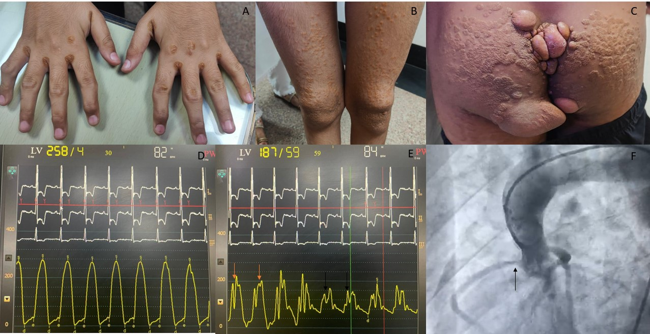
A teenage boy presented with multiple episodes of exertional syncope over the previous 3 months. Clinical examination revealed pulsus parvus et tardus and a late-peaking, ejection systolic, crescendo-decrescendo murmur in the aortic area with radiation to the carotids. There were visible tendon and intertriginous xanthomas on the hands (Figure A), tendon and eruptive xanthomas on the knees (Figure B), and tuberous xanthomas on the buttocks (Figure C). Twelve-lead electrocardiogram was suggestive of normal sinus rhythm with left axis deviation and left ventricular hypertrophy.
Two-dimensional transthoracic echocardiography revealed severe valvular aortic stenosis (AS) with a mean gradient of 80 mm Hg and severe supravalvular stenosis with a peak gradient of 80 mm Hg. Cardiac catheterization confirmed these findings, demonstrating significant pressure gradients and stenotic lesions at both valvular and supravalvular levels (Figure D and E). A pigtail angiogram in the aortic root revealed ostial stenosis of the right coronary artery (Figure F).
The patient had markedly elevated low-density lipoprotein cholesterol (LDL-C) (550 mg/dL) and total cholesterol (750 mg/dL). On genetic testing he was positive for LDL receptor mutation consistent with Type II familial hypercholesterolemia (FH). He was started on high-intensity statin therapy, bempedoic acid, and PCSK9 inhibitors, and was referred for aortic valve replacement and supravalvular AS repair.
This case illustrates a rare but critical cardiovascular complication of familial FH: combined valvular and supravalvular AS. While coronary artery disease is the most recognized and prevalent cardiovascular manifestation of FH, structural heart disease—particularly involving the aortic valve and ascending aorta—remains underappreciated and underreported.
Affiliations and Disclosures
Anbhigya Kumar Arya, MD1; Krishna Prasad Akkineni, MD, DM2; Shrividya Rao, MS, MCH3; Saurabh Kumar Singh, MD1; Souvik Sardar, MD, DM4; Anwar Hussain Ansari, MD, DM1; Devesh Kumar, MD, MRCP, DM1
From the 1Department of Cardiology, VMMC and Safdarjung Hospital, New Delhi, India; 2Department of Cardiology, Apollo Hospitals, Hyderabad, India; 3Department of Cardiothoracic Surgery, AIIMS, New Delhi, India; 4Department of Cardiology, Manorama Hospitex, Kolkata, India.
Disclosures: The authors report no financial relationships or conflicts of interest regarding the content herein.
Consent statement: The authors confirm that informed consent was obtained from the patient for the intervention described in the manuscript and for the publication thereof, including photographs.
Address for correspondence: Devesh Kumar, MD, MRCP(London), DM, Department of Cardiology, VMMC and Safdarjung Hospital, New Delhi, India. Email: devesh2.dk@gmail.com


