Cover photo, featuring the excel VTM system, courtesy of CUTERA, Inc.
Today, a cosmetic patient’s goal often is to improve the appearance of the hands with the least downtime and as few treatments as possible. If one treatment could treat both the hyperpigmentation as well as improve the appearance of the cutaneous atrophy of the hands, patients would be able achieve this goal. Some studies show improvement of both the pigment and the atrophy of the hands using non-ablative laser resurfacing.4
In this article, we present a case study of a 60-year-old female who presented for treatment of multiple lentigines on her dorsal hands. She was treated with 532-nm potassium titanyl phosphate (KTP) laser and achieved in addition to the expected improvement in the lentigines improvement in the cutaneous atrophy.
Therapeutic Alternatives
Treatment options for rejuvenation of the aging hand include topical agents, microdermabrasion, chemical peeling, intense pulsed light (IPL), ablative laser resurfacing, sclerotherapy and fillers.1 Some of these options improve hyperpigmentation, while others address the atrophy and other complaints. This section evaluates treatments that correct both pigmentary alterations and cutaneous atrophy.
Traditionally, topical agents such as tretinoin and hydroquinone have been used effectively for years to combat the superficial changes of photoaging on the hands.1 Tretinoin and other topical retinoids have been shown to increase epidermal thickness through stimulatory effects of keratinocytes and fibroblasts. It also reduces pigmentation abnormalities through dispersion of melanin granules and reduction in the rate of melansome transfer.5
Hydroquinone is a bleaching agent that is cytotoxic to melanocytes and inhibits tyrosinase decreasing melanosome formation.1 A concern with hydroquinone is that there have been reports of potential carcinogenic effects in animals.6 The principle advantage to topical treatments for the aging hand is the ease of home use. The downside is they are slow to work, although this can be helped by combination treatment using a retinoid and a bleaching agent.1 These are suitable options for mild photoaging for patients who do not want any invasive procedures and are satisfied with slow subtle improvement.
Microdermabrasion can be used as a minimally invasive treatment for the aging hand that has gained popularity recently. It provides superficial exfoliation with mild to complete abrasion of the stratum corneum following treatment. Studies have shown epidermal thickening, decreased melanization and increased elastin deposition after treatments.7 The main advantage of microdermabrasion is that there is little to no downtime. However, multiple microdermabrasion procedures on the hands are recommended and the end result is often minimal improvement in skin smoothness, texture and color.1
Chemical peeling can be performed on the hands to improve photoaging but must be done with extreme caution. The hands have fewer adnexal structures, which can lead to impaired wound healing and unpredictable results. Light to medium peels can provide significant improvement to photoaged hands but require multiple treatments and must be done conservatively. There have been several reports of scarring with use of medium peels.1 This procedure should be approached with caution and in the hands of someone very familiar with chemical peels.
IPL has been used as another non-invasive modality for photorejuvenation of the aging hand. IPL devices emit a wide spectrum of incoherent, pulsed, visible polychromatic light. IPL devices treat a wide range of complaints caused by photoaging including telangiectasias, hyperpigmentation, vascular alterations and lentigines. In addition, IPL can ameliorate abnormalities in texture and tone leading to photorejuvenation. Studies have found it good for both targeting melanin and for stimulating collagen.2 Additionally, IPL has different cutoff filters and pulse durations can be adjusted to treat different skin types.8 The disadvantages to IPL are that a number of treatments are needed and the modest improvement compared to more aggressive techniques.
Ablative laser resurfacing, like chemical peeling, must be approached cautiously and conservatively when used on non-facial skin due to the decreased density of adnexal structures, thinner epidermis and decreased vascularity.1 Laser resurfacing with ultrapulsed carbon dioxide laser and erbium:YAG laser have been performed safely on the dorsal of the hands with moderate improvement, but they must be approached conservatively. Scarring and infection after the use of ablative laser resurfacing on non-facial skin have been seen.1
Due to the need for caution with ablative resurfacing, improvement noted after treatment is not as great as that seen after facial ablative resurfacing.1 Risks from ablative laser resurfacing can be reduced by using the concept of fractional photothermolysis in ablative and non-ablative forms.9 There have been some case reports of using fractionated lasers on non-facial skin with less risk of scarring and permanent pigmentary changes than with the traditional ablative lasers.9
The skeletonization of the hands is best treated with fillers or fat augmentation. Fat augmentation has been used with good cosmetic outcomes on the dorsal hands.1 It has been shown to have slower reabsorption in hands than in the face, and could be a good long-term treatment option.10 Dermal fillers have been used in the dorsal hands as well with improvement. There are limited studies on the safety and efficacy of dermal fillers.1 It seems that both fat transfer and dermal fillers can improve the atrophy seen in the aging hand.

Case Study
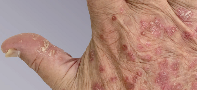
Our 60-year-old female patient with Fitzpatrick skin phototype II skin presented for evaluation of photoaging of her bilateral dorsal hands. Her main complaint was the lentigines on her dorsal hands that she believed gave her hands an aged appearance (Figures 1A and B). She was also concerned about the wrinkled appearance of her hands. The patient is a nurse who uses her hands everyday at work and could not be away from work for any significant amount of time. She also did not want to go through numerous procedures. Therefore, she was looking for a minimally invasive procedure to improve the look of her dorsal hands.
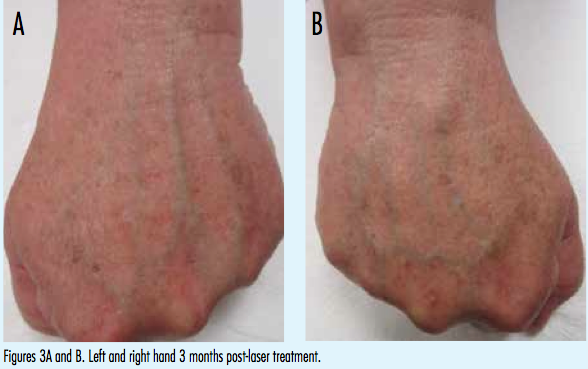
Despite having a scratch on each hand from her dog, the patient wanted to proceed with the treatment at the time of her appointment. The patient underwent 1 treatment with a Cutera Excel V 532-nm KTP laser on her dorsal hands. A fluence of 11 J/cm2 was used with a 5-mm spot size and pulse duration of 5 ms at 1 Hz. The patient had 2 passes done over the entire dorsal hand. One pass was done vertically and 1 pass was done horizontally. After the treatment, the nurses covered her hands bacitracin. The patient was advised to apply bacitracin ointment 2 times a day until her hands healed (Figures 2A and B). The procedure was well tolerated with minimal discomfort.
Discussion
As with our patient, the presence of lentigines on the hands is one of the most common complaints.1,2 The patients who develop these lesions are usually over the age of 40 with Fitzpatrick skin type I to III.2 The lentigines are found on the dorsal hands. Pathology shows hyperpigmentation of basal layer of the epidermis with elongation of the rete ridges as well as increased non-contiguous melanocytes.11 For years, these lesions have been treated with multiple modalities as discussed earlier, but recently the use of laser and other light sources have been increasingly used to treat these lesions.2
The concept of selective photothermolysis revolutionized the way lasers are used to treat skin conditions. It uses selective absorption of light pulses by pigmented targets (blood vessels, pigmented cells) to achieve selective thermally mediated injury.12 Short pulses are used to heat the target lesions before they can cool off resulting in extreme localized heating.12 This concept has allowed treatment of solar lentigines without damaging the surrounding skin which leads to shorter recovery time. Melanin absorbs light with the wavelengths of 250 to 1200 nm, which allows for the use multiple laser wavelengths in their treatment.13
In addition to the pigmentary changes noted in the aged hand, the atrophy seen is often of great concern to the patient. For example, 1 study found that the thickness of the skin on the hand at age 25 is 1.2 mm but decreases to 0.75 mm at the age of 70.14 Thus, treatments aimed at stimulating regeneration of collagen can markedly improve the appearance of the skin in the hands. Several laser systems can cause thermal injury to the dermis without injury to the epidermis, which stimulates fibroblasts to produce more collagen.1
Goldberg reported a clinical and histologic study of patients treated with a 1320-nm Nd:YAG laser with patients reporting clinical improvement in skin smoothness. However, in some of the patients these findings were short-lived.15 Other studies also showed mild-to-moderate improvement with the 1320-nm Nd:YAG laser, 1540-nm erbium glass, 585-nm flashlamp pulse dye, the 1450-nm diode and the combination 532-nm KTP and Nd:YAG lasers.1 The major advantage to these laser systems is the safety and the relative lack of downtown.
The 532-nm laser has been shown to improve both skin texture and smoothness as well as improve pigment abnormalities. One study in which 50 patients’ faces were treated with the 532-nm KTP alone after 3 to 6 treatments showed 70% to 80% improvement in redness and pigmentation, 30% to 50% improvement in skin tone/tightening, 30% to 40% improvement in skin texture and 20% to 30% improvement in rhytids.13 The study compared findings between the 532-nm KTP and the 1064-nm Nd:YAG laser and the combination of 2 lasers. The KTP was superior to the Nd:YAG, but the combination was only slightly superior to either laser alone.13 Additionally, biopsy specimens taken from the treated patients showed new collagen formation with all the treatment groups.13
As discussed, the non-ablative lasers for resurfacing and treatment of abnormal pigment are very safe treatment options. The 532-nm KTP laser showed only minimal transitory side effects in the Lee study. These included temporary swelling, temporary erythema, temporary hyperpigmentation and 1 case of blisters. None of the patients had any hypopigmentation or scarring.13 These findings are consistent with other studies of non-ablative lasers, which showed good safety profiles.16
Patient Outcome
Three months after 1 treatment with the Cutera Excel V 532-nm KTP laser, our patient noted moderate improvement in the multiple lentigines on her dorsal hands (Figures 3A and B). She also noticed impressive improvement in the texture and fullness with decreased rhytids on the dorsal hands. She had no complications from the procedure. The patient was very satisfied with the results and did not want further treatments at this time.
Conclusion
As the request for cosmetic procedures continues to rise, increasing numbers of patients may utilize hand rejuvenation to look younger. The most common hand complaint patients present with is the presence of lentigines but many are often bothered by the skeletonized look of the hands.1,2 Non-ablative laser resurfacing with the 532-nm KTP laser shows promise in treating both complaints, which is ideal for the on-the-go patient.
Dr. Pilcher is a fellow at Affiliated Dermatologists in Morristown, NJ.
Dr. Lee is a practicing dermatologist and the director of Procedural Dermatology of the ACGME-approved Procedural Dermatology Fellowship at Affiliated Dermatologists & Dermatologic Surgeons in Morristown, NJ.
Dr. Rogachefsky is a practicing dermatologist and the program director of the ACGME-approved Procedural Dermatology Fellowship at Affiliated Dermatologists & Dermatologist Surgeons in Morristown, NJ.
Disclosure: The authors report no relevant financial relationships.
References
1. Butterwick KJ. Rejuvenation of the aging hand. Dermatol Clin. 2005; 23(5):515-527.
2. Goldman A, Prati C, Rossato F. Hand rejuvenation using intense pulsed light. J Cutan Med Surg. 2008;12(3):107-113.
3. Fenske NA, Lober CW. Structural and functional changes of normal aging skin. J Am Acad Dermatol. 1986;15(4 Pt 1):571-585.
4. Sadik N, Schecter AK. Utilization of the 1320-nm Nd:YAG laser for the reduction of photoaging of the hands. Dermatol Surg. 2004;30(8):1140-1144.
5. Olson EA, Katz HI, Levine N, et al. Tretinoin emollient cream: A new therapy for photodamaged skin. J Am Acad Dermatol. 1992;26(2 Pt 1):215-244.
6. McGregor D. Hydroquinone: an evaluation of the human risks from its carcinogenic and mutagenic properties. Crit Rev Toxicol. 2007;37(10):887-914.
7. Shim EK, Barnette D, Hughs K, Greenway HT. Microdermabrasion: a clinical and histopathologic study. Dermatol Surg. 2001;27(6):524-530.
8. Raulin C, Greve B, Grema H. IPL technology: a review. Lasers Surg Med. 2003;32(2):78-87.
9. Janik JP, Markus JL, Al-Dujaili Z, Markus RF. Laser resurfacing. Semin Plast Surg. 2007;21(3):139-146.
10. Abrams HL, Lauber JS. Hand rejuvenation: the state of the art. Dermatol Clin. 1990;8(3):553-561.
11. Nolesses E, Nizard C, Cario-Andre M, et al. Skin ultrastructure in senile lentigo. Skin Pharmocol Physiol. 2006;19(2):95-100.
12. Sakamoto FH, Avram MM, Anderson RR. Lasers and other energy technologies — principles & skin interactions. In: Bolognia JL, Jorizzo JL, Schaffer JV, eds. Dermatology. 3rd ed. New York, NY: Elsevier; 2012:1885-1914.
13. Lee MC. Combination 532-nm and 1064-nm lasers for noninvasive skin rejuvenation and toning. Arch Dermatol. 2003;139(10):1265-1276.
14. Brodar V. Observations of skin thickness and subcutaneous tissue in man. Z Morph Anthrop. 1960;50:386.
15. Goldberg DJ. Full-face nonablative dermal remodeling with 1320 nm Nd:YAG laser. Dermatol Surg. 2000;26(10):915-918.
16. Alam M, Kakar R, Nodzenski M, et al. Multicenter prospective cohort study of the incidence of adverse events associated with cosmetic dermatologic procedures: lasers, energy devices, and injectable neurotoxins and fillers. JAMA Dermatol. 2015;151(3): 271-277.
Cover photo, featuring the excel VTM system, courtesy of CUTERA, Inc.
Today, a cosmetic patient’s goal often is to improve the appearance of the hands with the least downtime and as few treatments as possible. If one treatment could treat both the hyperpigmentation as well as improve the appearance of the cutaneous atrophy of the hands, patients would be able achieve this goal. Some studies show improvement of both the pigment and the atrophy of the hands using non-ablative laser resurfacing.4
In this article, we present a case study of a 60-year-old female who presented for treatment of multiple lentigines on her dorsal hands. She was treated with 532-nm potassium titanyl phosphate (KTP) laser and achieved in addition to the expected improvement in the lentigines improvement in the cutaneous atrophy.
Therapeutic Alternatives
Treatment options for rejuvenation of the aging hand include topical agents, microdermabrasion, chemical peeling, intense pulsed light (IPL), ablative laser resurfacing, sclerotherapy and fillers.1 Some of these options improve hyperpigmentation, while others address the atrophy and other complaints. This section evaluates treatments that correct both pigmentary alterations and cutaneous atrophy.
Traditionally, topical agents such as tretinoin and hydroquinone have been used effectively for years to combat the superficial changes of photoaging on the hands.1 Tretinoin and other topical retinoids have been shown to increase epidermal thickness through stimulatory effects of keratinocytes and fibroblasts. It also reduces pigmentation abnormalities through dispersion of melanin granules and reduction in the rate of melansome transfer.5
Hydroquinone is a bleaching agent that is cytotoxic to melanocytes and inhibits tyrosinase decreasing melanosome formation.1 A concern with hydroquinone is that there have been reports of potential carcinogenic effects in animals.6 The principle advantage to topical treatments for the aging hand is the ease of home use. The downside is they are slow to work, although this can be helped by combination treatment using a retinoid and a bleaching agent.1 These are suitable options for mild photoaging for patients who do not want any invasive procedures and are satisfied with slow subtle improvement.
Microdermabrasion can be used as a minimally invasive treatment for the aging hand that has gained popularity recently. It provides superficial exfoliation with mild to complete abrasion of the stratum corneum following treatment. Studies have shown epidermal thickening, decreased melanization and increased elastin deposition after treatments.7 The main advantage of microdermabrasion is that there is little to no downtime. However, multiple microdermabrasion procedures on the hands are recommended and the end result is often minimal improvement in skin smoothness, texture and color.1
Chemical peeling can be performed on the hands to improve photoaging but must be done with extreme caution. The hands have fewer adnexal structures, which can lead to impaired wound healing and unpredictable results. Light to medium peels can provide significant improvement to photoaged hands but require multiple treatments and must be done conservatively. There have been several reports of scarring with use of medium peels.1 This procedure should be approached with caution and in the hands of someone very familiar with chemical peels.
IPL has been used as another non-invasive modality for photorejuvenation of the aging hand. IPL devices emit a wide spectrum of incoherent, pulsed, visible polychromatic light. IPL devices treat a wide range of complaints caused by photoaging including telangiectasias, hyperpigmentation, vascular alterations and lentigines. In addition, IPL can ameliorate abnormalities in texture and tone leading to photorejuvenation. Studies have found it good for both targeting melanin and for stimulating collagen.2 Additionally, IPL has different cutoff filters and pulse durations can be adjusted to treat different skin types.8 The disadvantages to IPL are that a number of treatments are needed and the modest improvement compared to more aggressive techniques.
Ablative laser resurfacing, like chemical peeling, must be approached cautiously and conservatively when used on non-facial skin due to the decreased density of adnexal structures, thinner epidermis and decreased vascularity.1 Laser resurfacing with ultrapulsed carbon dioxide laser and erbium:YAG laser have been performed safely on the dorsal of the hands with moderate improvement, but they must be approached conservatively. Scarring and infection after the use of ablative laser resurfacing on non-facial skin have been seen.1
Due to the need for caution with ablative resurfacing, improvement noted after treatment is not as great as that seen after facial ablative resurfacing.1 Risks from ablative laser resurfacing can be reduced by using the concept of fractional photothermolysis in ablative and non-ablative forms.9 There have been some case reports of using fractionated lasers on non-facial skin with less risk of scarring and permanent pigmentary changes than with the traditional ablative lasers.9
The skeletonization of the hands is best treated with fillers or fat augmentation. Fat augmentation has been used with good cosmetic outcomes on the dorsal hands.1 It has been shown to have slower reabsorption in hands than in the face, and could be a good long-term treatment option.10 Dermal fillers have been used in the dorsal hands as well with improvement. There are limited studies on the safety and efficacy of dermal fillers.1 It seems that both fat transfer and dermal fillers can improve the atrophy seen in the aging hand.

Case Study
Our 60-year-old female patient with Fitzpatrick skin phototype II skin presented for evaluation of photoaging of her bilateral dorsal hands. Her main complaint was the lentigines on her dorsal hands that she believed gave her hands an aged appearance (Figures 1A and B). She was also concerned about the wrinkled appearance of her hands. The patient is a nurse who uses her hands everyday at work and could not be away from work for any significant amount of time. She also did not want to go through numerous procedures. Therefore, she was looking for a minimally invasive procedure to improve the look of her dorsal hands.
Despite having a scratch on each hand from her dog, the patient wanted to proceed with the treatment at the time of her appointment. The patient underwent 1 treatment with a Cutera Excel V 532-nm KTP laser on her dorsal hands. A fluence of 11 J/cm2 was used with a 5-mm spot size and pulse duration of 5 ms at 1 Hz. The patient had 2 passes done over the entire dorsal hand. One pass was done vertically and 1 pass was done horizontally. After the treatment, the nurses covered her hands bacitracin. The patient was advised to apply bacitracin ointment 2 times a day until her hands healed (Figures 2A and B). The procedure was well tolerated with minimal discomfort.
Discussion
As with our patient, the presence of lentigines on the hands is one of the most common complaints.1,2 The patients who develop these lesions are usually over the age of 40 with Fitzpatrick skin type I to III.2 The lentigines are found on the dorsal hands. Pathology shows hyperpigmentation of basal layer of the epidermis with elongation of the rete ridges as well as increased non-contiguous melanocytes.11 For years, these lesions have been treated with multiple modalities as discussed earlier, but recently the use of laser and other light sources have been increasingly used to treat these lesions.2
The concept of selective photothermolysis revolutionized the way lasers are used to treat skin conditions. It uses selective absorption of light pulses by pigmented targets (blood vessels, pigmented cells) to achieve selective thermally mediated injury.12 Short pulses are used to heat the target lesions before they can cool off resulting in extreme localized heating.12 This concept has allowed treatment of solar lentigines without damaging the surrounding skin which leads to shorter recovery time. Melanin absorbs light with the wavelengths of 250 to 1200 nm, which allows for the use multiple laser wavelengths in their treatment.13
In addition to the pigmentary changes noted in the aged hand, the atrophy seen is often of great concern to the patient. For example, 1 study found that the thickness of the skin on the hand at age 25 is 1.2 mm but decreases to 0.75 mm at the age of 70.14 Thus, treatments aimed at stimulating regeneration of collagen can markedly improve the appearance of the skin in the hands. Several laser systems can cause thermal injury to the dermis without injury to the epidermis, which stimulates fibroblasts to produce more collagen.1
Goldberg reported a clinical and histologic study of patients treated with a 1320-nm Nd:YAG laser with patients reporting clinical improvement in skin smoothness. However, in some of the patients these findings were short-lived.15 Other studies also showed mild-to-moderate improvement with the 1320-nm Nd:YAG laser, 1540-nm erbium glass, 585-nm flashlamp pulse dye, the 1450-nm diode and the combination 532-nm KTP and Nd:YAG lasers.1 The major advantage to these laser systems is the safety and the relative lack of downtown.
The 532-nm laser has been shown to improve both skin texture and smoothness as well as improve pigment abnormalities. One study in which 50 patients’ faces were treated with the 532-nm KTP alone after 3 to 6 treatments showed 70% to 80% improvement in redness and pigmentation, 30% to 50% improvement in skin tone/tightening, 30% to 40% improvement in skin texture and 20% to 30% improvement in rhytids.13 The study compared findings between the 532-nm KTP and the 1064-nm Nd:YAG laser and the combination of 2 lasers. The KTP was superior to the Nd:YAG, but the combination was only slightly superior to either laser alone.13 Additionally, biopsy specimens taken from the treated patients showed new collagen formation with all the treatment groups.13
As discussed, the non-ablative lasers for resurfacing and treatment of abnormal pigment are very safe treatment options. The 532-nm KTP laser showed only minimal transitory side effects in the Lee study. These included temporary swelling, temporary erythema, temporary hyperpigmentation and 1 case of blisters. None of the patients had any hypopigmentation or scarring.13 These findings are consistent with other studies of non-ablative lasers, which showed good safety profiles.16

Patient Outcome
Three months after 1 treatment with the Cutera Excel V 532-nm KTP laser, our patient noted moderate improvement in the multiple lentigines on her dorsal hands (Figures 3A and B). She also noticed impressive improvement in the texture and fullness with decreased rhytids on the dorsal hands. She had no complications from the procedure. The patient was very satisfied with the results and did not want further treatments at this time.
Conclusion
As the request for cosmetic procedures continues to rise, increasing numbers of patients may utilize hand rejuvenation to look younger. The most common hand complaint patients present with is the presence of lentigines but many are often bothered by the skeletonized look of the hands.1,2 Non-ablative laser resurfacing with the 532-nm KTP laser shows promise in treating both complaints, which is ideal for the on-the-go patient.
Dr. Pilcher is a fellow at Affiliated Dermatologists in Morristown, NJ.
Dr. Lee is a practicing dermatologist and the director of Procedural Dermatology of the ACGME-approved Procedural Dermatology Fellowship at Affiliated Dermatologists & Dermatologic Surgeons in Morristown, NJ.
Dr. Rogachefsky is a practicing dermatologist and the program director of the ACGME-approved Procedural Dermatology Fellowship at Affiliated Dermatologists & Dermatologist Surgeons in Morristown, NJ.
Disclosure: The authors report no relevant financial relationships.
References
1. Butterwick KJ. Rejuvenation of the aging hand. Dermatol Clin. 2005; 23(5):515-527.
2. Goldman A, Prati C, Rossato F. Hand rejuvenation using intense pulsed light. J Cutan Med Surg. 2008;12(3):107-113.
3. Fenske NA, Lober CW. Structural and functional changes of normal aging skin. J Am Acad Dermatol. 1986;15(4 Pt 1):571-585.
4. Sadik N, Schecter AK. Utilization of the 1320-nm Nd:YAG laser for the reduction of photoaging of the hands. Dermatol Surg. 2004;30(8):1140-1144.
5. Olson EA, Katz HI, Levine N, et al. Tretinoin emollient cream: A new therapy for photodamaged skin. J Am Acad Dermatol. 1992;26(2 Pt 1):215-244.
6. McGregor D. Hydroquinone: an evaluation of the human risks from its carcinogenic and mutagenic properties. Crit Rev Toxicol. 2007;37(10):887-914.
7. Shim EK, Barnette D, Hughs K, Greenway HT. Microdermabrasion: a clinical and histopathologic study. Dermatol Surg. 2001;27(6):524-530.
8. Raulin C, Greve B, Grema H. IPL technology: a review. Lasers Surg Med. 2003;32(2):78-87.
9. Janik JP, Markus JL, Al-Dujaili Z, Markus RF. Laser resurfacing. Semin Plast Surg. 2007;21(3):139-146.
10. Abrams HL, Lauber JS. Hand rejuvenation: the state of the art. Dermatol Clin. 1990;8(3):553-561.
11. Nolesses E, Nizard C, Cario-Andre M, et al. Skin ultrastructure in senile lentigo. Skin Pharmocol Physiol. 2006;19(2):95-100.
12. Sakamoto FH, Avram MM, Anderson RR. Lasers and other energy technologies — principles & skin interactions. In: Bolognia JL, Jorizzo JL, Schaffer JV, eds. Dermatology. 3rd ed. New York, NY: Elsevier; 2012:1885-1914.
13. Lee MC. Combination 532-nm and 1064-nm lasers for noninvasive skin rejuvenation and toning. Arch Dermatol. 2003;139(10):1265-1276.
14. Brodar V. Observations of skin thickness and subcutaneous tissue in man. Z Morph Anthrop. 1960;50:386.
15. Goldberg DJ. Full-face nonablative dermal remodeling with 1320 nm Nd:YAG laser. Dermatol Surg. 2000;26(10):915-918.
16. Alam M, Kakar R, Nodzenski M, et al. Multicenter prospective cohort study of the incidence of adverse events associated with cosmetic dermatologic procedures: lasers, energy devices, and injectable neurotoxins and fillers. JAMA Dermatol. 2015;151(3): 271-277.
Cover photo, featuring the excel VTM system, courtesy of CUTERA, Inc.
Today, a cosmetic patient’s goal often is to improve the appearance of the hands with the least downtime and as few treatments as possible. If one treatment could treat both the hyperpigmentation as well as improve the appearance of the cutaneous atrophy of the hands, patients would be able achieve this goal. Some studies show improvement of both the pigment and the atrophy of the hands using non-ablative laser resurfacing.4
In this article, we present a case study of a 60-year-old female who presented for treatment of multiple lentigines on her dorsal hands. She was treated with 532-nm potassium titanyl phosphate (KTP) laser and achieved in addition to the expected improvement in the lentigines improvement in the cutaneous atrophy.
Therapeutic Alternatives
Treatment options for rejuvenation of the aging hand include topical agents, microdermabrasion, chemical peeling, intense pulsed light (IPL), ablative laser resurfacing, sclerotherapy and fillers.1 Some of these options improve hyperpigmentation, while others address the atrophy and other complaints. This section evaluates treatments that correct both pigmentary alterations and cutaneous atrophy.
Traditionally, topical agents such as tretinoin and hydroquinone have been used effectively for years to combat the superficial changes of photoaging on the hands.1 Tretinoin and other topical retinoids have been shown to increase epidermal thickness through stimulatory effects of keratinocytes and fibroblasts. It also reduces pigmentation abnormalities through dispersion of melanin granules and reduction in the rate of melansome transfer.5
Hydroquinone is a bleaching agent that is cytotoxic to melanocytes and inhibits tyrosinase decreasing melanosome formation.1 A concern with hydroquinone is that there have been reports of potential carcinogenic effects in animals.6 The principle advantage to topical treatments for the aging hand is the ease of home use. The downside is they are slow to work, although this can be helped by combination treatment using a retinoid and a bleaching agent.1 These are suitable options for mild photoaging for patients who do not want any invasive procedures and are satisfied with slow subtle improvement.
Microdermabrasion can be used as a minimally invasive treatment for the aging hand that has gained popularity recently. It provides superficial exfoliation with mild to complete abrasion of the stratum corneum following treatment. Studies have shown epidermal thickening, decreased melanization and increased elastin deposition after treatments.7 The main advantage of microdermabrasion is that there is little to no downtime. However, multiple microdermabrasion procedures on the hands are recommended and the end result is often minimal improvement in skin smoothness, texture and color.1
Chemical peeling can be performed on the hands to improve photoaging but must be done with extreme caution. The hands have fewer adnexal structures, which can lead to impaired wound healing and unpredictable results. Light to medium peels can provide significant improvement to photoaged hands but require multiple treatments and must be done conservatively. There have been several reports of scarring with use of medium peels.1 This procedure should be approached with caution and in the hands of someone very familiar with chemical peels.
IPL has been used as another non-invasive modality for photorejuvenation of the aging hand. IPL devices emit a wide spectrum of incoherent, pulsed, visible polychromatic light. IPL devices treat a wide range of complaints caused by photoaging including telangiectasias, hyperpigmentation, vascular alterations and lentigines. In addition, IPL can ameliorate abnormalities in texture and tone leading to photorejuvenation. Studies have found it good for both targeting melanin and for stimulating collagen.2 Additionally, IPL has different cutoff filters and pulse durations can be adjusted to treat different skin types.8 The disadvantages to IPL are that a number of treatments are needed and the modest improvement compared to more aggressive techniques.
Ablative laser resurfacing, like chemical peeling, must be approached cautiously and conservatively when used on non-facial skin due to the decreased density of adnexal structures, thinner epidermis and decreased vascularity.1 Laser resurfacing with ultrapulsed carbon dioxide laser and erbium:YAG laser have been performed safely on the dorsal of the hands with moderate improvement, but they must be approached conservatively. Scarring and infection after the use of ablative laser resurfacing on non-facial skin have been seen.1
Due to the need for caution with ablative resurfacing, improvement noted after treatment is not as great as that seen after facial ablative resurfacing.1 Risks from ablative laser resurfacing can be reduced by using the concept of fractional photothermolysis in ablative and non-ablative forms.9 There have been some case reports of using fractionated lasers on non-facial skin with less risk of scarring and permanent pigmentary changes than with the traditional ablative lasers.9
The skeletonization of the hands is best treated with fillers or fat augmentation. Fat augmentation has been used with good cosmetic outcomes on the dorsal hands.1 It has been shown to have slower reabsorption in hands than in the face, and could be a good long-term treatment option.10 Dermal fillers have been used in the dorsal hands as well with improvement. There are limited studies on the safety and efficacy of dermal fillers.1 It seems that both fat transfer and dermal fillers can improve the atrophy seen in the aging hand.
Case Study
Our 60-year-old female patient with Fitzpatrick skin phototype II skin presented for evaluation of photoaging of her bilateral dorsal hands. Her main complaint was the lentigines on her dorsal hands that she believed gave her hands an aged appearance (Figures 1A and B). She was also concerned about the wrinkled appearance of her hands. The patient is a nurse who uses her hands everyday at work and could not be away from work for any significant amount of time. She also did not want to go through numerous procedures. Therefore, she was looking for a minimally invasive procedure to improve the look of her dorsal hands.
Despite having a scratch on each hand from her dog, the patient wanted to proceed with the treatment at the time of her appointment. The patient underwent 1 treatment with a Cutera Excel V 532-nm KTP laser on her dorsal hands. A fluence of 11 J/cm2 was used with a 5-mm spot size and pulse duration of 5 ms at 1 Hz. The patient had 2 passes done over the entire dorsal hand. One pass was done vertically and 1 pass was done horizontally. After the treatment, the nurses covered her hands bacitracin. The patient was advised to apply bacitracin ointment 2 times a day until her hands healed (Figures 2A and B). The procedure was well tolerated with minimal discomfort.
Discussion
As with our patient, the presence of lentigines on the hands is one of the most common complaints.1,2 The patients who develop these lesions are usually over the age of 40 with Fitzpatrick skin type I to III.2 The lentigines are found on the dorsal hands. Pathology shows hyperpigmentation of basal layer of the epidermis with elongation of the rete ridges as well as increased non-contiguous melanocytes.11 For years, these lesions have been treated with multiple modalities as discussed earlier, but recently the use of laser and other light sources have been increasingly used to treat these lesions.2
The concept of selective photothermolysis revolutionized the way lasers are used to treat skin conditions. It uses selective absorption of light pulses by pigmented targets (blood vessels, pigmented cells) to achieve selective thermally mediated injury.12 Short pulses are used to heat the target lesions before they can cool off resulting in extreme localized heating.12 This concept has allowed treatment of solar lentigines without damaging the surrounding skin which leads to shorter recovery time. Melanin absorbs light with the wavelengths of 250 to 1200 nm, which allows for the use multiple laser wavelengths in their treatment.13
In addition to the pigmentary changes noted in the aged hand, the atrophy seen is often of great concern to the patient. For example, 1 study found that the thickness of the skin on the hand at age 25 is 1.2 mm but decreases to 0.75 mm at the age of 70.14 Thus, treatments aimed at stimulating regeneration of collagen can markedly improve the appearance of the skin in the hands. Several laser systems can cause thermal injury to the dermis without injury to the epidermis, which stimulates fibroblasts to produce more collagen.1
Goldberg reported a clinical and histologic study of patients treated with a 1320-nm Nd:YAG laser with patients reporting clinical improvement in skin smoothness. However, in some of the patients these findings were short-lived.15 Other studies also showed mild-to-moderate improvement with the 1320-nm Nd:YAG laser, 1540-nm erbium glass, 585-nm flashlamp pulse dye, the 1450-nm diode and the combination 532-nm KTP and Nd:YAG lasers.1 The major advantage to these laser systems is the safety and the relative lack of downtown.
The 532-nm laser has been shown to improve both skin texture and smoothness as well as improve pigment abnormalities. One study in which 50 patients’ faces were treated with the 532-nm KTP alone after 3 to 6 treatments showed 70% to 80% improvement in redness and pigmentation, 30% to 50% improvement in skin tone/tightening, 30% to 40% improvement in skin texture and 20% to 30% improvement in rhytids.13 The study compared findings between the 532-nm KTP and the 1064-nm Nd:YAG laser and the combination of 2 lasers. The KTP was superior to the Nd:YAG, but the combination was only slightly superior to either laser alone.13 Additionally, biopsy specimens taken from the treated patients showed new collagen formation with all the treatment groups.13
As discussed, the non-ablative lasers for resurfacing and treatment of abnormal pigment are very safe treatment options. The 532-nm KTP laser showed only minimal transitory side effects in the Lee study. These included temporary swelling, temporary erythema, temporary hyperpigmentation and 1 case of blisters. None of the patients had any hypopigmentation or scarring.13 These findings are consistent with other studies of non-ablative lasers, which showed good safety profiles.16
Patient Outcome
Three months after 1 treatment with the Cutera Excel V 532-nm KTP laser, our patient noted moderate improvement in the multiple lentigines on her dorsal hands (Figures 3A and B). She also noticed impressive improvement in the texture and fullness with decreased rhytids on the dorsal hands. She had no complications from the procedure. The patient was very satisfied with the results and did not want further treatments at this time.
Conclusion
As the request for cosmetic procedures continues to rise, increasing numbers of patients may utilize hand rejuvenation to look younger. The most common hand complaint patients present with is the presence of lentigines but many are often bothered by the skeletonized look of the hands.1,2 Non-ablative laser resurfacing with the 532-nm KTP laser shows promise in treating both complaints, which is ideal for the on-the-go patient.
Dr. Pilcher is a fellow at Affiliated Dermatologists in Morristown, NJ.
Dr. Lee is a practicing dermatologist and the director of Procedural Dermatology of the ACGME-approved Procedural Dermatology Fellowship at Affiliated Dermatologists & Dermatologic Surgeons in Morristown, NJ.
Dr. Rogachefsky is a practicing dermatologist and the program director of the ACGME-approved Procedural Dermatology Fellowship at Affiliated Dermatologists & Dermatologist Surgeons in Morristown, NJ.
Disclosure: The authors report no relevant financial relationships.
References
1. Butterwick KJ. Rejuvenation of the aging hand. Dermatol Clin. 2005; 23(5):515-527.
2. Goldman A, Prati C, Rossato F. Hand rejuvenation using intense pulsed light. J Cutan Med Surg. 2008;12(3):107-113.
3. Fenske NA, Lober CW. Structural and functional changes of normal aging skin. J Am Acad Dermatol. 1986;15(4 Pt 1):571-585.
4. Sadik N, Schecter AK. Utilization of the 1320-nm Nd:YAG laser for the reduction of photoaging of the hands. Dermatol Surg. 2004;30(8):1140-1144.
5. Olson EA, Katz HI, Levine N, et al. Tretinoin emollient cream: A new therapy for photodamaged skin. J Am Acad Dermatol. 1992;26(2 Pt 1):215-244.
6. McGregor D. Hydroquinone: an evaluation of the human risks from its carcinogenic and mutagenic properties. Crit Rev Toxicol. 2007;37(10):887-914.
7. Shim EK, Barnette D, Hughs K, Greenway HT. Microdermabrasion: a clinical and histopathologic study. Dermatol Surg. 2001;27(6):524-530.
8. Raulin C, Greve B, Grema H. IPL technology: a review. Lasers Surg Med. 2003;32(2):78-87.
9. Janik JP, Markus JL, Al-Dujaili Z, Markus RF. Laser resurfacing. Semin Plast Surg. 2007;21(3):139-146.
10. Abrams HL, Lauber JS. Hand rejuvenation: the state of the art. Dermatol Clin. 1990;8(3):553-561.
11. Nolesses E, Nizard C, Cario-Andre M, et al. Skin ultrastructure in senile lentigo. Skin Pharmocol Physiol. 2006;19(2):95-100.
12. Sakamoto FH, Avram MM, Anderson RR. Lasers and other energy technologies — principles & skin interactions. In: Bolognia JL, Jorizzo JL, Schaffer JV, eds. Dermatology. 3rd ed. New York, NY: Elsevier; 2012:1885-1914.
13. Lee MC. Combination 532-nm and 1064-nm lasers for noninvasive skin rejuvenation and toning. Arch Dermatol. 2003;139(10):1265-1276.
14. Brodar V. Observations of skin thickness and subcutaneous tissue in man. Z Morph Anthrop. 1960;50:386.
15. Goldberg DJ. Full-face nonablative dermal remodeling with 1320 nm Nd:YAG laser. Dermatol Surg. 2000;26(10):915-918.
16. Alam M, Kakar R, Nodzenski M, et al. Multicenter prospective cohort study of the incidence of adverse events associated with cosmetic dermatologic procedures: lasers, energy devices, and injectable neurotoxins and fillers. JAMA Dermatol. 2015;151(3): 271-277.