The ubiquity of laser therapy in medicine has allowed for the broad treatment of skin disorders with light-based treatments. In the past, scar revision was often relegated to surgical treatment, whereas current standards allow for more conservative non-surgical treatment modalities. Among the many laser options currently available, the pulsed dye laser stands out as one of the most effective in the early treatment of visible scars.
On initial examination, patients often express a willingness to “improve” a scar, but are often unaware of the many different aspects of scar formation that can make a scar visibly unappealing. Scar formation can involve changes in color (hypopigmentation or hyperpigmentation), texture, surface topography, volume, width, thickness, tissue rigidity and erythema. Scars can also cause symptoms such as pain, itching, burning, and changes in sensation. By explaining these different aspects of a scar, patients and physicians are better able to manage scar revision treatments.
The pulsed dye laser is a vascular-specific technology that allows for the treatment of scars that display erythema. The mechanism of action is unknown, but is thought to relate to laser-induced tissue hypoxia, decreased microvascular perfusion, collagen fiber heating and destruction or alteration in fibroblast proliferation and viability.
In clinical practice, scar treatment with the pulsed dye laser is broken down into 2 modalities: volumetric improvement in surface topography and reduction in surface erythema.
Scar Evaluation
The treatment of visible scars should begin with a thorough examination that goes beyond the scar in question. By observing variations in scar formation throughout a patient’s body, the clinician and patient are able to better understand the scope of the scar treatment. In short, the characteristics of other scars on the body can help identify patterns of scar formation that are specific to that individual.
The discussion of non-surgical options for scar revision should be directed to the individual characteristics of the specific scar, as each scar will vary in morphology. Patients are often more apt to appreciate that a ‘cure-all’ laser does not exist for all scars, but different lasers can be used for different aspects of the scar. The pulsed dye laser can be used for active scars that demonstrate erythema with or without hypertrophy.
Patients should also be aware of other scar features that may require future treatment, such as surface texture, scar widening, hypopigmentation or hyperpigmentation. Treatments for post-inflammatory hyperpigmentation can often be performed simultaneously with laser in order to improve surface color and reduce laser-induced pigmentation. Topical melanin-inhibitors are often used as first-line therapy.
Targeted Treatment
A potentially successful approach to pulsed dye laser therapy would be to direct the treatments to 2 specific aspects of a scar, namely the thickness and the redness. The approach taken in the author’s practice is to begin with pulsed dye laser treatments to address scar volume and thickness and then proceed to interval laser sessions to reduce surface erythema. Based on experience, initial treatments with a short pulse width often cause purpura and may further aggravate surface erythema in the short term.
Initial treatments utilize the pulsed dye laser with a short pulse width of 0.45 ms, which have been shown to induce purpura and cause a decrease in scar volume, thickness and rigidity. Patients should be aware that the widened scars will exhibit no changes in scar width or visibility of track marks. Treatments are usually spaced 3-4 weeks apart, giving enough time for tissue healing and collagen resorption. The use of intralesional corticosteroids varies from practitioner to practitioner, and has variable responsiveness depending on scar density.
Once volume changes have been observed and the scar has achieved an improvement in surface topography, a second phase of pulsed dye laser is utilized to decrease surface erythema. In the author’s practice, a pulse width of 10 ms is used on the lesion at an interval of about 3-4 weeks. The longer pulse width is subpurpuric, and patients will begin to notice a gradual change in surface erythema. It is prudent to take interval photographs of the improvement as patients are often unable to appreciate changes in surface color between visits.
Pulsed dye laser remains one of many treatment options for non-surgical scar revision. Optimal results begin with a thorough understanding of scar physiology and scar morphology. Patient expectations should be discussed with respect to genetic factors, patterns of scar formation and the specific morphology of the scar in question.
Treatment with the pulsed dye laser should generally follow, rather than precede, surgical intervention. If surgery is planned, patients should be advised on the need to perform post-surgical pulsed dye laser to minimize hypertrophy and surface erythema. n

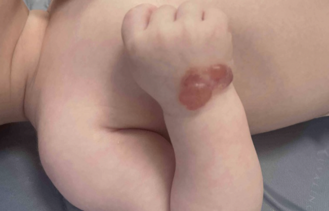
Figure 1. Before - Scar complex A" Flat with inflammation and redness.
Scar Complex B: Raised scar eith inflammation and redness.
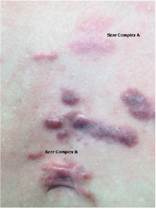
Figure 2. After - Scar Complex A: Subpurpuric pulsed dye laser
Scar Complex B: Purpuric pulsed dye laser.
Dr. Karamanoukian is the surgical director of Kare Plastic Surgery in Santa Monica, CA, and clinical assistant professor in the University of California, Irvine Department of Plastic Surgery.
Disclosure: The author has no conflicts of interest to report.
To read more news, trends and case studies about lasers and light therapies, visit.the-dermatologist.com/laser-light-therapies.
The ubiquity of laser therapy in medicine has allowed for the broad treatment of skin disorders with light-based treatments. In the past, scar revision was often relegated to surgical treatment, whereas current standards allow for more conservative non-surgical treatment modalities. Among the many laser options currently available, the pulsed dye laser stands out as one of the most effective in the early treatment of visible scars.
On initial examination, patients often express a willingness to “improve” a scar, but are often unaware of the many different aspects of scar formation that can make a scar visibly unappealing. Scar formation can involve changes in color (hypopigmentation or hyperpigmentation), texture, surface topography, volume, width, thickness, tissue rigidity and erythema. Scars can also cause symptoms such as pain, itching, burning, and changes in sensation. By explaining these different aspects of a scar, patients and physicians are better able to manage scar revision treatments.
The pulsed dye laser is a vascular-specific technology that allows for the treatment of scars that display erythema. The mechanism of action is unknown, but is thought to relate to laser-induced tissue hypoxia, decreased microvascular perfusion, collagen fiber heating and destruction or alteration in fibroblast proliferation and viability.
In clinical practice, scar treatment with the pulsed dye laser is broken down into 2 modalities: volumetric improvement in surface topography and reduction in surface erythema.
Scar Evaluation
The treatment of visible scars should begin with a thorough examination that goes beyond the scar in question. By observing variations in scar formation throughout a patient’s body, the clinician and patient are able to better understand the scope of the scar treatment. In short, the characteristics of other scars on the body can help identify patterns of scar formation that are specific to that individual.
The discussion of non-surgical options for scar revision should be directed to the individual characteristics of the specific scar, as each scar will vary in morphology. Patients are often more apt to appreciate that a ‘cure-all’ laser does not exist for all scars, but different lasers can be used for different aspects of the scar. The pulsed dye laser can be used for active scars that demonstrate erythema with or without hypertrophy.
Patients should also be aware of other scar features that may require future treatment, such as surface texture, scar widening, hypopigmentation or hyperpigmentation. Treatments for post-inflammatory hyperpigmentation can often be performed simultaneously with laser in order to improve surface color and reduce laser-induced pigmentation. Topical melanin-inhibitors are often used as first-line therapy.
Targeted Treatment
A potentially successful approach to pulsed dye laser therapy would be to direct the treatments to 2 specific aspects of a scar, namely the thickness and the redness. The approach taken in the author’s practice is to begin with pulsed dye laser treatments to address scar volume and thickness and then proceed to interval laser sessions to reduce surface erythema. Based on experience, initial treatments with a short pulse width often cause purpura and may further aggravate surface erythema in the short term.
Initial treatments utilize the pulsed dye laser with a short pulse width of 0.45 ms, which have been shown to induce purpura and cause a decrease in scar volume, thickness and rigidity. Patients should be aware that the widened scars will exhibit no changes in scar width or visibility of track marks. Treatments are usually spaced 3-4 weeks apart, giving enough time for tissue healing and collagen resorption. The use of intralesional corticosteroids varies from practitioner to practitioner, and has variable responsiveness depending on scar density.
Once volume changes have been observed and the scar has achieved an improvement in surface topography, a second phase of pulsed dye laser is utilized to decrease surface erythema. In the author’s practice, a pulse width of 10 ms is used on the lesion at an interval of about 3-4 weeks. The longer pulse width is subpurpuric, and patients will begin to notice a gradual change in surface erythema. It is prudent to take interval photographs of the improvement as patients are often unable to appreciate changes in surface color between visits.
Pulsed dye laser remains one of many treatment options for non-surgical scar revision. Optimal results begin with a thorough understanding of scar physiology and scar morphology. Patient expectations should be discussed with respect to genetic factors, patterns of scar formation and the specific morphology of the scar in question.
Treatment with the pulsed dye laser should generally follow, rather than precede, surgical intervention. If surgery is planned, patients should be advised on the need to perform post-surgical pulsed dye laser to minimize hypertrophy and surface erythema. n
Figure 1. Before - Scar complex A" Flat with inflammation and redness.
Scar Complex B: Raised scar eith inflammation and redness.
Figure 2. After - Scar Complex A: Subpurpuric pulsed dye laser
Scar Complex B: Purpuric pulsed dye laser.
Dr. Karamanoukian is the surgical director of Kare Plastic Surgery in Santa Monica, CA, and clinical assistant professor in the University of California, Irvine Department of Plastic Surgery.
Disclosure: The author has no conflicts of interest to report.
To read more news, trends and case studies about lasers and light therapies, visit.the-dermatologist.com/laser-light-therapies.