PATIENT PRESENTATION
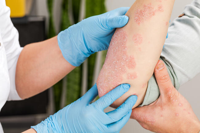
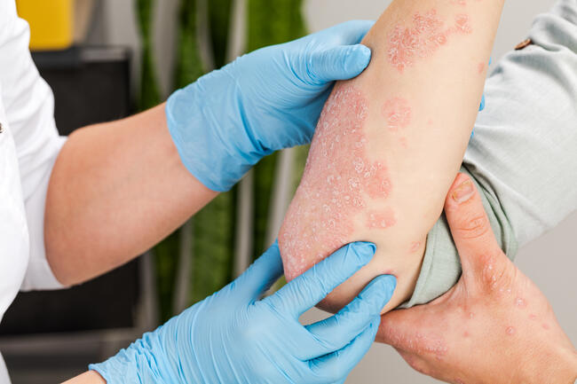
Patient 1 is a 39-year-old Caucasian man who initially noticed a cluster of small, skin-colored, scaly, asymptomatic lesions on his right dorsal hand at the age of 9 years, with slow progression to the dorsal third and fifth digits over the following 25 years (Top). Prior treatment included electrodessication, cryotherapy, and topical salicylic acid, all of which only temporarily eliminated the lesions. They rapidly recurred in the same distribution. His past medical history was negative for cutaneous malignancy. Physical examination was significant for plaques of coalescing flat hyperkeratotic, flesh-colored papules on the dorsal hand, third, and fifth digits. The rest of the skin examination revealed no abnormalities. Patient 2 is the 9-year-old daughter of patient 1. She presented with similar right dorsal hand lesions, which developed at the age of 8. Physical examination was significant for plaques of coalescing flat hyperkeratotic, flesh-colored papules on the dorsal hand, and first and third digits (Bottom).
What is Your Diagnosis?
___________________________
Diagnosis: An Unusual Presentation of Verrucae Planae
We report an unusual case of verrucae planae in a father and daughter, both developing in childhood and presenting with lesions on the right dorsal hand. While verrucae planae can present within families and at an early age, this particular presentation was unique in that it followed an autosomal dominant inheritance pattern with nearly identical history, distribution and morphology.
Background
Verrucae are among the most common skin lesions, occurring in 10% of children and young adults. Verrucae planae appear as sharply defined, flat-topped, flesh-colored to light brown papules, 2 mm to 4 mm in diameter.1,2 They are most often seen on the face and dorsal hands of children. These lesions represent a clinical manifestation of human papillomavirus (HPV) infection. Verrucae planae are most commonly associated with HPV serotypes 3 and 10, but may also be caused by serotypes 27, 38, 41, 49, 75, or 76.3
Clinical diagnosis and Treatment Options
The two main entities to be considered in the differential diagnosis of verrucae planae are epidermodysplasia verruciformis (EV) and acrokeratosis verruciformis of Hopf (AKV). 1. Epidermodysplasia Verruciformis EV is a rare genodermatosis that presents as flat, pink to brown, disseminated, wart-like lesions, often refractory to treatment.4-8
Clinical Presentation
EV presents in childhood as pityriasis versicolor-like macules that begin on the dorsa of the hands and forehead, then spread to the limbs, neck and trunk.8-12 By adulthood, the macules progress to hyperkeratotic papules, resembling flat warts. There is a predisposition for early malignant changes in sun-exposed areas;4-12 50% of EV patients develop non-melanoma skin cancers and invasive malignancies, including squamous cell carcinomas and sweat gland neoplasms by 30 to 40 years of age.4,5,9,13-17
Pathogenesis
EV is commonly inherited in an autosomal recessive pattern.4,5,18 Single-family occurrences have been described in X-linked recessive19 and autosomal dominant inheritance patterns.20 Approximately 50% of the patients carry mutations in the EVER1,2 genes on chromosome 17q25. There is a resulting defect in cell-mediated immunity, leading to an abnormal susceptibility to HPV infections21-23 and a propensity to developing skin cancer. Implicated HPV strains include serotypes 3, 5, 8-10, 12, 14, 15, 17, 19 to 25, and 47.4,24-27
Treatment
EV treatment includes careful surveillance, prompt biopsy of uncertain or changing lesions, and treatment of pre-malignant and malignant lesions.15,28,29 2. Acrokeratosis verruciformis of Hopf AKV is inherited in an autosomal dominant manner, although sporadic cases have been described.30,31 Lesions are usually present at birth or early childhood, but may not become apparent until the 5th decade.32
Clinical Presentation
AKV presents as multiple flat-topped or convex, flesh-colored to brown, papules, commonly located on the dorsal hands and feet, with possible extension onto the forearms and lower legs.32 Lesions tend to persist throughout life and become more prominent after prolonged sun exposure.32 Punctate hyperkeratosis of the palms and nail abnormalities33 are commonly seen.32
Pathogenesis
AKV may arise from a missense mutation on chromosome 12 at the ATP2A2 gene, encoding the sarcoendoplasmic reticulum calcium-ATPase pump, which is also defective in Darier’s disease.34 It is thought to be a localized disorder of keratinization, however, the exact pathogenesis remains unknown.
Treatment
Topical retinoids, as well as cryotherapy and laser ablation have shown to be effective forms of therapy.31
DIAGNOSTIC CONSIDERATIONS IN THIS CASE
Histologically, EV displays similar changes to verrucae planae, but with prominent, swollen keratinocytes containing abundant basophilic cytoplasm. AKV papules most notably show papillomatosis resembling church spires, without koilocytosis. Diseases with verruca-like clinical morphology were considered, including EV and AKV. However, histologic examination eliminated EV and AKV as possible etiologies. The lesions described in our case show similar clinical morphology to verrucae planae, EV and AKV. The location is consistent with the clinical presentation of EV in adulthood. However, unlike most cases of EV, the lesions failed to disseminate. In addition, most cases of EV are autosomal recessive. The lesions of our patients also failed to demonstrate early clinical signs of malignant changes, a concern in EV. The location, inheritance and evolution are consistent with AKV. However, neither patient described palmar pitting or nail changes. The histology of both patients’ lesions showed hyperkeratosis, slight papillomatosis, koilocytosis and regular acanthosis, most consistent with verrucae planae. No dysplastic or malignant changes were seen. In situ hybridization for HPV subtypes 6, 11, 16, 18, 31, 33 and 51 were negative in both tissue samples. This test did not help to narrow the diagnosis, since testing for HPV subtypes specific for verrucae plana and EV was not available.
Making the Diagnosis
Given the clinical and histologic picture, we support a diagnosis of an atypical, familial presentation of verrucae planae. Due to the limitations of HPV subtype analysis, it is important to also consider a diagnosis of EV and to follow these patients closely for possible malignant degeneration. A 4-mm punch biopsy was performed on the dorsum of the hand. The histology of the lesion revealed hyperkeratosis, slight papillomatosis, koilocytosis and regular acanthosis, most consistent with verruca plana (Figure 3). In situ hybridization for human papillomavirus (HPV) subtypes 6, 11, 16, 18, 31, 33 and 51 displayed negative staining.
RESOLUTION
After histologic diagnosis of verrucae planea was made, cryotherapy was initiated once again with successful results. Both patients were free from recurrence at 6 month follow-up.
Dr. Sherrod is with the David Geffen School of Medicine at the University of California, Los Angeles, Division of Dermatology. Ms. Senavsky is with the David Geffen School of Medicine at the University of California, Los Angeles, Division of Dermatology. Dr. Gethner is with the David Geffen School of Medicine at the University of California, Los Angeles, Division of Dermatology. Dr. Binder is with the David Geffen School of Medicine at the University of California, Los Angeles, Department of Pathology, Division of Dermatopathology. Dr. Young is with the David Geffen School of Medicine at the University of California, Los Angeles, Division of Dermatology. Dr. Khachemoune, the Section Editor of Derm Dx, is with Department of Dermatology, State University of New York, Brooklyn, NY. Disclosure: The authors have no conflicts of interest to disclose. ___________________________
CALL FOR CASES
If you have a case you’d like to see published, send a write-up (1,200 to 1,500 words) and an image of the patient’s condition to: Dr. Amor Khachemoune Department of Dermatology SUNY Downstate Medical Center 450 Clarkson Avenue Brooklyn, NY 11203 Or, e-mail them to amorkh@pol.net.
PATIENT PRESENTATION
Patient 1 is a 39-year-old Caucasian man who initially noticed a cluster of small, skin-colored, scaly, asymptomatic lesions on his right dorsal hand at the age of 9 years, with slow progression to the dorsal third and fifth digits over the following 25 years (Top). Prior treatment included electrodessication, cryotherapy, and topical salicylic acid, all of which only temporarily eliminated the lesions. They rapidly recurred in the same distribution. His past medical history was negative for cutaneous malignancy. Physical examination was significant for plaques of coalescing flat hyperkeratotic, flesh-colored papules on the dorsal hand, third, and fifth digits. The rest of the skin examination revealed no abnormalities. Patient 2 is the 9-year-old daughter of patient 1. She presented with similar right dorsal hand lesions, which developed at the age of 8. Physical examination was significant for plaques of coalescing flat hyperkeratotic, flesh-colored papules on the dorsal hand, and first and third digits (Bottom).
What is Your Diagnosis?
___________________________
Diagnosis: An Unusual Presentation of Verrucae Planae
We report an unusual case of verrucae planae in a father and daughter, both developing in childhood and presenting with lesions on the right dorsal hand. While verrucae planae can present within families and at an early age, this particular presentation was unique in that it followed an autosomal dominant inheritance pattern with nearly identical history, distribution and morphology.
Background
Verrucae are among the most common skin lesions, occurring in 10% of children and young adults. Verrucae planae appear as sharply defined, flat-topped, flesh-colored to light brown papules, 2 mm to 4 mm in diameter.1,2 They are most often seen on the face and dorsal hands of children. These lesions represent a clinical manifestation of human papillomavirus (HPV) infection. Verrucae planae are most commonly associated with HPV serotypes 3 and 10, but may also be caused by serotypes 27, 38, 41, 49, 75, or 76.3
Clinical diagnosis and Treatment Options
The two main entities to be considered in the differential diagnosis of verrucae planae are epidermodysplasia verruciformis (EV) and acrokeratosis verruciformis of Hopf (AKV). 1. Epidermodysplasia Verruciformis EV is a rare genodermatosis that presents as flat, pink to brown, disseminated, wart-like lesions, often refractory to treatment.4-8
Clinical Presentation
EV presents in childhood as pityriasis versicolor-like macules that begin on the dorsa of the hands and forehead, then spread to the limbs, neck and trunk.8-12 By adulthood, the macules progress to hyperkeratotic papules, resembling flat warts. There is a predisposition for early malignant changes in sun-exposed areas;4-12 50% of EV patients develop non-melanoma skin cancers and invasive malignancies, including squamous cell carcinomas and sweat gland neoplasms by 30 to 40 years of age.4,5,9,13-17
Pathogenesis
EV is commonly inherited in an autosomal recessive pattern.4,5,18 Single-family occurrences have been described in X-linked recessive19 and autosomal dominant inheritance patterns.20 Approximately 50% of the patients carry mutations in the EVER1,2 genes on chromosome 17q25. There is a resulting defect in cell-mediated immunity, leading to an abnormal susceptibility to HPV infections21-23 and a propensity to developing skin cancer. Implicated HPV strains include serotypes 3, 5, 8-10, 12, 14, 15, 17, 19 to 25, and 47.4,24-27
Treatment
EV treatment includes careful surveillance, prompt biopsy of uncertain or changing lesions, and treatment of pre-malignant and malignant lesions.15,28,29 2. Acrokeratosis verruciformis of Hopf AKV is inherited in an autosomal dominant manner, although sporadic cases have been described.30,31 Lesions are usually present at birth or early childhood, but may not become apparent until the 5th decade.32
Clinical Presentation
AKV presents as multiple flat-topped or convex, flesh-colored to brown, papules, commonly located on the dorsal hands and feet, with possible extension onto the forearms and lower legs.32 Lesions tend to persist throughout life and become more prominent after prolonged sun exposure.32 Punctate hyperkeratosis of the palms and nail abnormalities33 are commonly seen.32
Pathogenesis
AKV may arise from a missense mutation on chromosome 12 at the ATP2A2 gene, encoding the sarcoendoplasmic reticulum calcium-ATPase pump, which is also defective in Darier’s disease.34 It is thought to be a localized disorder of keratinization, however, the exact pathogenesis remains unknown.
Treatment
Topical retinoids, as well as cryotherapy and laser ablation have shown to be effective forms of therapy.31
DIAGNOSTIC CONSIDERATIONS IN THIS CASE
Histologically, EV displays similar changes to verrucae planae, but with prominent, swollen keratinocytes containing abundant basophilic cytoplasm. AKV papules most notably show papillomatosis resembling church spires, without koilocytosis. Diseases with verruca-like clinical morphology were considered, including EV and AKV. However, histologic examination eliminated EV and AKV as possible etiologies. The lesions described in our case show similar clinical morphology to verrucae planae, EV and AKV. The location is consistent with the clinical presentation of EV in adulthood. However, unlike most cases of EV, the lesions failed to disseminate. In addition, most cases of EV are autosomal recessive. The lesions of our patients also failed to demonstrate early clinical signs of malignant changes, a concern in EV. The location, inheritance and evolution are consistent with AKV. However, neither patient described palmar pitting or nail changes. The histology of both patients’ lesions showed hyperkeratosis, slight papillomatosis, koilocytosis and regular acanthosis, most consistent with verrucae planae. No dysplastic or malignant changes were seen. In situ hybridization for HPV subtypes 6, 11, 16, 18, 31, 33 and 51 were negative in both tissue samples. This test did not help to narrow the diagnosis, since testing for HPV subtypes specific for verrucae plana and EV was not available.
Making the Diagnosis
Given the clinical and histologic picture, we support a diagnosis of an atypical, familial presentation of verrucae planae. Due to the limitations of HPV subtype analysis, it is important to also consider a diagnosis of EV and to follow these patients closely for possible malignant degeneration. A 4-mm punch biopsy was performed on the dorsum of the hand. The histology of the lesion revealed hyperkeratosis, slight papillomatosis, koilocytosis and regular acanthosis, most consistent with verruca plana (Figure 3). In situ hybridization for human papillomavirus (HPV) subtypes 6, 11, 16, 18, 31, 33 and 51 displayed negative staining.
RESOLUTION
After histologic diagnosis of verrucae planea was made, cryotherapy was initiated once again with successful results. Both patients were free from recurrence at 6 month follow-up.
Dr. Sherrod is with the David Geffen School of Medicine at the University of California, Los Angeles, Division of Dermatology. Ms. Senavsky is with the David Geffen School of Medicine at the University of California, Los Angeles, Division of Dermatology. Dr. Gethner is with the David Geffen School of Medicine at the University of California, Los Angeles, Division of Dermatology. Dr. Binder is with the David Geffen School of Medicine at the University of California, Los Angeles, Department of Pathology, Division of Dermatopathology. Dr. Young is with the David Geffen School of Medicine at the University of California, Los Angeles, Division of Dermatology. Dr. Khachemoune, the Section Editor of Derm Dx, is with Department of Dermatology, State University of New York, Brooklyn, NY. Disclosure: The authors have no conflicts of interest to disclose. ___________________________
CALL FOR CASES
If you have a case you’d like to see published, send a write-up (1,200 to 1,500 words) and an image of the patient’s condition to: Dr. Amor Khachemoune Department of Dermatology SUNY Downstate Medical Center 450 Clarkson Avenue Brooklyn, NY 11203 Or, e-mail them to amorkh@pol.net.
PATIENT PRESENTATION
Patient 1 is a 39-year-old Caucasian man who initially noticed a cluster of small, skin-colored, scaly, asymptomatic lesions on his right dorsal hand at the age of 9 years, with slow progression to the dorsal third and fifth digits over the following 25 years (Top). Prior treatment included electrodessication, cryotherapy, and topical salicylic acid, all of which only temporarily eliminated the lesions. They rapidly recurred in the same distribution. His past medical history was negative for cutaneous malignancy. Physical examination was significant for plaques of coalescing flat hyperkeratotic, flesh-colored papules on the dorsal hand, third, and fifth digits. The rest of the skin examination revealed no abnormalities. Patient 2 is the 9-year-old daughter of patient 1. She presented with similar right dorsal hand lesions, which developed at the age of 8. Physical examination was significant for plaques of coalescing flat hyperkeratotic, flesh-colored papules on the dorsal hand, and first and third digits (Bottom).
What is Your Diagnosis?
___________________________
Diagnosis: An Unusual Presentation of Verrucae Planae
We report an unusual case of verrucae planae in a father and daughter, both developing in childhood and presenting with lesions on the right dorsal hand. While verrucae planae can present within families and at an early age, this particular presentation was unique in that it followed an autosomal dominant inheritance pattern with nearly identical history, distribution and morphology.
Background
Verrucae are among the most common skin lesions, occurring in 10% of children and young adults. Verrucae planae appear as sharply defined, flat-topped, flesh-colored to light brown papules, 2 mm to 4 mm in diameter.1,2 They are most often seen on the face and dorsal hands of children. These lesions represent a clinical manifestation of human papillomavirus (HPV) infection. Verrucae planae are most commonly associated with HPV serotypes 3 and 10, but may also be caused by serotypes 27, 38, 41, 49, 75, or 76.3
Clinical diagnosis and Treatment Options
The two main entities to be considered in the differential diagnosis of verrucae planae are epidermodysplasia verruciformis (EV) and acrokeratosis verruciformis of Hopf (AKV). 1. Epidermodysplasia Verruciformis EV is a rare genodermatosis that presents as flat, pink to brown, disseminated, wart-like lesions, often refractory to treatment.4-8
Clinical Presentation
EV presents in childhood as pityriasis versicolor-like macules that begin on the dorsa of the hands and forehead, then spread to the limbs, neck and trunk.8-12 By adulthood, the macules progress to hyperkeratotic papules, resembling flat warts. There is a predisposition for early malignant changes in sun-exposed areas;4-12 50% of EV patients develop non-melanoma skin cancers and invasive malignancies, including squamous cell carcinomas and sweat gland neoplasms by 30 to 40 years of age.4,5,9,13-17
Pathogenesis
EV is commonly inherited in an autosomal recessive pattern.4,5,18 Single-family occurrences have been described in X-linked recessive19 and autosomal dominant inheritance patterns.20 Approximately 50% of the patients carry mutations in the EVER1,2 genes on chromosome 17q25. There is a resulting defect in cell-mediated immunity, leading to an abnormal susceptibility to HPV infections21-23 and a propensity to developing skin cancer. Implicated HPV strains include serotypes 3, 5, 8-10, 12, 14, 15, 17, 19 to 25, and 47.4,24-27
Treatment
EV treatment includes careful surveillance, prompt biopsy of uncertain or changing lesions, and treatment of pre-malignant and malignant lesions.15,28,29 2. Acrokeratosis verruciformis of Hopf AKV is inherited in an autosomal dominant manner, although sporadic cases have been described.30,31 Lesions are usually present at birth or early childhood, but may not become apparent until the 5th decade.32
Clinical Presentation
AKV presents as multiple flat-topped or convex, flesh-colored to brown, papules, commonly located on the dorsal hands and feet, with possible extension onto the forearms and lower legs.32 Lesions tend to persist throughout life and become more prominent after prolonged sun exposure.32 Punctate hyperkeratosis of the palms and nail abnormalities33 are commonly seen.32
Pathogenesis
AKV may arise from a missense mutation on chromosome 12 at the ATP2A2 gene, encoding the sarcoendoplasmic reticulum calcium-ATPase pump, which is also defective in Darier’s disease.34 It is thought to be a localized disorder of keratinization, however, the exact pathogenesis remains unknown.
Treatment
Topical retinoids, as well as cryotherapy and laser ablation have shown to be effective forms of therapy.31
DIAGNOSTIC CONSIDERATIONS IN THIS CASE
Histologically, EV displays similar changes to verrucae planae, but with prominent, swollen keratinocytes containing abundant basophilic cytoplasm. AKV papules most notably show papillomatosis resembling church spires, without koilocytosis. Diseases with verruca-like clinical morphology were considered, including EV and AKV. However, histologic examination eliminated EV and AKV as possible etiologies. The lesions described in our case show similar clinical morphology to verrucae planae, EV and AKV. The location is consistent with the clinical presentation of EV in adulthood. However, unlike most cases of EV, the lesions failed to disseminate. In addition, most cases of EV are autosomal recessive. The lesions of our patients also failed to demonstrate early clinical signs of malignant changes, a concern in EV. The location, inheritance and evolution are consistent with AKV. However, neither patient described palmar pitting or nail changes. The histology of both patients’ lesions showed hyperkeratosis, slight papillomatosis, koilocytosis and regular acanthosis, most consistent with verrucae planae. No dysplastic or malignant changes were seen. In situ hybridization for HPV subtypes 6, 11, 16, 18, 31, 33 and 51 were negative in both tissue samples. This test did not help to narrow the diagnosis, since testing for HPV subtypes specific for verrucae plana and EV was not available.
Making the Diagnosis
Given the clinical and histologic picture, we support a diagnosis of an atypical, familial presentation of verrucae planae. Due to the limitations of HPV subtype analysis, it is important to also consider a diagnosis of EV and to follow these patients closely for possible malignant degeneration. A 4-mm punch biopsy was performed on the dorsum of the hand. The histology of the lesion revealed hyperkeratosis, slight papillomatosis, koilocytosis and regular acanthosis, most consistent with verruca plana (Figure 3). In situ hybridization for human papillomavirus (HPV) subtypes 6, 11, 16, 18, 31, 33 and 51 displayed negative staining.
RESOLUTION
After histologic diagnosis of verrucae planea was made, cryotherapy was initiated once again with successful results. Both patients were free from recurrence at 6 month follow-up.
Dr. Sherrod is with the David Geffen School of Medicine at the University of California, Los Angeles, Division of Dermatology. Ms. Senavsky is with the David Geffen School of Medicine at the University of California, Los Angeles, Division of Dermatology. Dr. Gethner is with the David Geffen School of Medicine at the University of California, Los Angeles, Division of Dermatology. Dr. Binder is with the David Geffen School of Medicine at the University of California, Los Angeles, Department of Pathology, Division of Dermatopathology. Dr. Young is with the David Geffen School of Medicine at the University of California, Los Angeles, Division of Dermatology. Dr. Khachemoune, the Section Editor of Derm Dx, is with Department of Dermatology, State University of New York, Brooklyn, NY. Disclosure: The authors have no conflicts of interest to disclose. ___________________________
CALL FOR CASES
If you have a case you’d like to see published, send a write-up (1,200 to 1,500 words) and an image of the patient’s condition to: Dr. Amor Khachemoune Department of Dermatology SUNY Downstate Medical Center 450 Clarkson Avenue Brooklyn, NY 11203 Or, e-mail them to amorkh@pol.net.