 Features that are balanced and symmetric define facial beauty. Alterations in the underlying facial structures resulting in facial asymmetry can be disfiguring and can cause psychological distress.1,2 Loss of subcutaneous facial fat can result in depressions leading to facial asymmetry. This lipoatrophy can be acquired or genetic. It has multiple causes including familial lipoatrophy and panniculitis, HIV infection, aging and idiopathic.3 The idiopathic form is rare, and facial involvement is its rarest form.1
A variety of agents have been used to treat facial asymmetry resulting from lipoatrophy, but no ideal filling agent has been found. The search began more than 100 years ago when Neuber described the free autologous fat graft for soft-tissue augmentation.4 The ideal filler should be safe (non-immunogenic, non-carcinogenic, non-teratogenic, non-infectious and low abuse potential), efficacious (natural look and feel with long-term benefit) and practical (cost-effective, easy to use and removable).4 In many ways, fat fulfills these criteria, but its predictability and persistence over time is still debated.5
The use of autologous fat transfer for lipoatrophy has increased in popularity over the past several years, especially for larger defects.4 When a defect requires larger volumes, the cost of synthetic fillers can be prohibitive due to the number of syringes needed to correct the defect.5 In these cases, autologous fat is a popular choice for volume correction.
In this article, we present a case study of a patient with idiopathic hemifacial lipoatrophy treated successfully with autologous fat transfer.
Therapeutic Alternatives
Lipoatrophy has been treated with multiple other modalities, including silicone oil, collagen, calcium hydroxlyapatite, hyaluronic acid and poly-L-lactic acid fillers.6 These treatments are synthetic and are often more expensive than autologous fat transfer, but offer increased ease of use.
Poly-L-lactic acid and hyaluronic acid fillers are the most common synthetic fillers used to treat facial lipoatrophy. A meta-analysis of 19 studies comparing poly-L-lactic acid, hyaluronic acid and autologous fat transfer showed long-term stability of both dermal fillers and autologous fat transfer in HIV-associated facial lipoatrophy.7 Patient satisfaction scores and long-term thickness scales were significantly increased with both the autologous fat transfer and the filler groups.7 This demonstrates that both fillers and autologous fat transfer are superb options for facial lipoatrophy.
In this review article, the autologous fat transfer group did not experience any adverse events other than suboptimal aesthetic outcomes.7 In contrast, 27% of patients in the dermal filler groups experienced bruising and pain. Additionally, complications based on the foreign body nature of the filler were reported in 15% of the poly-L-lactic acid patients, including nodule and papule formation and rare anaphylactic reactions.7
Rates of repeated injections were discussed in 12 studies reviewed in the meta-analysis. Patients treated with poly-L-lactic acid received more sets of injections than those treated with fat transfer or hyaluronic acid. The majority of patients treated with poly-L-lactic acid had 3 or more injections, while those treated with hyaluronic acid required a maximum of 2 injections.7 In a separate study, autologous fat transfer required 2 treatments in only 46% of patients.8 Additionally, all synthetic fillers are made of biologic materials that are subject to degradation by the body, requiring the need for possible future injections.9 The synthetic fillers’ cost is based on the amount of product used, and multiple injections increase cost dramatically making synthetic fillers less desirable for large facial defects.
Another treatment option for facial lipoatrophy is the use of microdroplet liquid silicone. While not FDA approved, it is used legally off-label as a permanent treatment for HIV-associated lipoatrophy.10 It has a high success rate with few side effects and high patient satisfaction.10 Despite these benefits, it requires multiple injections over the course of several months and is not commonly used for lipoatrophy not associated with HIV.
Case Study
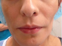
Our 35-year-old female patient presented for evaluation of facial asymmetry. Upon examination, a significant depression was noted on the left cheek (Figure 1). Palpation revealed an area of left mid-facial lipoatrophy. She said that the defect had been present since birth. She has never had the area treated. Options for treatment were discussed, and due to the size of the defect the patient chose to undergo autologous fat transfer.
Figure 1. Facial asymmetry prior to fat transfer.
The patient was brought to the surgical suite. Her outer thigh was prepped with chlorhexidine and draped in sterile fashion. Tumescent anesthesia was prepared (1 L of 0.9% normal saline with a 2 mL epinephrine 1:1000, 25 mL 2% plain lidocaine, 1 mL triamcinolone 10 mg/mL and 10 mL of 8.4% bicarbonate). After being anesthetized with 2% lidocaine, a small incision was made in the patient’s outer thigh with an 11-blade scalpel. The tumescent anesthesia was infiltrated until anesthesia was obtained. Using a 4-mm sterile liposuction cannula, approximately 15 mL of fat aspirate was removed. The procedure was repeated on the abdomen to obtain another 5 mL of fat aspirate.
The area to be filled on the left cheek was marked. After anesthesia was obtained with 2% lidocaine, a small incision in the preauricular sulcus was made. Multiple passes were used to infiltrate the 20 mL of fat aspirate into the defect. The defect was slightly overcorrected to account for expected fat loss. All of the incisions were sutured closed using 5-0 vicryl dermal sutures and 5-0 prolene sutures to approximate the skin edges. Compression dressings were used over the fat donor sites and the area on the cheek was dressed. The procedure was well-tolerated by the patient.
Discussion
Autologous fat transfer is a popular and effective method to restore volume to facial defects. Most patients have some reservoir of fat that can be easily obtained for use in autologous fat transfer and for subsequent touch-up procedures if needed. The availability of the filler makes it more useful and cost-effective than synthetic fillers in repairing large defects.5
The use of autologous fat is controversial. Autologous fat transfer is a multistep procedure, and each step has been debated over the years. The most debated aspect is graft resorption. One study evaluating mid-face volume augmentation with fat transfer showed that 32% of the initial volume injected remained at approximately 1.5 years. This study used both clinical evaluation and 3-dimensional imaging using a Canfield Scientific Vectra camera and software.11
All of the study participants were treated using the same technique. Despite this, it was noted that there was variability in the degree of volume retention between patients and between sides of the face in the same patients. Only 8 of 33 patients in this study required a touch-up procedure. The study noted that a waiting period of 6 months is recommended prior to touch-up procedures to allow for initial swelling and resorption to subside.11
These results have been confirmed in other studies including animal studies that document resorption rates of 60% to 70%.9 A survey of surgeons showed that the majority of respondents believed overcorrection is necessary. This survey also indicated that most physicians correctly estimate the amount of required overcorrection because only 31% reported that more than half of the their patients required reinjection.9 Therefore, due to this resorption of fat, we recommend overcorrecting by 50% when performing a fat transfer.
The complication rate for fat augmentation is very low.5 Rare complications include fat hypertrophy after weight gain, fat necrosis and infection. Overcorrection is probably the most common complication. It is most commonly found in the infraorbital area, and this area should be approached with caution if at all.5
Additionally, this area, especially medial to the mid-pupillary line, is high risk for vascular occlusion caused by inadvertent intravascular injection, which is the most serious complication of fat augmentation. Blindness and middle cerebral artery occlusion can be caused by this accidental intravascular injection.5 This risk can be mitigated by using blunt tipped cannulas, retrograde injection and avoidance of the infraorbital area. Because of these risks, in our practice, we do not treat the infraorbital area with fat transfer.
Patient Outcome
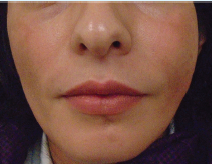
Our patient denied any significant pain or bruising after the procedure. She had no adverse events. At 1-month follow-up, the patient had minimal swelling with marked improvement of the facial asymmetry. (Figure 2). She was very happy with the results.
Figure 2. One month after fat transfer.
Conclusion
Despite the large amount of volume loss, patients are overwhelming pleased with the procedure.11 Several studies have confirmed that patients with facial asymmetry have a high level of satisfaction with the procedure.4 Additionally, it has a number of potential advantages even though it is a more invasive procedure. This autologous procedure is associated with rare complications, and unlike the fillers discussed requires fewer touch-up procedures and decreased cost.7 We recommend the use of autologous fat transfer for large facial defects requiring more than 3 mL of filler except in the infraorbital area.
Dr. Pilcher is a fellow at Affiliated Dermatologists in Morristown, NJ.
Dr. Torres is a practicing dermatologist at Affiliated Dermatologists in Morristown, NJ.
Dr. Rogachefsky is a practicing dermatologist and the Program Director of the ACGME-approved Procedural Dermatology Fellowship at Affiliated Dermatologists & Dermatologist Surgeons in Morristown, NJ.
Disclosure: The authors report no relevant financial relationships..
References
1. Anbarasi K, Sathasivasubramanian S, Krithika CL, Venkatasai PM. Focal lipoatrophy of face: a rare esthetic complaint. J Clin Imaging Sci. 2012;2:14.
2. Echaves M, Hoestman W. Relationship between lipoatrophy and quality of life. AIDS Read. 2005;15(7):369-375.
3. Liu SW, Cohen GF. Idiopathic hemi-facial lipoatrophy treated with autologous fat transfer. J Cosmet Dermatol. 2010;9(3):226-229.
4. Guijarro-Martinez R, Alba LM, Marqués Mateo M, Puche Torres M, Pascual Gil JV. Autologous fat transfer to the cranio-maxillofacial region: updates and controversies. J Craniomaxillofac Surg. 2011;39(5):359-363.
5. Butterwick KJ. Autologous fat transfer: evolving concepts and techniques. In: Robinson JK, Hanke CW, Siegel DM, Fatila A, eds. Surgery of the Skin. 2nd ed. Philadelphia, PA: Elsevier, Inc.; 2010:chap 28.
6. Tierney E, Sengelmann R, Sattler G, Hanke CW. Soft tissue augmentation. In: Robinson JK, Hanke CW, Siegel DM, Fatila A, eds. Surgery of the Skin. 2nd ed. Philadelphia, PA: Elsevier, Inc.; 2010:chap 23.
7. Shuck J, Iorio ML, Hung R, Davison SP. Autologous fat grafting and injectable dermal fillers for human immunodeficiency virus-associated facial lipodystrophy: a comparison of safety, efficacy and long-term treatment outcomes. Plast Reconstr Surg. 2012;131(3):499-506.
8. Serra-Renom JM, Fontdevila J. Treatment of facial fat atrophy related to treatment with protease inhibitors by autologous fat injection in patients with human immunodeficiency virus infection. Plast Reconstr Surg. 2004;114(2):551-555.
9. Kaufman MR, Bradley JP, Dickinson B, et al. Autologous fat transfer national consensus survey: trends in techniques for harvest, preparation and application, and perception of short- and long-term results. Plast Reconstr Surg. 2007;119(1):323-331.
10. Jones DH, Carruthers A, Orentreich D, et al. Highly purified 1000-cSt silicone oil for treatment of human immunodeficiency virus-associated facial lipoatrophy: an open pilot trial. Dermatol Surg. 2004;30(10):1279-1286.
11. Meier JD, Glasgold, RA, Glasgold MJ. Autologous fat grafting long-term evidence of its efficacy in midfacial rejuvenation. Arch Facial Plast Surg. 2009;11(1):24-28.
 Features that are balanced and symmetric define facial beauty. Alterations in the underlying facial structures resulting in facial asymmetry can be disfiguring and can cause psychological distress.1,2 Loss of subcutaneous facial fat can result in depressions leading to facial asymmetry. This lipoatrophy can be acquired or genetic. It has multiple causes including familial lipoatrophy and panniculitis, HIV infection, aging and idiopathic.3 The idiopathic form is rare, and facial involvement is its rarest form.1
A variety of agents have been used to treat facial asymmetry resulting from lipoatrophy, but no ideal filling agent has been found. The search began more than 100 years ago when Neuber described the free autologous fat graft for soft-tissue augmentation.4 The ideal filler should be safe (non-immunogenic, non-carcinogenic, non-teratogenic, non-infectious and low abuse potential), efficacious (natural look and feel with long-term benefit) and practical (cost-effective, easy to use and removable).4 In many ways, fat fulfills these criteria, but its predictability and persistence over time is still debated.5
The use of autologous fat transfer for lipoatrophy has increased in popularity over the past several years, especially for larger defects.4 When a defect requires larger volumes, the cost of synthetic fillers can be prohibitive due to the number of syringes needed to correct the defect.5 In these cases, autologous fat is a popular choice for volume correction.
In this article, we present a case study of a patient with idiopathic hemifacial lipoatrophy treated successfully with autologous fat transfer.
Therapeutic Alternatives
Lipoatrophy has been treated with multiple other modalities, including silicone oil, collagen, calcium hydroxlyapatite, hyaluronic acid and poly-L-lactic acid fillers.6 These treatments are synthetic and are often more expensive than autologous fat transfer, but offer increased ease of use.
Poly-L-lactic acid and hyaluronic acid fillers are the most common synthetic fillers used to treat facial lipoatrophy. A meta-analysis of 19 studies comparing poly-L-lactic acid, hyaluronic acid and autologous fat transfer showed long-term stability of both dermal fillers and autologous fat transfer in HIV-associated facial lipoatrophy.7 Patient satisfaction scores and long-term thickness scales were significantly increased with both the autologous fat transfer and the filler groups.7 This demonstrates that both fillers and autologous fat transfer are superb options for facial lipoatrophy.
In this review article, the autologous fat transfer group did not experience any adverse events other than suboptimal aesthetic outcomes.7 In contrast, 27% of patients in the dermal filler groups experienced bruising and pain. Additionally, complications based on the foreign body nature of the filler were reported in 15% of the poly-L-lactic acid patients, including nodule and papule formation and rare anaphylactic reactions.7
Rates of repeated injections were discussed in 12 studies reviewed in the meta-analysis. Patients treated with poly-L-lactic acid received more sets of injections than those treated with fat transfer or hyaluronic acid. The majority of patients treated with poly-L-lactic acid had 3 or more injections, while those treated with hyaluronic acid required a maximum of 2 injections.7 In a separate study, autologous fat transfer required 2 treatments in only 46% of patients.8 Additionally, all synthetic fillers are made of biologic materials that are subject to degradation by the body, requiring the need for possible future injections.9 The synthetic fillers’ cost is based on the amount of product used, and multiple injections increase cost dramatically making synthetic fillers less desirable for large facial defects.
Another treatment option for facial lipoatrophy is the use of microdroplet liquid silicone. While not FDA approved, it is used legally off-label as a permanent treatment for HIV-associated lipoatrophy.10 It has a high success rate with few side effects and high patient satisfaction.10 Despite these benefits, it requires multiple injections over the course of several months and is not commonly used for lipoatrophy not associated with HIV.
Case Study
Our 35-year-old female patient presented for evaluation of facial asymmetry. Upon examination, a significant depression was noted on the left cheek (Figure 1). Palpation revealed an area of left mid-facial lipoatrophy. She said that the defect had been present since birth. She has never had the area treated. Options for treatment were discussed, and due to the size of the defect the patient chose to undergo autologous fat transfer.
Figure 1. Facial asymmetry prior to fat transfer.
The patient was brought to the surgical suite. Her outer thigh was prepped with chlorhexidine and draped in sterile fashion. Tumescent anesthesia was prepared (1 L of 0.9% normal saline with a 2 mL epinephrine 1:1000, 25 mL 2% plain lidocaine, 1 mL triamcinolone 10 mg/mL and 10 mL of 8.4% bicarbonate). After being anesthetized with 2% lidocaine, a small incision was made in the patient’s outer thigh with an 11-blade scalpel. The tumescent anesthesia was infiltrated until anesthesia was obtained. Using a 4-mm sterile liposuction cannula, approximately 15 mL of fat aspirate was removed. The procedure was repeated on the abdomen to obtain another 5 mL of fat aspirate.
The area to be filled on the left cheek was marked. After anesthesia was obtained with 2% lidocaine, a small incision in the preauricular sulcus was made. Multiple passes were used to infiltrate the 20 mL of fat aspirate into the defect. The defect was slightly overcorrected to account for expected fat loss. All of the incisions were sutured closed using 5-0 vicryl dermal sutures and 5-0 prolene sutures to approximate the skin edges. Compression dressings were used over the fat donor sites and the area on the cheek was dressed. The procedure was well-tolerated by the patient.
Discussion
Autologous fat transfer is a popular and effective method to restore volume to facial defects. Most patients have some reservoir of fat that can be easily obtained for use in autologous fat transfer and for subsequent touch-up procedures if needed. The availability of the filler makes it more useful and cost-effective than synthetic fillers in repairing large defects.5
The use of autologous fat is controversial. Autologous fat transfer is a multistep procedure, and each step has been debated over the years. The most debated aspect is graft resorption. One study evaluating mid-face volume augmentation with fat transfer showed that 32% of the initial volume injected remained at approximately 1.5 years. This study used both clinical evaluation and 3-dimensional imaging using a Canfield Scientific Vectra camera and software.11
All of the study participants were treated using the same technique. Despite this, it was noted that there was variability in the degree of volume retention between patients and between sides of the face in the same patients. Only 8 of 33 patients in this study required a touch-up procedure. The study noted that a waiting period of 6 months is recommended prior to touch-up procedures to allow for initial swelling and resorption to subside.11
These results have been confirmed in other studies including animal studies that document resorption rates of 60% to 70%.9 A survey of surgeons showed that the majority of respondents believed overcorrection is necessary. This survey also indicated that most physicians correctly estimate the amount of required overcorrection because only 31% reported that more than half of the their patients required reinjection.9 Therefore, due to this resorption of fat, we recommend overcorrecting by 50% when performing a fat transfer.
The complication rate for fat augmentation is very low.5 Rare complications include fat hypertrophy after weight gain, fat necrosis and infection. Overcorrection is probably the most common complication. It is most commonly found in the infraorbital area, and this area should be approached with caution if at all.5
Additionally, this area, especially medial to the mid-pupillary line, is high risk for vascular occlusion caused by inadvertent intravascular injection, which is the most serious complication of fat augmentation. Blindness and middle cerebral artery occlusion can be caused by this accidental intravascular injection.5 This risk can be mitigated by using blunt tipped cannulas, retrograde injection and avoidance of the infraorbital area. Because of these risks, in our practice, we do not treat the infraorbital area with fat transfer.
Patient Outcome
Our patient denied any significant pain or bruising after the procedure. She had no adverse events. At 1-month follow-up, the patient had minimal swelling with marked improvement of the facial asymmetry. (Figure 2). She was very happy with the results.
Figure 2. One month after fat transfer.
Conclusion
Despite the large amount of volume loss, patients are overwhelming pleased with the procedure.11 Several studies have confirmed that patients with facial asymmetry have a high level of satisfaction with the procedure.4 Additionally, it has a number of potential advantages even though it is a more invasive procedure. This autologous procedure is associated with rare complications, and unlike the fillers discussed requires fewer touch-up procedures and decreased cost.7 We recommend the use of autologous fat transfer for large facial defects requiring more than 3 mL of filler except in the infraorbital area.
Dr. Pilcher is a fellow at Affiliated Dermatologists in Morristown, NJ.
Dr. Torres is a practicing dermatologist at Affiliated Dermatologists in Morristown, NJ.
Dr. Rogachefsky is a practicing dermatologist and the Program Director of the ACGME-approved Procedural Dermatology Fellowship at Affiliated Dermatologists & Dermatologist Surgeons in Morristown, NJ.
Disclosure: The authors report no relevant financial relationships..
References
1. Anbarasi K, Sathasivasubramanian S, Krithika CL, Venkatasai PM. Focal lipoatrophy of face: a rare esthetic complaint. J Clin Imaging Sci. 2012;2:14.
2. Echaves M, Hoestman W. Relationship between lipoatrophy and quality of life. AIDS Read. 2005;15(7):369-375.
3. Liu SW, Cohen GF. Idiopathic hemi-facial lipoatrophy treated with autologous fat transfer. J Cosmet Dermatol. 2010;9(3):226-229.
4. Guijarro-Martinez R, Alba LM, Marqués Mateo M, Puche Torres M, Pascual Gil JV. Autologous fat transfer to the cranio-maxillofacial region: updates and controversies. J Craniomaxillofac Surg. 2011;39(5):359-363.
5. Butterwick KJ. Autologous fat transfer: evolving concepts and techniques. In: Robinson JK, Hanke CW, Siegel DM, Fatila A, eds. Surgery of the Skin. 2nd ed. Philadelphia, PA: Elsevier, Inc.; 2010:chap 28.
6. Tierney E, Sengelmann R, Sattler G, Hanke CW. Soft tissue augmentation. In: Robinson JK, Hanke CW, Siegel DM, Fatila A, eds. Surgery of the Skin. 2nd ed. Philadelphia, PA: Elsevier, Inc.; 2010:chap 23.
7. Shuck J, Iorio ML, Hung R, Davison SP. Autologous fat grafting and injectable dermal fillers for human immunodeficiency virus-associated facial lipodystrophy: a comparison of safety, efficacy and long-term treatment outcomes. Plast Reconstr Surg. 2012;131(3):499-506.
8. Serra-Renom JM, Fontdevila J. Treatment of facial fat atrophy related to treatment with protease inhibitors by autologous fat injection in patients with human immunodeficiency virus infection. Plast Reconstr Surg. 2004;114(2):551-555.
9. Kaufman MR, Bradley JP, Dickinson B, et al. Autologous fat transfer national consensus survey: trends in techniques for harvest, preparation and application, and perception of short- and long-term results. Plast Reconstr Surg. 2007;119(1):323-331.
10. Jones DH, Carruthers A, Orentreich D, et al. Highly purified 1000-cSt silicone oil for treatment of human immunodeficiency virus-associated facial lipoatrophy: an open pilot trial. Dermatol Surg. 2004;30(10):1279-1286.
11. Meier JD, Glasgold, RA, Glasgold MJ. Autologous fat grafting long-term evidence of its efficacy in midfacial rejuvenation. Arch Facial Plast Surg. 2009;11(1):24-28.
Features that are balanced and symmetric define facial beauty. Alterations in the underlying facial structures resulting in facial asymmetry can be disfiguring and can cause psychological distress.1,2 Loss of subcutaneous facial fat can result in depressions leading to facial asymmetry. This lipoatrophy can be acquired or genetic. It has multiple causes including familial lipoatrophy and panniculitis, HIV infection, aging and idiopathic.3 The idiopathic form is rare, and facial involvement is its rarest form.1
A variety of agents have been used to treat facial asymmetry resulting from lipoatrophy, but no ideal filling agent has been found. The search began more than 100 years ago when Neuber described the free autologous fat graft for soft-tissue augmentation.4 The ideal filler should be safe (non-immunogenic, non-carcinogenic, non-teratogenic, non-infectious and low abuse potential), efficacious (natural look and feel with long-term benefit) and practical (cost-effective, easy to use and removable).4 In many ways, fat fulfills these criteria, but its predictability and persistence over time is still debated.5
The use of autologous fat transfer for lipoatrophy has increased in popularity over the past several years, especially for larger defects.4 When a defect requires larger volumes, the cost of synthetic fillers can be prohibitive due to the number of syringes needed to correct the defect.5 In these cases, autologous fat is a popular choice for volume correction.
In this article, we present a case study of a patient with idiopathic hemifacial lipoatrophy treated successfully with autologous fat transfer.
Therapeutic Alternatives
Lipoatrophy has been treated with multiple other modalities, including silicone oil, collagen, calcium hydroxlyapatite, hyaluronic acid and poly-L-lactic acid fillers.6 These treatments are synthetic and are often more expensive than autologous fat transfer, but offer increased ease of use.
Poly-L-lactic acid and hyaluronic acid fillers are the most common synthetic fillers used to treat facial lipoatrophy. A meta-analysis of 19 studies comparing poly-L-lactic acid, hyaluronic acid and autologous fat transfer showed long-term stability of both dermal fillers and autologous fat transfer in HIV-associated facial lipoatrophy.7 Patient satisfaction scores and long-term thickness scales were significantly increased with both the autologous fat transfer and the filler groups.7 This demonstrates that both fillers and autologous fat transfer are superb options for facial lipoatrophy.
In this review article, the autologous fat transfer group did not experience any adverse events other than suboptimal aesthetic outcomes.7 In contrast, 27% of patients in the dermal filler groups experienced bruising and pain. Additionally, complications based on the foreign body nature of the filler were reported in 15% of the poly-L-lactic acid patients, including nodule and papule formation and rare anaphylactic reactions.7
Rates of repeated injections were discussed in 12 studies reviewed in the meta-analysis. Patients treated with poly-L-lactic acid received more sets of injections than those treated with fat transfer or hyaluronic acid. The majority of patients treated with poly-L-lactic acid had 3 or more injections, while those treated with hyaluronic acid required a maximum of 2 injections.7 In a separate study, autologous fat transfer required 2 treatments in only 46% of patients.8 Additionally, all synthetic fillers are made of biologic materials that are subject to degradation by the body, requiring the need for possible future injections.9 The synthetic fillers’ cost is based on the amount of product used, and multiple injections increase cost dramatically making synthetic fillers less desirable for large facial defects.
Another treatment option for facial lipoatrophy is the use of microdroplet liquid silicone. While not FDA approved, it is used legally off-label as a permanent treatment for HIV-associated lipoatrophy.10 It has a high success rate with few side effects and high patient satisfaction.10 Despite these benefits, it requires multiple injections over the course of several months and is not commonly used for lipoatrophy not associated with HIV.
Case Study
Our 35-year-old female patient presented for evaluation of facial asymmetry. Upon examination, a significant depression was noted on the left cheek (Figure 1). Palpation revealed an area of left mid-facial lipoatrophy. She said that the defect had been present since birth. She has never had the area treated. Options for treatment were discussed, and due to the size of the defect the patient chose to undergo autologous fat transfer.
Figure 1. Facial asymmetry prior to fat transfer.
The patient was brought to the surgical suite. Her outer thigh was prepped with chlorhexidine and draped in sterile fashion. Tumescent anesthesia was prepared (1 L of 0.9% normal saline with a 2 mL epinephrine 1:1000, 25 mL 2% plain lidocaine, 1 mL triamcinolone 10 mg/mL and 10 mL of 8.4% bicarbonate). After being anesthetized with 2% lidocaine, a small incision was made in the patient’s outer thigh with an 11-blade scalpel. The tumescent anesthesia was infiltrated until anesthesia was obtained. Using a 4-mm sterile liposuction cannula, approximately 15 mL of fat aspirate was removed. The procedure was repeated on the abdomen to obtain another 5 mL of fat aspirate.
The area to be filled on the left cheek was marked. After anesthesia was obtained with 2% lidocaine, a small incision in the preauricular sulcus was made. Multiple passes were used to infiltrate the 20 mL of fat aspirate into the defect. The defect was slightly overcorrected to account for expected fat loss. All of the incisions were sutured closed using 5-0 vicryl dermal sutures and 5-0 prolene sutures to approximate the skin edges. Compression dressings were used over the fat donor sites and the area on the cheek was dressed. The procedure was well-tolerated by the patient.
Discussion
Autologous fat transfer is a popular and effective method to restore volume to facial defects. Most patients have some reservoir of fat that can be easily obtained for use in autologous fat transfer and for subsequent touch-up procedures if needed. The availability of the filler makes it more useful and cost-effective than synthetic fillers in repairing large defects.5
The use of autologous fat is controversial. Autologous fat transfer is a multistep procedure, and each step has been debated over the years. The most debated aspect is graft resorption. One study evaluating mid-face volume augmentation with fat transfer showed that 32% of the initial volume injected remained at approximately 1.5 years. This study used both clinical evaluation and 3-dimensional imaging using a Canfield Scientific Vectra camera and software.11
All of the study participants were treated using the same technique. Despite this, it was noted that there was variability in the degree of volume retention between patients and between sides of the face in the same patients. Only 8 of 33 patients in this study required a touch-up procedure. The study noted that a waiting period of 6 months is recommended prior to touch-up procedures to allow for initial swelling and resorption to subside.11
These results have been confirmed in other studies including animal studies that document resorption rates of 60% to 70%.9 A survey of surgeons showed that the majority of respondents believed overcorrection is necessary. This survey also indicated that most physicians correctly estimate the amount of required overcorrection because only 31% reported that more than half of the their patients required reinjection.9 Therefore, due to this resorption of fat, we recommend overcorrecting by 50% when performing a fat transfer.
The complication rate for fat augmentation is very low.5 Rare complications include fat hypertrophy after weight gain, fat necrosis and infection. Overcorrection is probably the most common complication. It is most commonly found in the infraorbital area, and this area should be approached with caution if at all.5
Additionally, this area, especially medial to the mid-pupillary line, is high risk for vascular occlusion caused by inadvertent intravascular injection, which is the most serious complication of fat augmentation. Blindness and middle cerebral artery occlusion can be caused by this accidental intravascular injection.5 This risk can be mitigated by using blunt tipped cannulas, retrograde injection and avoidance of the infraorbital area. Because of these risks, in our practice, we do not treat the infraorbital area with fat transfer.
Patient Outcome
Our patient denied any significant pain or bruising after the procedure. She had no adverse events. At 1-month follow-up, the patient had minimal swelling with marked improvement of the facial asymmetry. (Figure 2). She was very happy with the results.
Figure 2. One month after fat transfer.
Conclusion
Despite the large amount of volume loss, patients are overwhelming pleased with the procedure.11 Several studies have confirmed that patients with facial asymmetry have a high level of satisfaction with the procedure.4 Additionally, it has a number of potential advantages even though it is a more invasive procedure. This autologous procedure is associated with rare complications, and unlike the fillers discussed requires fewer touch-up procedures and decreased cost.7 We recommend the use of autologous fat transfer for large facial defects requiring more than 3 mL of filler except in the infraorbital area.
Dr. Pilcher is a fellow at Affiliated Dermatologists in Morristown, NJ.
Dr. Torres is a practicing dermatologist at Affiliated Dermatologists in Morristown, NJ.
Dr. Rogachefsky is a practicing dermatologist and the Program Director of the ACGME-approved Procedural Dermatology Fellowship at Affiliated Dermatologists & Dermatologist Surgeons in Morristown, NJ.
Disclosure: The authors report no relevant financial relationships..
References
1. Anbarasi K, Sathasivasubramanian S, Krithika CL, Venkatasai PM. Focal lipoatrophy of face: a rare esthetic complaint. J Clin Imaging Sci. 2012;2:14.
2. Echaves M, Hoestman W. Relationship between lipoatrophy and quality of life. AIDS Read. 2005;15(7):369-375.
3. Liu SW, Cohen GF. Idiopathic hemi-facial lipoatrophy treated with autologous fat transfer. J Cosmet Dermatol. 2010;9(3):226-229.
4. Guijarro-Martinez R, Alba LM, Marqués Mateo M, Puche Torres M, Pascual Gil JV. Autologous fat transfer to the cranio-maxillofacial region: updates and controversies. J Craniomaxillofac Surg. 2011;39(5):359-363.
5. Butterwick KJ. Autologous fat transfer: evolving concepts and techniques. In: Robinson JK, Hanke CW, Siegel DM, Fatila A, eds. Surgery of the Skin. 2nd ed. Philadelphia, PA: Elsevier, Inc.; 2010:chap 28.
6. Tierney E, Sengelmann R, Sattler G, Hanke CW. Soft tissue augmentation. In: Robinson JK, Hanke CW, Siegel DM, Fatila A, eds. Surgery of the Skin. 2nd ed. Philadelphia, PA: Elsevier, Inc.; 2010:chap 23.
7. Shuck J, Iorio ML, Hung R, Davison SP. Autologous fat grafting and injectable dermal fillers for human immunodeficiency virus-associated facial lipodystrophy: a comparison of safety, efficacy and long-term treatment outcomes. Plast Reconstr Surg. 2012;131(3):499-506.
8. Serra-Renom JM, Fontdevila J. Treatment of facial fat atrophy related to treatment with protease inhibitors by autologous fat injection in patients with human immunodeficiency virus infection. Plast Reconstr Surg. 2004;114(2):551-555.
9. Kaufman MR, Bradley JP, Dickinson B, et al. Autologous fat transfer national consensus survey: trends in techniques for harvest, preparation and application, and perception of short- and long-term results. Plast Reconstr Surg. 2007;119(1):323-331.
10. Jones DH, Carruthers A, Orentreich D, et al. Highly purified 1000-cSt silicone oil for treatment of human immunodeficiency virus-associated facial lipoatrophy: an open pilot trial. Dermatol Surg. 2004;30(10):1279-1286.
11. Meier JD, Glasgold, RA, Glasgold MJ. Autologous fat grafting long-term evidence of its efficacy in midfacial rejuvenation. Arch Facial Plast Surg. 2009;11(1):24-28.