PATIENT PRESENTATION
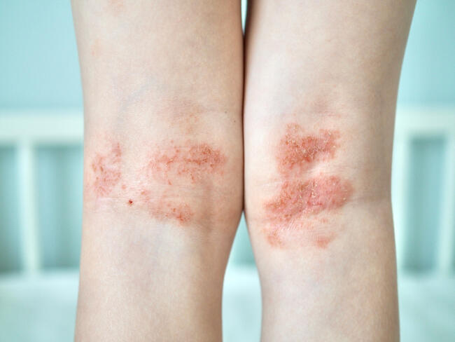
A 68-year-old Bengali male admitted to the hospital for acute coronary syndrome was evaluated in dermatologic consultation for longstanding skin-colored 4 cm to 5 cm hyperpigmented, hyperkeratotic large round nodules just inferior to the knees bilaterally. These lesions were intermittently uncomfortable to the patient, whose past medical history was significant for type II diabetes mellitus controlled with metformin. The social history was notable for the patient’s being a devout Muslim who prayed five times a day. The remainder of his personal, family, medical or surgical histories were non-contributory. What is Your Diagnosis? The skin has numerous functions, one of which is to protect the body from external damage by mechanical forces and various other external stimuli. Many activities, including religious and cultural practices, can be the cause of skin lesions, which are, in essence, the equivalent of occupational calluses. Calluses are well-circumscribed, hyperkeratotic plaques induced by persistent and repeated trauma, and their location usually suggests the diagnosis and etiology.1 Hyperkeratosis is a normal protective response of the skin, which becomes pathologic when the callus grows so large that it becomes symptomatic, such as in our patient.2 Calluses of knees, ankles and foreheads secondary to repeat trauma sustained during Muslim praying have been occasionally reported in the dermatologic literature.3,4 During prayer, the worshipper kneels and bows, with his forehead and knees touching the ground. This motion can occur over 40 times during the five daily prayers, resulting in repeated trauma to the skin and subsequent callus formation.5 In the Quran, there exists a probable reference to such calluses in the following verse, “Their (the believers) foreheads show the mark left by frequent prostration,” Surah Al-Fath (Quran 48:29).6 There are also examples of prayer calluses in Christianity, the most famous of which being James the apostle. First documented by Jerome in the 4th century, James’s knees were said to be calloused similar to a camel’s knees secondary to his religious devotion.7 Additionally, calluses of the knees have been documented in individuals who work in kneeling positions such as miners, plumbers and floor scrubbers. Ankle calluses have been reported in people who work with their ankles crossed, such as linoleum layers. Finally, calluses in the middle of both shins have been documented in painters and paperhangers due to the constant pressure of one rung of the ladder on an area of skin with minimal subcutaneous fat.3 Our patient’s calluses were pigmented calluses, which have been reported in Fitzpatrick skin type IV and V patients. Moreover, in patients suffering from sensory neuropathy, such as those with diabetes (our patient) and leprosy, stimuli that might normally elicit pain or discomfort are attenuated, thereby predisposing the skin to repetitive trauma and subsequent callus formation. Sensory deprivation can also lead to an exaggerated form of these calluses (ie, ulcerated calluses).1
Diagnosis
The diagnosis of calluses is essentially clinical, and it should focus on the etiology, including those developing from cultural and devotional practices in addition to those due to repeat trauma alone or in combination with neuropathies. Oftentimes, there is an association of calluses with orthopedic foot problems, predisposing occupations or particular hobbies.3 Differential Diagnosis While we propose that our patient’s distinctly located calluses are the result of his religious practices, warts should be included in the differential diagnosis as they are frequently mistaken for calluses and vice versa.2 Other differential diagnoses may include corn/heloma, which unlike calluses are inflamed and tender, and prurigo nodules. Differentiating a Callus from a Wart Debridement — If it is necessary to differentiate a callus from a wart, paring with a #15 surgical blade may be performed. Warts, unlike mechanical lesions, tend to bleed on sharp debridement. If small black or brown dots are visible after removal of the hyperkeratotic tissue, thrombosed blood vessels have become entrapped in the cylindrical projections formed by the virus.8 Dermoscopy —Epiluminescent microscopy (ELM), also known as dermatoscopy, can also be useful in differentiating warts from calluses. Under ELM, homogeneous black to red dots and globules representing thrombosed capillaries as well as papilliform surfaces or interrupted skin lines will be more clearly visible if the lesion is a viral wart. In addition, trimming the surface keratin will make the capillaries more prominent and subsequent ELM may reveal accompanying red linear vessels. In contrast to warts, calluses under ELM will appear as a homogeneous opacity. 9
Histopathology
Although skin biopsies are generally not necessary for the diagnosis of this condition, histopathologically calluses are characterized by compact hyperkeratosis, hypergranulosis, acanthosis, and potentially a mild inflammatory infiltrate in the upper dermis.3
Management
Treatment focuses upon conservative management by providing symptomatic relief via debridement and keratolytics, as well as formulating a treatment plan that includes avoidance of further mechanical injury to the area. If conservative measures fail to provide symptomatic control, surgical excision may be considered. Debridement — The primary treatment method for calluses is sharp debridement to reduce the amount of hyperkeratotic tissue, providing almost complete relief to the area. Keratolytics — Topical keratolyics, such as those containing either salicylic acid or urea, may also be employed. These agents should be used with caution, however, especially in neuropathic and immunocompromised patients because they may cause damage to surrounding normal tissues. Behavioral modification — As calluses are reactive lesions secondary to increased mechanical pressure or friction, once the injury-causing behavior has been identified, the patient may be able to institute behavioral modifications to reduce pressure or friction on the area. One such behavioral modification is the use of therapeutic padding, which comes in many forms such as silicone and felt, and will reduce the amount of mechanical irritation to the site of the callus and thereby help to alleviate symptoms. Surgery — If the conservative measures outlined above fail, surgery may be considered to correct abnormal mechanical stresses. Surgical correction is usually reserved for calluses of the foot, where orthopedic deformities result in increased pressure to certain areas and subsequent callus formation.6 Analgesics — The role for analgesics in the management of calluses has not been well delineated in the literature but may also be worth considering in patient with significant levels of pain. Moreover, although oral as well as topical and transdermal agents, such as gabapentin and lidocaine, are currently commonly prescribed for neuropathic pain, there are no reports in the literature for these medications being used specifically for neuropathic pain related to calluses. Our Patient The patient was offered keratolytics, as well as surgical excision for symptomatic treatment of his lesions. However, upon learning these lesions were secondary to his religious devotion, he declined further treatment.
Summary
This case highlights the effects of repetitive motions or activities in causing callus formation, which may be exacerbated by neuropathy. Diagnosis of callus is mainly clinical, and may be aided by ELM. Skin biopsy may also be performed if necessary. Differential diagnosis includes warts, corns, and purigo nodules. Treatment options include behavioral modifications, surgical debridement, or keratolytics. Dr. Pothiawala is a graduate student at Harvard School of Public Health, Boston, MA. Dr. Ibrahimi is a resident, Department of Dermatology, Harvard Medical School, and Massachusetts General Hospital, Boston, MA. Dr. Kroshinsky is Instructor in Dermatology, Department of Dermatology, Harvard Medical School, and Massachusetts General Hospital, Boston, MA. Dr. Khachemoune, the Section Editor of Derm Dx, is with the Department of Dermatology, State University of New York, Brooklyn, NY. Disclosure: The authors have no conflict of interest with any material presented in this month’s column.
PATIENT PRESENTATION
A 68-year-old Bengali male admitted to the hospital for acute coronary syndrome was evaluated in dermatologic consultation for longstanding skin-colored 4 cm to 5 cm hyperpigmented, hyperkeratotic large round nodules just inferior to the knees bilaterally. These lesions were intermittently uncomfortable to the patient, whose past medical history was significant for type II diabetes mellitus controlled with metformin. The social history was notable for the patient’s being a devout Muslim who prayed five times a day. The remainder of his personal, family, medical or surgical histories were non-contributory. What is Your Diagnosis? The skin has numerous functions, one of which is to protect the body from external damage by mechanical forces and various other external stimuli. Many activities, including religious and cultural practices, can be the cause of skin lesions, which are, in essence, the equivalent of occupational calluses. Calluses are well-circumscribed, hyperkeratotic plaques induced by persistent and repeated trauma, and their location usually suggests the diagnosis and etiology.1 Hyperkeratosis is a normal protective response of the skin, which becomes pathologic when the callus grows so large that it becomes symptomatic, such as in our patient.2 Calluses of knees, ankles and foreheads secondary to repeat trauma sustained during Muslim praying have been occasionally reported in the dermatologic literature.3,4 During prayer, the worshipper kneels and bows, with his forehead and knees touching the ground. This motion can occur over 40 times during the five daily prayers, resulting in repeated trauma to the skin and subsequent callus formation.5 In the Quran, there exists a probable reference to such calluses in the following verse, “Their (the believers) foreheads show the mark left by frequent prostration,” Surah Al-Fath (Quran 48:29).6 There are also examples of prayer calluses in Christianity, the most famous of which being James the apostle. First documented by Jerome in the 4th century, James’s knees were said to be calloused similar to a camel’s knees secondary to his religious devotion.7 Additionally, calluses of the knees have been documented in individuals who work in kneeling positions such as miners, plumbers and floor scrubbers. Ankle calluses have been reported in people who work with their ankles crossed, such as linoleum layers. Finally, calluses in the middle of both shins have been documented in painters and paperhangers due to the constant pressure of one rung of the ladder on an area of skin with minimal subcutaneous fat.3 Our patient’s calluses were pigmented calluses, which have been reported in Fitzpatrick skin type IV and V patients. Moreover, in patients suffering from sensory neuropathy, such as those with diabetes (our patient) and leprosy, stimuli that might normally elicit pain or discomfort are attenuated, thereby predisposing the skin to repetitive trauma and subsequent callus formation. Sensory deprivation can also lead to an exaggerated form of these calluses (ie, ulcerated calluses).1
Diagnosis
The diagnosis of calluses is essentially clinical, and it should focus on the etiology, including those developing from cultural and devotional practices in addition to those due to repeat trauma alone or in combination with neuropathies. Oftentimes, there is an association of calluses with orthopedic foot problems, predisposing occupations or particular hobbies.3 Differential Diagnosis While we propose that our patient’s distinctly located calluses are the result of his religious practices, warts should be included in the differential diagnosis as they are frequently mistaken for calluses and vice versa.2 Other differential diagnoses may include corn/heloma, which unlike calluses are inflamed and tender, and prurigo nodules. Differentiating a Callus from a Wart Debridement — If it is necessary to differentiate a callus from a wart, paring with a #15 surgical blade may be performed. Warts, unlike mechanical lesions, tend to bleed on sharp debridement. If small black or brown dots are visible after removal of the hyperkeratotic tissue, thrombosed blood vessels have become entrapped in the cylindrical projections formed by the virus.8 Dermoscopy —Epiluminescent microscopy (ELM), also known as dermatoscopy, can also be useful in differentiating warts from calluses. Under ELM, homogeneous black to red dots and globules representing thrombosed capillaries as well as papilliform surfaces or interrupted skin lines will be more clearly visible if the lesion is a viral wart. In addition, trimming the surface keratin will make the capillaries more prominent and subsequent ELM may reveal accompanying red linear vessels. In contrast to warts, calluses under ELM will appear as a homogeneous opacity. 9
Histopathology
Although skin biopsies are generally not necessary for the diagnosis of this condition, histopathologically calluses are characterized by compact hyperkeratosis, hypergranulosis, acanthosis, and potentially a mild inflammatory infiltrate in the upper dermis.3
Management
Treatment focuses upon conservative management by providing symptomatic relief via debridement and keratolytics, as well as formulating a treatment plan that includes avoidance of further mechanical injury to the area. If conservative measures fail to provide symptomatic control, surgical excision may be considered. Debridement — The primary treatment method for calluses is sharp debridement to reduce the amount of hyperkeratotic tissue, providing almost complete relief to the area. Keratolytics — Topical keratolyics, such as those containing either salicylic acid or urea, may also be employed. These agents should be used with caution, however, especially in neuropathic and immunocompromised patients because they may cause damage to surrounding normal tissues. Behavioral modification — As calluses are reactive lesions secondary to increased mechanical pressure or friction, once the injury-causing behavior has been identified, the patient may be able to institute behavioral modifications to reduce pressure or friction on the area. One such behavioral modification is the use of therapeutic padding, which comes in many forms such as silicone and felt, and will reduce the amount of mechanical irritation to the site of the callus and thereby help to alleviate symptoms. Surgery — If the conservative measures outlined above fail, surgery may be considered to correct abnormal mechanical stresses. Surgical correction is usually reserved for calluses of the foot, where orthopedic deformities result in increased pressure to certain areas and subsequent callus formation.6 Analgesics — The role for analgesics in the management of calluses has not been well delineated in the literature but may also be worth considering in patient with significant levels of pain. Moreover, although oral as well as topical and transdermal agents, such as gabapentin and lidocaine, are currently commonly prescribed for neuropathic pain, there are no reports in the literature for these medications being used specifically for neuropathic pain related to calluses. Our Patient The patient was offered keratolytics, as well as surgical excision for symptomatic treatment of his lesions. However, upon learning these lesions were secondary to his religious devotion, he declined further treatment.
Summary
This case highlights the effects of repetitive motions or activities in causing callus formation, which may be exacerbated by neuropathy. Diagnosis of callus is mainly clinical, and may be aided by ELM. Skin biopsy may also be performed if necessary. Differential diagnosis includes warts, corns, and purigo nodules. Treatment options include behavioral modifications, surgical debridement, or keratolytics. Dr. Pothiawala is a graduate student at Harvard School of Public Health, Boston, MA. Dr. Ibrahimi is a resident, Department of Dermatology, Harvard Medical School, and Massachusetts General Hospital, Boston, MA. Dr. Kroshinsky is Instructor in Dermatology, Department of Dermatology, Harvard Medical School, and Massachusetts General Hospital, Boston, MA. Dr. Khachemoune, the Section Editor of Derm Dx, is with the Department of Dermatology, State University of New York, Brooklyn, NY. Disclosure: The authors have no conflict of interest with any material presented in this month’s column.