PATIENT PRESENTATION
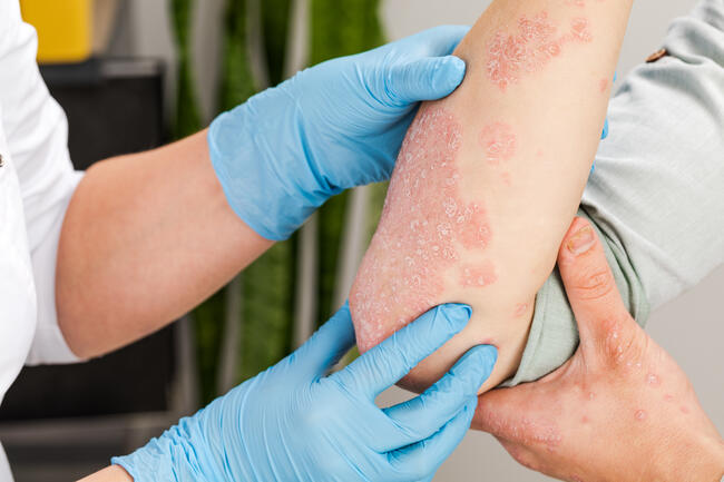
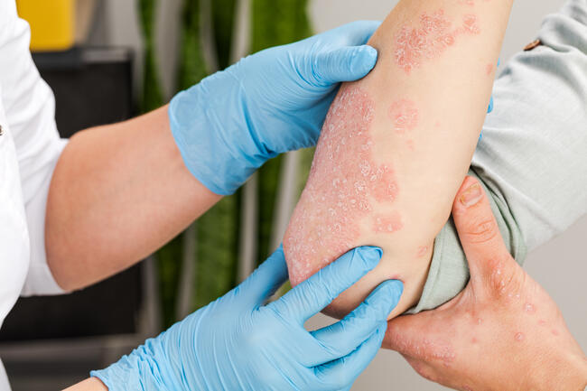
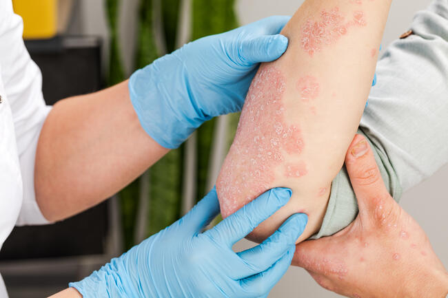
A 64-year-old woman presented with multiple painful blisters on her hands. She noted these after gardening 1 week prior. The patient complained of exquisite pain and tenderness from the sites. She was unaware of any caustic substance that she was exposed to. Her medical history included heart disease, chronic obstructive pulmonary disease, and a history of breast cancer. Aside from these chronic conditions, she was otherwise doing well. She denied any new medications or over-the-counter treatments. On physical examination, she had numerous tense, violaceous pustules and bullae on her dorsal hands and fingers. No other body sites were involved. The patient had no palpable anterior cervical, supraclavicular, or axillary lymphadenopathy. Multiple biopsies were obtained, which revealed dense neutrophilic infiltrates.
_______________________________
Diagnosis: Neutrophilic Dermatosis of the Dorsal Hands
Robert Douglas Sweet first described acute febrile neutrophilic dermatosis, (later renamed Sweet’s Syndrome) in 1964.1 Characteristic skin lesions include painful yet non-pruritic erythematous papules, plaques, pustules, purpura and vesicles primarily involving the face and upper body including the hands.2 Middle-aged females are predominantly affected.3 Associated findings may include fever, arthralgias, pathergy, peripheral blood neutrophilia, elevated erythrocyte sedimentation rate (ESR), slight elevation in alkaline phosphatase and liver transaminases.4 These lesions are often misdiagnosed as infectious in nature and treated unsuccessfully with antibiotics. Palmoplantar pustulosis (PPP) and neutrophilic dermatosis of the dorsal hands (NDDH) may represent subclasses of Sweet’s syndrome, as they both fulfill histologic criteria for diagnosis.5 The former is characterized by erythematous shiny scales involving the palms and soles. Associations with psoriasis and SAPHO (synovitis, acne, pustulosis, hyperostosis and osteomyelitis) syndrome have been described with PPP. NDDH may appear morphologically identical to those of patients with classic Sweet’s syndrome.3 In 1995 Strutton et al6 reported six women with lesions similar to Sweet’s syndrome limited to the dorsal hands but with definite leukocytoclastic vasculitis. The terms “pustular vasculitis” or “pustular vasculitis of the dorsal hands” are used to describe these eruptions.3 Whether or not the vasculitis is itself pathogenic or an epiphenomenon remains an issue of debate.
Histology
A cutaneous biopsy of Sweet’s syndrome classically reveals bandlike dermal neutrophilic invasion, dermal papillary edema — often with epidermal sparing leukocytoclasia, neutrophil karyorrhexis and a characteristic lack of leukocytoclastic vasculitis.2,5,7
Pathogenesis/Immunology
Pathogenesis is felt to be the result of cytokine dysregulation.8,9 Granulocyte colony stimulating factor (G-CSF), granulocyte-macrophage colony stimulating factor (GM-CSF), interferon gamma, and interleukins (IL1, IL3, IL6, IL8) are thought to be key participants. Sweet’s syndrome may represent a cutaneous marker of systemic disease.10 About 85% of malignancies associated with Sweet’s syndrome are hematopoetic (myelodysplastic syndromes, acute myelogenous leukemia, hairy cell leukemia, etc.). Solid tumors account for 15% (breast, gastrointestinal and genitourinary).11,12,13 Bacterial infections (URI, UTI, chronic osteomyelitis), autoimmune/collagen vascular disease, inflammatory bowel disease and pregnancy have all been associated.1,14,15,16 Drug-induced Sweet’s syndrome has also been described. Offending medications include: G-CSF, GM-CSF, trimethoprim-sulfamethoxazole, lithium, oral contraceptives, furosemide, minocycline, imatinib, bortezomib, and all-trans retinoic acid.2 The anti-tumor necrosis factor antibody, infliximab, has been implicated as a possible inciting agent during the treatment of ankylosing spondylitis.15
Differential Diagnosis
The differential diagnosis may include pyoderma gangrenosum, pustular psoriasis, reactive arthritis, SAPHO syndrome, palmoplantar pustulosis, subcorneal pustular dermatosis, bowel-associated dermatosis-arthritis syndrome, Behcet’s disease, neutrophilic dermatosis associated with rheumatoid arthritis, neutrophilic eccrine hidradenitis, erythema multiforme, urticarial vasculitis, erythema elevatum diutinum, lymphoma cutis, and deep bacterial or fungal infections. Biopsy and culture when appropriate can be used to differentiate these.
Criteria to Assist Diagnosis
In addition, Su and Liu proposed major and minor criteria to assist in the diagnosis.17 For this classification, two major and two minor criteria are required to make the diagnosis of Sweet’s syndrome. The major criteria include:
1. abrupt onset of typical cutaneous lesions and
2. histology supportive of Sweet’s syndrome.
The minor criteria include:
1. association with specific infection or vaccination,
2. associated malignancy or inflammatory disorder, associated drug exposure,
3. presence of fever and constitutional signs/symptoms, 3) leukocytosis, and 4) response to systemic corticosteroids. Neutrophilic dermatosis of the dorsal hands is a specific subset of Sweet’s syndrome. Its differentiation is clear with its specific anatomic involvement.
Management/Treatment
Sweet’ syndrome lesions can remit without treatment, although glucocorticoids are often necessary. Tar, retinoids, PUVA and methotrexate have been used with variable success in persistent cases.3,4,6 NDDH may also be treated with steroids and/or dapsone. Dapsone may be used for steroid resistant cases and in patients with recurrent flares. Low doses (50 mg/day to 75 mg/day) have been successful as maintenance therapy.3,6 The patient described worsened after treatment with topical corticosteroids and an oral corticosteroid taper. The addition of dapsone, however, did resolve her cutaneous eruption.
Outcomes
Early diagnosis and treatment can avoid unnecessary antibiotic therapy in affected patients. Prompt recognition of Sweet’s syndrome or its variants can clue the physician to an undiagnosed systemic disorder or drug reaction.
Dr. Persad is with the Division of Dermatology in the Department of Internal Medicine at Mercer University School of Medicine, Macon, GA. Dr. Lane is with the Division of Dermatology in the Departments of Internal Medicine and Surgery at Mercer University School of Medicine, Macon, GA, as well as the Medical College of Georgia, Augusta, GA. He is also with the Department of Dermatology at Emory University School of Medicine, Atlanta, GA. Dr. Khachemoune, the Section Editor of Derm Dx, is with the Department of Dermatology, State University of New York, Brooklyn, NY. Disclosure: The authors have no conflict of interest with any material presented in this column.
________________________________
CALL FOR CASES
f you have a case you’d like to see published, send a write-up (1,200 to 1,500 words) and an image of the patient’s condition to: Dr. Amor Khachemoune Department of Dermatology SUNY Downstate Medical Center 450 Clarkson Avenue Brooklyn, NY 11203 Or, e-mail them to amorkh@pol.net.
PATIENT PRESENTATION
A 64-year-old woman presented with multiple painful blisters on her hands. She noted these after gardening 1 week prior. The patient complained of exquisite pain and tenderness from the sites. She was unaware of any caustic substance that she was exposed to. Her medical history included heart disease, chronic obstructive pulmonary disease, and a history of breast cancer. Aside from these chronic conditions, she was otherwise doing well. She denied any new medications or over-the-counter treatments. On physical examination, she had numerous tense, violaceous pustules and bullae on her dorsal hands and fingers. No other body sites were involved. The patient had no palpable anterior cervical, supraclavicular, or axillary lymphadenopathy. Multiple biopsies were obtained, which revealed dense neutrophilic infiltrates.
_______________________________
Diagnosis: Neutrophilic Dermatosis of the Dorsal Hands
Robert Douglas Sweet first described acute febrile neutrophilic dermatosis, (later renamed Sweet’s Syndrome) in 1964.1 Characteristic skin lesions include painful yet non-pruritic erythematous papules, plaques, pustules, purpura and vesicles primarily involving the face and upper body including the hands.2 Middle-aged females are predominantly affected.3 Associated findings may include fever, arthralgias, pathergy, peripheral blood neutrophilia, elevated erythrocyte sedimentation rate (ESR), slight elevation in alkaline phosphatase and liver transaminases.4 These lesions are often misdiagnosed as infectious in nature and treated unsuccessfully with antibiotics. Palmoplantar pustulosis (PPP) and neutrophilic dermatosis of the dorsal hands (NDDH) may represent subclasses of Sweet’s syndrome, as they both fulfill histologic criteria for diagnosis.5 The former is characterized by erythematous shiny scales involving the palms and soles. Associations with psoriasis and SAPHO (synovitis, acne, pustulosis, hyperostosis and osteomyelitis) syndrome have been described with PPP. NDDH may appear morphologically identical to those of patients with classic Sweet’s syndrome.3 In 1995 Strutton et al6 reported six women with lesions similar to Sweet’s syndrome limited to the dorsal hands but with definite leukocytoclastic vasculitis. The terms “pustular vasculitis” or “pustular vasculitis of the dorsal hands” are used to describe these eruptions.3 Whether or not the vasculitis is itself pathogenic or an epiphenomenon remains an issue of debate.
Histology
A cutaneous biopsy of Sweet’s syndrome classically reveals bandlike dermal neutrophilic invasion, dermal papillary edema — often with epidermal sparing leukocytoclasia, neutrophil karyorrhexis and a characteristic lack of leukocytoclastic vasculitis.2,5,7
Pathogenesis/Immunology
Pathogenesis is felt to be the result of cytokine dysregulation.8,9 Granulocyte colony stimulating factor (G-CSF), granulocyte-macrophage colony stimulating factor (GM-CSF), interferon gamma, and interleukins (IL1, IL3, IL6, IL8) are thought to be key participants. Sweet’s syndrome may represent a cutaneous marker of systemic disease.10 About 85% of malignancies associated with Sweet’s syndrome are hematopoetic (myelodysplastic syndromes, acute myelogenous leukemia, hairy cell leukemia, etc.). Solid tumors account for 15% (breast, gastrointestinal and genitourinary).11,12,13 Bacterial infections (URI, UTI, chronic osteomyelitis), autoimmune/collagen vascular disease, inflammatory bowel disease and pregnancy have all been associated.1,14,15,16 Drug-induced Sweet’s syndrome has also been described. Offending medications include: G-CSF, GM-CSF, trimethoprim-sulfamethoxazole, lithium, oral contraceptives, furosemide, minocycline, imatinib, bortezomib, and all-trans retinoic acid.2 The anti-tumor necrosis factor antibody, infliximab, has been implicated as a possible inciting agent during the treatment of ankylosing spondylitis.15
Differential Diagnosis
The differential diagnosis may include pyoderma gangrenosum, pustular psoriasis, reactive arthritis, SAPHO syndrome, palmoplantar pustulosis, subcorneal pustular dermatosis, bowel-associated dermatosis-arthritis syndrome, Behcet’s disease, neutrophilic dermatosis associated with rheumatoid arthritis, neutrophilic eccrine hidradenitis, erythema multiforme, urticarial vasculitis, erythema elevatum diutinum, lymphoma cutis, and deep bacterial or fungal infections. Biopsy and culture when appropriate can be used to differentiate these.
Criteria to Assist Diagnosis
In addition, Su and Liu proposed major and minor criteria to assist in the diagnosis.17 For this classification, two major and two minor criteria are required to make the diagnosis of Sweet’s syndrome. The major criteria include:
1. abrupt onset of typical cutaneous lesions and
2. histology supportive of Sweet’s syndrome.
The minor criteria include:
1. association with specific infection or vaccination,
2. associated malignancy or inflammatory disorder, associated drug exposure,
3. presence of fever and constitutional signs/symptoms, 3) leukocytosis, and 4) response to systemic corticosteroids. Neutrophilic dermatosis of the dorsal hands is a specific subset of Sweet’s syndrome. Its differentiation is clear with its specific anatomic involvement.
Management/Treatment
Sweet’ syndrome lesions can remit without treatment, although glucocorticoids are often necessary. Tar, retinoids, PUVA and methotrexate have been used with variable success in persistent cases.3,4,6 NDDH may also be treated with steroids and/or dapsone. Dapsone may be used for steroid resistant cases and in patients with recurrent flares. Low doses (50 mg/day to 75 mg/day) have been successful as maintenance therapy.3,6 The patient described worsened after treatment with topical corticosteroids and an oral corticosteroid taper. The addition of dapsone, however, did resolve her cutaneous eruption.
Outcomes
Early diagnosis and treatment can avoid unnecessary antibiotic therapy in affected patients. Prompt recognition of Sweet’s syndrome or its variants can clue the physician to an undiagnosed systemic disorder or drug reaction.
Dr. Persad is with the Division of Dermatology in the Department of Internal Medicine at Mercer University School of Medicine, Macon, GA. Dr. Lane is with the Division of Dermatology in the Departments of Internal Medicine and Surgery at Mercer University School of Medicine, Macon, GA, as well as the Medical College of Georgia, Augusta, GA. He is also with the Department of Dermatology at Emory University School of Medicine, Atlanta, GA. Dr. Khachemoune, the Section Editor of Derm Dx, is with the Department of Dermatology, State University of New York, Brooklyn, NY. Disclosure: The authors have no conflict of interest with any material presented in this column.
________________________________
CALL FOR CASES
f you have a case you’d like to see published, send a write-up (1,200 to 1,500 words) and an image of the patient’s condition to: Dr. Amor Khachemoune Department of Dermatology SUNY Downstate Medical Center 450 Clarkson Avenue Brooklyn, NY 11203 Or, e-mail them to amorkh@pol.net.
PATIENT PRESENTATION
A 64-year-old woman presented with multiple painful blisters on her hands. She noted these after gardening 1 week prior. The patient complained of exquisite pain and tenderness from the sites. She was unaware of any caustic substance that she was exposed to. Her medical history included heart disease, chronic obstructive pulmonary disease, and a history of breast cancer. Aside from these chronic conditions, she was otherwise doing well. She denied any new medications or over-the-counter treatments. On physical examination, she had numerous tense, violaceous pustules and bullae on her dorsal hands and fingers. No other body sites were involved. The patient had no palpable anterior cervical, supraclavicular, or axillary lymphadenopathy. Multiple biopsies were obtained, which revealed dense neutrophilic infiltrates.
_______________________________
Diagnosis: Neutrophilic Dermatosis of the Dorsal Hands
Robert Douglas Sweet first described acute febrile neutrophilic dermatosis, (later renamed Sweet’s Syndrome) in 1964.1 Characteristic skin lesions include painful yet non-pruritic erythematous papules, plaques, pustules, purpura and vesicles primarily involving the face and upper body including the hands.2 Middle-aged females are predominantly affected.3 Associated findings may include fever, arthralgias, pathergy, peripheral blood neutrophilia, elevated erythrocyte sedimentation rate (ESR), slight elevation in alkaline phosphatase and liver transaminases.4 These lesions are often misdiagnosed as infectious in nature and treated unsuccessfully with antibiotics. Palmoplantar pustulosis (PPP) and neutrophilic dermatosis of the dorsal hands (NDDH) may represent subclasses of Sweet’s syndrome, as they both fulfill histologic criteria for diagnosis.5 The former is characterized by erythematous shiny scales involving the palms and soles. Associations with psoriasis and SAPHO (synovitis, acne, pustulosis, hyperostosis and osteomyelitis) syndrome have been described with PPP. NDDH may appear morphologically identical to those of patients with classic Sweet’s syndrome.3 In 1995 Strutton et al6 reported six women with lesions similar to Sweet’s syndrome limited to the dorsal hands but with definite leukocytoclastic vasculitis. The terms “pustular vasculitis” or “pustular vasculitis of the dorsal hands” are used to describe these eruptions.3 Whether or not the vasculitis is itself pathogenic or an epiphenomenon remains an issue of debate.
Histology
A cutaneous biopsy of Sweet’s syndrome classically reveals bandlike dermal neutrophilic invasion, dermal papillary edema — often with epidermal sparing leukocytoclasia, neutrophil karyorrhexis and a characteristic lack of leukocytoclastic vasculitis.2,5,7
Pathogenesis/Immunology
Pathogenesis is felt to be the result of cytokine dysregulation.8,9 Granulocyte colony stimulating factor (G-CSF), granulocyte-macrophage colony stimulating factor (GM-CSF), interferon gamma, and interleukins (IL1, IL3, IL6, IL8) are thought to be key participants. Sweet’s syndrome may represent a cutaneous marker of systemic disease.10 About 85% of malignancies associated with Sweet’s syndrome are hematopoetic (myelodysplastic syndromes, acute myelogenous leukemia, hairy cell leukemia, etc.). Solid tumors account for 15% (breast, gastrointestinal and genitourinary).11,12,13 Bacterial infections (URI, UTI, chronic osteomyelitis), autoimmune/collagen vascular disease, inflammatory bowel disease and pregnancy have all been associated.1,14,15,16 Drug-induced Sweet’s syndrome has also been described. Offending medications include: G-CSF, GM-CSF, trimethoprim-sulfamethoxazole, lithium, oral contraceptives, furosemide, minocycline, imatinib, bortezomib, and all-trans retinoic acid.2 The anti-tumor necrosis factor antibody, infliximab, has been implicated as a possible inciting agent during the treatment of ankylosing spondylitis.15
Differential Diagnosis
The differential diagnosis may include pyoderma gangrenosum, pustular psoriasis, reactive arthritis, SAPHO syndrome, palmoplantar pustulosis, subcorneal pustular dermatosis, bowel-associated dermatosis-arthritis syndrome, Behcet’s disease, neutrophilic dermatosis associated with rheumatoid arthritis, neutrophilic eccrine hidradenitis, erythema multiforme, urticarial vasculitis, erythema elevatum diutinum, lymphoma cutis, and deep bacterial or fungal infections. Biopsy and culture when appropriate can be used to differentiate these.
Criteria to Assist Diagnosis
In addition, Su and Liu proposed major and minor criteria to assist in the diagnosis.17 For this classification, two major and two minor criteria are required to make the diagnosis of Sweet’s syndrome. The major criteria include:
1. abrupt onset of typical cutaneous lesions and
2. histology supportive of Sweet’s syndrome.
The minor criteria include:
1. association with specific infection or vaccination,
2. associated malignancy or inflammatory disorder, associated drug exposure,
3. presence of fever and constitutional signs/symptoms, 3) leukocytosis, and 4) response to systemic corticosteroids. Neutrophilic dermatosis of the dorsal hands is a specific subset of Sweet’s syndrome. Its differentiation is clear with its specific anatomic involvement.
Management/Treatment
Sweet’ syndrome lesions can remit without treatment, although glucocorticoids are often necessary. Tar, retinoids, PUVA and methotrexate have been used with variable success in persistent cases.3,4,6 NDDH may also be treated with steroids and/or dapsone. Dapsone may be used for steroid resistant cases and in patients with recurrent flares. Low doses (50 mg/day to 75 mg/day) have been successful as maintenance therapy.3,6 The patient described worsened after treatment with topical corticosteroids and an oral corticosteroid taper. The addition of dapsone, however, did resolve her cutaneous eruption.
Outcomes
Early diagnosis and treatment can avoid unnecessary antibiotic therapy in affected patients. Prompt recognition of Sweet’s syndrome or its variants can clue the physician to an undiagnosed systemic disorder or drug reaction.
Dr. Persad is with the Division of Dermatology in the Department of Internal Medicine at Mercer University School of Medicine, Macon, GA. Dr. Lane is with the Division of Dermatology in the Departments of Internal Medicine and Surgery at Mercer University School of Medicine, Macon, GA, as well as the Medical College of Georgia, Augusta, GA. He is also with the Department of Dermatology at Emory University School of Medicine, Atlanta, GA. Dr. Khachemoune, the Section Editor of Derm Dx, is with the Department of Dermatology, State University of New York, Brooklyn, NY. Disclosure: The authors have no conflict of interest with any material presented in this column.
________________________________
CALL FOR CASES
f you have a case you’d like to see published, send a write-up (1,200 to 1,500 words) and an image of the patient’s condition to: Dr. Amor Khachemoune Department of Dermatology SUNY Downstate Medical Center 450 Clarkson Avenue Brooklyn, NY 11203 Or, e-mail them to amorkh@pol.net.