Figure 1. Close-up of patient’s mouth at initial presentation.
An 8-year-old girl presented with a history of recent appearance of scaly lesions on her lower lip. Her mother reports that these began to appear approximately 10 days prior to their visit and sometimes minor trauma, such as rubbing, will cause them to bleed. The patient is healthy otherwise and attends school regularly. Physical examination demonstrated to linear symmetric patches with
hyperpigmented well-defined borders involving the perioral skin of the lower lip
with sparing of the midline and surrounding scale formation (Figure 1).
WHAT IS YOUR DIAGNOSIS?
Answer on page 2
{{pagebreak}}
DIAGNOSIS: LIP LICKER DERMATITIS
Lip licker dermatitis is a form of irritant contact dermatitis (ICD) that occurs secondary to persistent lip licking as the name suggests, seen most frequently in children.1-3 The underlying mechanism of lip licker dermatitis is from the habitual, repeated reintroduction of saliva to the perioral skin and vermillion surfaces where its chronic presence acts as an irritant.3
Clinical Presentation
Patients may initially present with a circumferential, symmetric, erythematous macule of the lips and perioral skin with sharp borders as defined by the extent of the tongue’s reach with or without scale formation.1-3 Prolonged disease may also develop more advanced findings including edema, crust formation, pigment changes, fissuring, vesicles, weeping, burning, tenderness or pain.4-8 The severity of presentation can vary considerably from one individual to the next, and even within the same patient, depending on the skin integrity and the accrued effects of successive re-exposure1 (Figures 2 and 3).
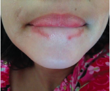
Figure 2. Patient’s mouth with lower lip curled, illustrating full extent of the rash.
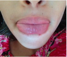
Figure 3. Patient’s mouth with tongue partially extruded and exhibiting typical reach when lip-licking.
Although the vermillion zone of the lips depends on salivary spread for moisture, prolonged salivary contact produces an eczematous eruption of the lips and perioral area likely due to the irritant effects of digestive enzymes present in saliva such as amylase, maltase and others.4,5 This irritant mechanism is similarly illustrated in infants during the teething/eating stage where chronic drooling causes a chin-and-cheek dermatitis and also in toddlers with circumoral dermatitis secondary to salivary trapping from thumb sucking or pacifier usage for extended periods of time.1
Lip licker dermatitis occurs with greater frequency in the winter due to environmental factors. More specifically, the low humidity of the winter months combined with indoor heating systems promotes xerosis and dermatitis.1,4 In these instances, the lips may become chapped and lip licking provides temporary relief. Children with a history of atopy are particularly sensitive to the irritating effects of saliva and have an amplified risk for developing lip licker dermatitis.1
It is also worth noting that saliva-induced dermatitis is observed in the geriatric population when seepage of saliva into the overhanging skin folds at the labial commissures causes a localized dermatitis termed perlèche.4,5
Differential Diagnosis
Lip licking is one of the most frequently cited causes of ICD cheilitis with eczematous changes especially in the pediatric population.9,10 Other entities, either alone or in combination, may produce similar symptomatology with more common causes being contact with allergens and irritants other than saliva; mechanical insults, infection or systemic diseases remain less common.5,8,11
A thorough history and careful examination will greatly narrow the differential and guide further examination or testing as needed.5 Discussion with patients about atopy, lip licking habits and exposures should be done while keeping in mind that irritants come in the form of a variety of agents including foods, dental hygiene products, sunscreens, flavorings, fragrances, metals, topical medications and mouthpieces of musical instruments.5,8 Citrus fruits are sometimes implicated in the development of phytophotodermatitis with associated cheilitis upon exposure to ultraviolet light in susceptible individuals.5,12
The pattern of the affected area also provides clues. For example, contact dermatitis due to toothpaste may present asymmetrically in the perioral area because the irritant is spread by the toothbrush held in the patient’s preferred hand (ie, right-sided perioral dermatitis in right-handed patients).3 Another striking pattern can occur in musicians who develop a hypersensitivity to their wind instruments with lesions reflecting the surfaces of their mouthpiece or reed.3,5 In the latter case, a number of reports exist where saxophonists and clarinetists have developed irritant and allergic dermatitis to cane reeds made of Arundo donax.13 In these cases, the distribution of eczematous changes developed in the median lower lip reflecting the area where frequent contact with the reed occurred.14,15 Distribution of lip licker dermatitis is typically symmetric with eczematous changes limited to the extent of the patient’s tongue.3
Discussion with patients regarding recent lifestyle changes or new habits that bring the lips into contact with a new irritant is also revealing. In the pediatric population, introduction of new foods to the diet may produce a new perioral contact dermatitis.1 Similarly, the condition may develop in adolescent girls who begin using cosmetics for the first time applied to the face and lips.8,10
Actinic cheilitis is a precursor to squamous cell carcinoma of the lip that typically presents in older patients with a long history of sun exposure. Physical examination may show hyperkeratotosis and desquamation of the lips with erythema, scaling, crusting and erosion formation.5
Mechanical causes of cheilitis should be considered and can often be deduced by the history and pattern of distribution.3,5 Angular cheilitis is a reactive process with several causes including lip licking, but can occur secondary to dental trauma such as flossing.5 A less common pathology, exfoliativa cheilitis, is a chronic inflammatory condition characterized by hyperkeratiziation and crusting of the lips. Its cause can be idiopathic, however, studies have shown that self-mutilating behaviors are a major cause.11 “Le tic de lèvres” is a French term that describes patients with a preoccupation of the lips that produces a factitious exfoliativa cheilitis secondary to self-inflicted lip injury by repetitive picking, biting or licking.6-11
Cheilitis glandularis is an uncommon, chronic, inflammatory disease of the labial salivary glands where duct ectasia, hypersecretion and squamous metaplasia occurs secondary to chronic insult to the lip. The lip may have crusts, swelling and lip eversion typically affecting the lower lip. Eversion demonstrates the punctae of salivary glands, which release a sticky substance that forms crust on the lips as it dries. A rarer entity is cheilitis granulomatosa, an inflammatory disorder of the lips that is caused by lymph obstruction secondary to granuloma formation presenting with swelling of the lips. Although granuloma formation may be idiopathic, it is also seen in Crohn’s disease, sarcoidosis and a component of Melkersson-Rosenthal syndrome. Presentation is episodic with swelling of one or both lips — the upper lip occurring with greater frequency. Concerning for this condition is permanent alteration of the lips’ normal architecture and impaired lymph drainage, which can also cause buccal, palatal and lingual edema. Facial distortion can occur as well as reduced eating and speaking functionality.5
Plasma cell cheilitis is another rare benign idiopathic disorder that mainly affects the lower lip with oral mucosal involvement.5 It presents with a shiny, erythematous vermillion with thickening, fissure formation and tenderness.5,16
Management
Successful treatment of lip licker dermatitis relies on eliminating the offending behavior.1 Patients may be unaware of their habit and it may occur unconsciously as a relieving measure for chronically chapped lips, which actually prolongs and worsen the symptoms.1,17 Cessation of lip licking will greatly improve the dermatitis while a simple emollient such as petrolatum jelly can be used as a moisturizer and protective agent in more advanced cases.1,3 A low-potency corticosteroid can be used initially to calm inflammation but atrophy can occur with long-term use.5
Our Patient
Patient and parents were informed of the diagnosis and educated regarding its benign nature and link to the causative behavior. They were instructed to use moisturizer and mild steroids as needed and emphasis was placed on discontinuing lip licking behavior. Patient and her family were compliant with the dermatologist’s recommendations, which resulted in significant improvement.
Conclusion
Lip licker dermatitis is one of the most frequently occurring forms of ICD in children presenting primarily with a symmetric, well-defined, circumoral, erythematous plaque with cheilitis. Diagnosis is based on history and clinical presentation, but similar lesion patterns may occur secondary to a wide host of etiologies. Therefore, a complete patient history remains paramount in elucidating the causative agent while the pattern of lesion distribution may offer clues to possible etiologies.
Mr. Longhurst is with the University of North Dakota School of Medicine and Health Sciences in Grand Forks, ND.
Dr. Khachemoune, the Section Editor of Derm DX, is with the Department of Dermatology at the State University of New York Downstate in Brooklyn, NY.
Disclosure: The authors have no conflicts of interest to report.
References
1. Paller A, Mancini A. Eczematous eruptions in childhood. In: Bonnet C, Gabbedy R, Mortimer A, eds. Hurwitz Clinical Pediatric Dermatology: A Textbook of Skin Disorders of Childhood and Adolescence. 4th ed. New York, NY: Elsevier Saunders; 2011:37-70.
2. Cohen D, Souza A. Irritant contact dermatitis. In: Bolognia J, Jorrizo JL, Schaffer J, et al, eds. Dermatology; vol 1. St. Louis, MO: Mosby Elsevier; 2008:chap 15.
3. Sheehan MP, Huynh M, Chung M, Zirwas M, Feldman SR. Regional atlas of contact dermatitis: mouth, lips and perioral region. The Dermatologist. 2013;21(5):1-3.
4. Rietschel R, Fowler J. Contact stomatitis and cheilitis. In Rietschel R, Fowler J, eds. Fisher’s Contact Dermatitis. Hamilton, Ontario: BC Decker; 2008:700-721.
5. Rogers RS 3rd, Bekic M. Diseases of the lips. Semin Cutan Med Surg. 1997;16(4):328-336.
6. James W, Berger T, Elston D. Disorders of the mucous membranes. In: Gabbedy R, Pinczewski S, eds. Andrew’s Diseases of the Skin: Clinical Dermatology. 11th ed. New York, NY: Elsevier Saunders; 2006:chapter 34.
7. Lipozencic J, Ljubojevic S. Perioral dermatitis. Clin Dermatol. 2011;29(2):157-161.
8. Zug KA, Kornik R, Belsito DV, et al. Patch-testing North American lip dermatitis patients: data from the North American Contact Dermatitis Group, 2001 to 2004. Dermatitis. 2008;19(4):202-208.
9. Freeman S, Stephens R. Cheilitis: analysis of 75 cases referred to a contact dermatitis clinic. Am J Contact Dermat. 1999;10(4):198-200.
10. Lim SW, Goh CL. Epidemiology of eczematous cheilitis at a tertiary dermatological referral centre in Singapore. Contact Dermatitis. 2000;43(6):322-326.
11. Taniguchi S, Kono T. Exfoliative cheilitis: a case report and review of the literature. Dermatology. 1998;196(2):253-255.
12. Bowers AG. Phytophotodermatitis. Am J Contact Dermat. 1999;10(2):89-93.
13. Gambichler T, Uzun A, Boms S, Altmeyer P, Altenmüller E. Skin conditions in instrumental musicians: a self-reported survey. Contact Dermatitis. 2008;58(4):217-222.
14. Inoue A, Shoji A, Yashiro K. Saxophonist’s cane reed cheilitis. Contact Dermatitis. 1998;39(1):37.
15. Ruiz-Hornillos FJ, Alonso E, Zapatero L, Pérez C, Martínez-Molero I. Clarinetist’s cheilitis caused by immediate-type allergy to cane reed. Contact Dermatitis. 2007;56(4):243-245.
16. Farrier JN, Perkins CS. Plasma cell cheilitis. Br J Oral Maxillofac Surg. 2008;46(8):679-680.
17. Watt CJ, Hong HC. Dermacase. Lip licker’s dermatitis. Can Fam Physician. 2002;48:1051, 1059.
Figure 1. Close-up of patient’s mouth at initial presentation.
An 8-year-old girl presented with a history of recent appearance of scaly lesions on her lower lip. Her mother reports that these began to appear approximately 10 days prior to their visit and sometimes minor trauma, such as rubbing, will cause them to bleed. The patient is healthy otherwise and attends school regularly. Physical examination demonstrated to linear symmetric patches with
hyperpigmented well-defined borders involving the perioral skin of the lower lip
with sparing of the midline and surrounding scale formation (Figure 1).
WHAT IS YOUR DIAGNOSIS?
Answer on page 2
{{pagebreak}}
DIAGNOSIS: LIP LICKER DERMATITIS
Lip licker dermatitis is a form of irritant contact dermatitis (ICD) that occurs secondary to persistent lip licking as the name suggests, seen most frequently in children.1-3 The underlying mechanism of lip licker dermatitis is from the habitual, repeated reintroduction of saliva to the perioral skin and vermillion surfaces where its chronic presence acts as an irritant.3
Clinical Presentation
Patients may initially present with a circumferential, symmetric, erythematous macule of the lips and perioral skin with sharp borders as defined by the extent of the tongue’s reach with or without scale formation.1-3 Prolonged disease may also develop more advanced findings including edema, crust formation, pigment changes, fissuring, vesicles, weeping, burning, tenderness or pain.4-8 The severity of presentation can vary considerably from one individual to the next, and even within the same patient, depending on the skin integrity and the accrued effects of successive re-exposure1 (Figures 2 and 3).
Figure 2. Patient’s mouth with lower lip curled, illustrating full extent of the rash.
Figure 3. Patient’s mouth with tongue partially extruded and exhibiting typical reach when lip-licking.
Although the vermillion zone of the lips depends on salivary spread for moisture, prolonged salivary contact produces an eczematous eruption of the lips and perioral area likely due to the irritant effects of digestive enzymes present in saliva such as amylase, maltase and others.4,5 This irritant mechanism is similarly illustrated in infants during the teething/eating stage where chronic drooling causes a chin-and-cheek dermatitis and also in toddlers with circumoral dermatitis secondary to salivary trapping from thumb sucking or pacifier usage for extended periods of time.1
Lip licker dermatitis occurs with greater frequency in the winter due to environmental factors. More specifically, the low humidity of the winter months combined with indoor heating systems promotes xerosis and dermatitis.1,4 In these instances, the lips may become chapped and lip licking provides temporary relief. Children with a history of atopy are particularly sensitive to the irritating effects of saliva and have an amplified risk for developing lip licker dermatitis.1
It is also worth noting that saliva-induced dermatitis is observed in the geriatric population when seepage of saliva into the overhanging skin folds at the labial commissures causes a localized dermatitis termed perlèche.4,5
Differential Diagnosis
Lip licking is one of the most frequently cited causes of ICD cheilitis with eczematous changes especially in the pediatric population.9,10 Other entities, either alone or in combination, may produce similar symptomatology with more common causes being contact with allergens and irritants other than saliva; mechanical insults, infection or systemic diseases remain less common.5,8,11
A thorough history and careful examination will greatly narrow the differential and guide further examination or testing as needed.5 Discussion with patients about atopy, lip licking habits and exposures should be done while keeping in mind that irritants come in the form of a variety of agents including foods, dental hygiene products, sunscreens, flavorings, fragrances, metals, topical medications and mouthpieces of musical instruments.5,8 Citrus fruits are sometimes implicated in the development of phytophotodermatitis with associated cheilitis upon exposure to ultraviolet light in susceptible individuals.5,12
The pattern of the affected area also provides clues. For example, contact dermatitis due to toothpaste may present asymmetrically in the perioral area because the irritant is spread by the toothbrush held in the patient’s preferred hand (ie, right-sided perioral dermatitis in right-handed patients).3 Another striking pattern can occur in musicians who develop a hypersensitivity to their wind instruments with lesions reflecting the surfaces of their mouthpiece or reed.3,5 In the latter case, a number of reports exist where saxophonists and clarinetists have developed irritant and allergic dermatitis to cane reeds made of Arundo donax.13 In these cases, the distribution of eczematous changes developed in the median lower lip reflecting the area where frequent contact with the reed occurred.14,15 Distribution of lip licker dermatitis is typically symmetric with eczematous changes limited to the extent of the patient’s tongue.3
Discussion with patients regarding recent lifestyle changes or new habits that bring the lips into contact with a new irritant is also revealing. In the pediatric population, introduction of new foods to the diet may produce a new perioral contact dermatitis.1 Similarly, the condition may develop in adolescent girls who begin using cosmetics for the first time applied to the face and lips.8,10
Actinic cheilitis is a precursor to squamous cell carcinoma of the lip that typically presents in older patients with a long history of sun exposure. Physical examination may show hyperkeratotosis and desquamation of the lips with erythema, scaling, crusting and erosion formation.5
Mechanical causes of cheilitis should be considered and can often be deduced by the history and pattern of distribution.3,5 Angular cheilitis is a reactive process with several causes including lip licking, but can occur secondary to dental trauma such as flossing.5 A less common pathology, exfoliativa cheilitis, is a chronic inflammatory condition characterized by hyperkeratiziation and crusting of the lips. Its cause can be idiopathic, however, studies have shown that self-mutilating behaviors are a major cause.11 “Le tic de lèvres” is a French term that describes patients with a preoccupation of the lips that produces a factitious exfoliativa cheilitis secondary to self-inflicted lip injury by repetitive picking, biting or licking.6-11
Cheilitis glandularis is an uncommon, chronic, inflammatory disease of the labial salivary glands where duct ectasia, hypersecretion and squamous metaplasia occurs secondary to chronic insult to the lip. The lip may have crusts, swelling and lip eversion typically affecting the lower lip. Eversion demonstrates the punctae of salivary glands, which release a sticky substance that forms crust on the lips as it dries. A rarer entity is cheilitis granulomatosa, an inflammatory disorder of the lips that is caused by lymph obstruction secondary to granuloma formation presenting with swelling of the lips. Although granuloma formation may be idiopathic, it is also seen in Crohn’s disease, sarcoidosis and a component of Melkersson-Rosenthal syndrome. Presentation is episodic with swelling of one or both lips — the upper lip occurring with greater frequency. Concerning for this condition is permanent alteration of the lips’ normal architecture and impaired lymph drainage, which can also cause buccal, palatal and lingual edema. Facial distortion can occur as well as reduced eating and speaking functionality.5
Plasma cell cheilitis is another rare benign idiopathic disorder that mainly affects the lower lip with oral mucosal involvement.5 It presents with a shiny, erythematous vermillion with thickening, fissure formation and tenderness.5,16
Management
Successful treatment of lip licker dermatitis relies on eliminating the offending behavior.1 Patients may be unaware of their habit and it may occur unconsciously as a relieving measure for chronically chapped lips, which actually prolongs and worsen the symptoms.1,17 Cessation of lip licking will greatly improve the dermatitis while a simple emollient such as petrolatum jelly can be used as a moisturizer and protective agent in more advanced cases.1,3 A low-potency corticosteroid can be used initially to calm inflammation but atrophy can occur with long-term use.5
Our Patient
Patient and parents were informed of the diagnosis and educated regarding its benign nature and link to the causative behavior. They were instructed to use moisturizer and mild steroids as needed and emphasis was placed on discontinuing lip licking behavior. Patient and her family were compliant with the dermatologist’s recommendations, which resulted in significant improvement.
Conclusion
Lip licker dermatitis is one of the most frequently occurring forms of ICD in children presenting primarily with a symmetric, well-defined, circumoral, erythematous plaque with cheilitis. Diagnosis is based on history and clinical presentation, but similar lesion patterns may occur secondary to a wide host of etiologies. Therefore, a complete patient history remains paramount in elucidating the causative agent while the pattern of lesion distribution may offer clues to possible etiologies.
Mr. Longhurst is with the University of North Dakota School of Medicine and Health Sciences in Grand Forks, ND.
Dr. Khachemoune, the Section Editor of Derm DX, is with the Department of Dermatology at the State University of New York Downstate in Brooklyn, NY.
Disclosure: The authors have no conflicts of interest to report.
References
1. Paller A, Mancini A. Eczematous eruptions in childhood. In: Bonnet C, Gabbedy R, Mortimer A, eds. Hurwitz Clinical Pediatric Dermatology: A Textbook of Skin Disorders of Childhood and Adolescence. 4th ed. New York, NY: Elsevier Saunders; 2011:37-70.
2. Cohen D, Souza A. Irritant contact dermatitis. In: Bolognia J, Jorrizo JL, Schaffer J, et al, eds. Dermatology; vol 1. St. Louis, MO: Mosby Elsevier; 2008:chap 15.
3. Sheehan MP, Huynh M, Chung M, Zirwas M, Feldman SR. Regional atlas of contact dermatitis: mouth, lips and perioral region. The Dermatologist. 2013;21(5):1-3.
4. Rietschel R, Fowler J. Contact stomatitis and cheilitis. In Rietschel R, Fowler J, eds. Fisher’s Contact Dermatitis. Hamilton, Ontario: BC Decker; 2008:700-721.
5. Rogers RS 3rd, Bekic M. Diseases of the lips. Semin Cutan Med Surg. 1997;16(4):328-336.
6. James W, Berger T, Elston D. Disorders of the mucous membranes. In: Gabbedy R, Pinczewski S, eds. Andrew’s Diseases of the Skin: Clinical Dermatology. 11th ed. New York, NY: Elsevier Saunders; 2006:chapter 34.
7. Lipozencic J, Ljubojevic S. Perioral dermatitis. Clin Dermatol. 2011;29(2):157-161.
8. Zug KA, Kornik R, Belsito DV, et al. Patch-testing North American lip dermatitis patients: data from the North American Contact Dermatitis Group, 2001 to 2004. Dermatitis. 2008;19(4):202-208.
9. Freeman S, Stephens R. Cheilitis: analysis of 75 cases referred to a contact dermatitis clinic. Am J Contact Dermat. 1999;10(4):198-200.
10. Lim SW, Goh CL. Epidemiology of eczematous cheilitis at a tertiary dermatological referral centre in Singapore. Contact Dermatitis. 2000;43(6):322-326.
11. Taniguchi S, Kono T. Exfoliative cheilitis: a case report and review of the literature. Dermatology. 1998;196(2):253-255.
12. Bowers AG. Phytophotodermatitis. Am J Contact Dermat. 1999;10(2):89-93.
13. Gambichler T, Uzun A, Boms S, Altmeyer P, Altenmüller E. Skin conditions in instrumental musicians: a self-reported survey. Contact Dermatitis. 2008;58(4):217-222.
14. Inoue A, Shoji A, Yashiro K. Saxophonist’s cane reed cheilitis. Contact Dermatitis. 1998;39(1):37.
15. Ruiz-Hornillos FJ, Alonso E, Zapatero L, Pérez C, Martínez-Molero I. Clarinetist’s cheilitis caused by immediate-type allergy to cane reed. Contact Dermatitis. 2007;56(4):243-245.
16. Farrier JN, Perkins CS. Plasma cell cheilitis. Br J Oral Maxillofac Surg. 2008;46(8):679-680.
17. Watt CJ, Hong HC. Dermacase. Lip licker’s dermatitis. Can Fam Physician. 2002;48:1051, 1059.