The Next Era of Left Atrial Appendage Occlusion: One Size Does Not Fit All
© 2026 HMP Global. All Rights Reserved.
Any views and opinions expressed are those of the author(s) and/or participants and do not necessarily reflect the views, policy, or position of EP Lab Digest or HMP Global, their employees, and affiliates.
EP LAB DIGEST. 2026;26(10):10-13.
Paul C Weber, DO,1 and Dipak P Shah, MD, FACC, FHRS2
1Department of Cardiology, Henry Ford Providence Southfield Hospital, Southfield, Michigan; 2Medical Director, Atrial Fibrillation Clinic; Co-Director, Structural Heart, Southfield, Michigan; Co-Chief of Electrophysiology, Henry Ford Health System
Case Presentation: When a “4-mm Leak” Is Not Really a 4-mm Leak
A 74-year-old man with permanent atrial fibrillation (AF) underwent Watchman (Boston Scientific) implantation at an outside hospital and was advised to discontinue oral anticoagulation (OAC) in favor of aspirin monotherapy after outside imaging reported “only” a 4-mm peri-device leak (PDL).
On detailed reevaluation with computed tomography (CT), the residual communication was not a pinpoint defect at the device margin. The leak was crescentic and extended beyond the device lobe, leaving the distal left atrial appendage (LAA) substantially patent with an uncovered posterior lobe (Figure 1A). The residual flow through a crescentic channel created a broad zone of persistent flow despite an “acceptable” maximal linear measurement where OAC could be discontinued (Figure 1B). Transcatheter closure of the residual PDL was subsequently performed using coils and the Amplatzer Ductal Occluder II (Abbott) (Figures 1C and 1D).
This case illustrates a fundamental limitation of relying on a single maximal width threshold for PDL characterization. Crescentic leaks expose a potentially substantially larger thrombogenic risk than any linear dimension implies. Residual surface area, uncovered lobes, a patent LAA, and flow characteristics may prove more clinically relevant than maximal diameter alone.
Introduction
Left atrial appendage occlusion (LAAO) has transitioned from a limited indication for patients contraindicated to OAC to a more expanded stroke prevention strategy for AF. The recently published CHAMPION-AF trial concluded that LAAO was noninferior to direct oral anticoagulant (DOAC) therapy in patients who were candidates for OAC.1 As indications potentially broaden to include healthier and younger patients for LAAO, procedural endpoints related to PDL, incomplete endothelialization, and device-related thrombus (DRT) must improve. The addition of newer trial data suggests that selecting the appropriate patient for LAAO is critical to the long-term success of this therapy.
Recent AF Trials
Recent trials have largely supported the expanded indication for LAAO. The OPTION trial randomized 1600 patients undergoing AF ablation to Watchman FLX (Boston Scientific) implantation or continued OAC. At 36 months, LAAO met the criterion for noninferiority for the primary efficacy endpoint of all-cause death, stroke, or systemic embolism, and was superior for nonprocedure-related major or clinically relevant nonmajor bleeding.2 CHAMPION-AF randomized 3000 anticoagulation-eligible patients to LAAO versus non-vitamin K oral anticoagulant (NOAC) therapy. At 3 years, LAAO was noninferior for cardiovascular death, stroke, or systemic embolism (5.7% versus 4.8%; 95% confidence interval [CI] for the difference, -0.8 to 2.6) and superior for nonprocedure-related bleeding (10.9% versus 19.0%; hazard ratio [HR] 0.55; 95% CI, 0.45-0.67).1 Although the OCEAN trial was not an LAAO study, it provides important insight into the evolving management of anticoagulation in lower-risk patients with AF. OCEAN evaluated patients who remained free of recurrent AF 1 year after successful catheter ablation and randomized them to continued rivaroxaban, 15 mg daily, or aspirin, 81 mg daily. The trial demonstrated no significant difference in ischemic outcomes between the 2 groups, whereas patients assigned to aspirin experienced fewer bleeding events.3 These findings suggest that, in carefully selected lower-risk patients, long-term OAC may not be necessary following successful rhythm control with ablation, further emphasizing the need to carefully define the patient population most likely to benefit from LAAO.
Another trial providing valuable insight and suggesting potential harm with LAAO in high-risk patients is CLOSURE-AF. This nonindustry-funded trial enrolled a high-risk population with a mean age of 77.9 years, a mean CHA2DS2-VASc score of 5.2, and a mean HAS-BLED score of 3.0; enrollment required a history of bleeding or an estimated high bleeding risk. Multiple device types were used.4 LAAO did not meet the criterion for noninferiority compared with physician-directed medical therapy for the composite endpoint of stroke, systemic embolism, major bleeding, or cardiovascular death (16.8 vs 13.3 events per 100 patient-years; difference in restricted mean survival time, -0.36 years; 95% CI, -0.70 to -0.01; P = 0.44 for noninferiority).4 Rather than validating the procedure, CLOSURE-AF highlights its persistent limitations. In elderly and frail patients, even modest complication rates can offset long-term reduction in stroke risk. As LAAO becomes more widely adopted, a deeper understanding of the evidence surrounding PDL and DRT is essential to optimize patient selection, particularly as this therapy expands to younger, healthier populations.
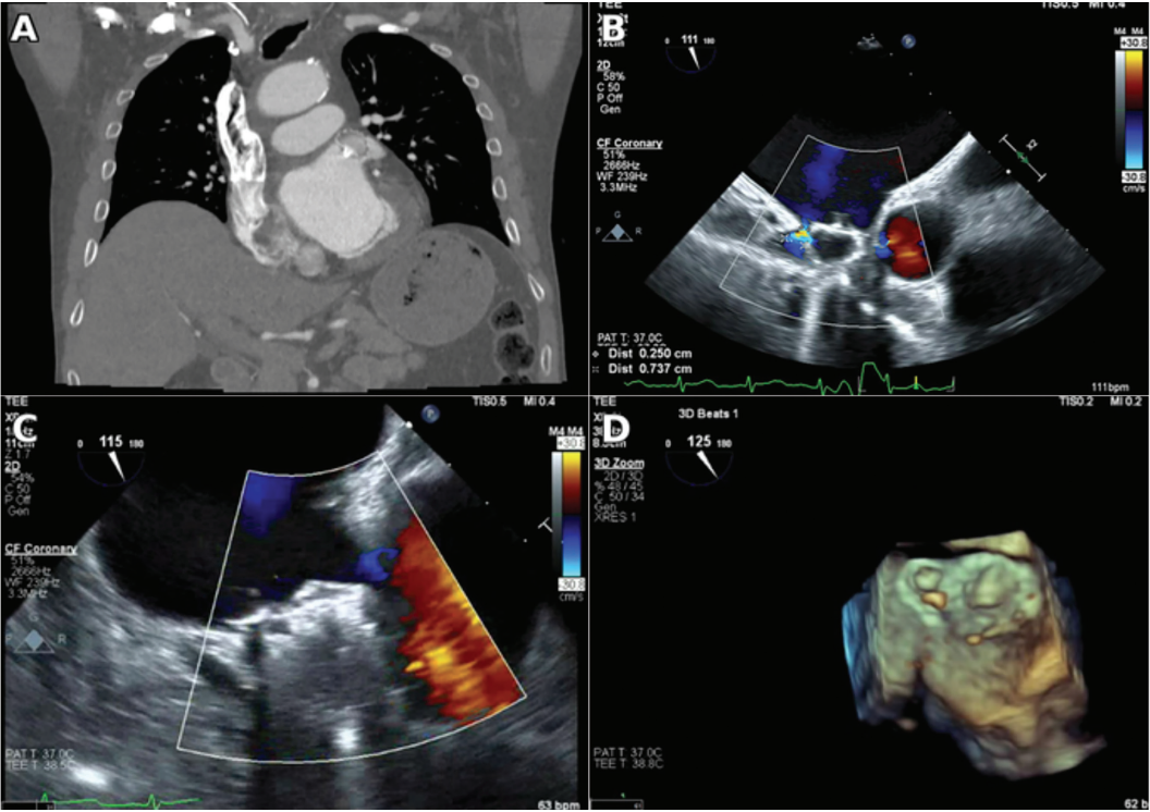
Abbreviations: ADO, Amplatzer Ductal Occluder II; CT, computed tomography; LAA, left atrial appendage; LAAO, left atrial appendage occlusion; PDL, peri-device leak; TEE, transesophageal echocardiography.
The Persistent Challenge of PDL
Historically, a leak measuring less than 5 mm was considered a successful endpoint; however, recent evidence suggests a new goal of less than 3 mm.5 It is now clear that a PDL of any size increases the risk of stroke and thromboemebolism.6 The difficulty of PDL is that the current classification system is relatively crude, with substantial variability across imaging modalities and device types.
Much of this risk stems from anatomic features that linear measurements cannot adequately capture. The LAA is morphologically heterogeneous, with configurations including the chicken wing, cactus, windsock, and cauliflower morphologies. Oval landing zones, multilobed anatomy, and variable ostial geometry all create a mismatch with circular devices, generating persistent flow channels that may exist despite acceptable device compression on imaging. As Dukkipati et al6 pointed out, a 4-mm PDL covering a small arc of the device circumference would be classified as more severe than a crescent-shaped 2-mm PDL spanning half the perimeter, yet the latter exposes far more thrombogenic surface area to blood.
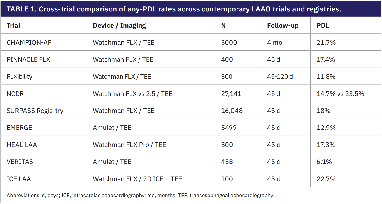
In CHAMPION-AF, the Watchman device demonstrated a postprocedural rate of any leak at 10.9%; however, at 4-month follow-up imaging, this increased to 21.7%.1 These findings are consistent with the Watchman data from PINNACLE-FLX and SURPASS, which reported any leak rates of 17.4% and 18% at 45 days, respectively.7,8 These results were based on transesophageal echocardiography (TEE), and despite the expanded use of intracardiac echocardiography (ICE) during these procedures, PDL outcomes have not improved. Two-dimensional ICE in the ICE LAA trial resulted in a PDL rate of 22.7%, whereas 3D ICE in ICE WATCHMAN demonstrated a rate of 18%.9,10
A single-lobe device implanted in a complex structure such as the LAA has intrinsic constraints that a dual-closure device with a disc and lobe could theoretically address. However, the Amulet IDE trial reported leak rates of 37.0% at 45 days.11 As operators gained implant experience, these outcomes improved, with the EMERGE postapproval study reporting a PDL rate of 12.9%.12 With the investigational Amulet 360 device, which features a more conformable lobe, the rate of any PDL decreased to 6.1% in the VERITAS trial.13
Although TEE has been the gold standard imaging modality in many of these trials, cardiac CT is more sensitive and provides a more comprehensive evaluation of the LAA and device interaction.14 The SWISS-APERO data were sobering, demonstrating combined side-gap and mixed leak rates of 26.7% with Amulet and 48% with Watchman by CT.15 In this study, approximately three-quarters of patients received the Watchman FLX device, whereas the remainder received the Watchman 2.5. If the goal is to exclude the LAA entirely from the blood pool, any contrast in the LAA could theoretically be considered a procedural failure. In the SWISS-APERO study at 45 days, 67.6% of Amulet and 70% of Watchman devices demonstrated a patent LAA.15
Early after device placement, some patent appendages are likely attributable to intradevice leaks resulting from incomplete endothelialization. However, the 1-year SWISS-APERO data continued to demonstrate a substantial rate of patent LAA, with rates of 53.6% for Amulet and 48.8% for Watchman.16 Although endothelialization likely progressed over the first year, the PDL rate at 3 years remained substantial, at 35.1% with Amulet and 42.7% with Watchman.16 These PDL rates did not account for specific leak patterns unique to dual-closure devices, in which a leak around the lobe is likely quite different from a leak between the disc and the lobe. Ultimately, PDL assessment will need to account for residual patent appendage volume, uncovered lobes, flow characteristics and surface area, and type of leak in dual-closure devices.
DRT
DRT is the other major limitation of LAAO. A pooled meta-analysis of 66 studies involving more than 10,000 patients undergoing surveillance imaging estimated an overall DRT incidence of approximately 3.8%, although rates varied across individual trials.17 In CHAMPION-AF, DRT was identified in 4.8% of patients at 4 months, with 1.8% meeting criteria for clinically relevant thrombus requiring resumption of anticoagulation.1 Across registries, DRT is associated with an approximately 3.5-fold increased risk of stroke and systemic embolism (adjusted rate ratio, 3.55; 95% CI, 2.18-5.79).18 About one-third of DRT cases persist or recur despite anticoagulation, and an initial thrombus measuring greater than 7 mm is the strongest predictor of an unfavorable clinical course.19 Established risk factors for DRT include prior stroke or transient ischemic attack (TIA), persistent AF, reduced ejection fraction, larger LAA orifice diameter, greater LAAO implantation depth, and interruption of anticoagulation during the healing period.18,20
The timing of DRT differs by device. Most Amulet-related DRT occurs within 45 days, whereas most Watchman-related DRT presents later, likely reflecting differences in endothelialization kinetics and device-specific features.21 The imaging modality used during post-LAAO surveillance is also an important consideration. Cardiac CT identifies substantially more DRT than TEE, often visualized as hypoattenuated thickening (HAT) on the device surface. A CT-based HAT grading system helps distinguish incidental findings from true thrombus: grades 0 and 1 require no change in treatment, whereas grades 2 and 3 warrant prompt therapeutic anticoagulation. Current guidelines recommend repeat imaging every 45 to 90 days until higher-grade thrombus resolves.21,22 In the 3-year SWISS-APERO data, the combined rate of definite and possible DRT was significantly lower with Amulet than with Watchman (3.7% vs 11.8%; P = 0.028), although adjudicated definite DRT alone did not differ significantly between devices (3.6% vs 6.4%; P = 0.35).16
Post-LAAO antithrombotic regimens are being refined to address DRT and bleeding. The ongoing SIMPLAAFY trial is comparing aspirin monotherapy against dual antiplatelet therapy (DAPT) or reduced-dose DOAC therapy. Observational data suggest that half-dose DOAC therapy may reduce the combined incidence of DRT and thromboembolic events compared with standard antiplatelet therapy.22,23 Further investigation is needed to determine optimal strategies in periprocedural anticoagulant management.
LAAO is also competing against a moving pharmacologic target. Factor XI inhibition represents a promising therapeutic strategy by playing a central role in pathologic thrombosis while contributing relatively little to normal hemostasis, raising the possibility of reducing bleeding without compromising stroke prevention. The OCEANIC-STROKE trial evaluated patients with a recent noncardioembolic ischemic stroke or TIA and demonstrated that the addition of asundexian to antiplatelet therapy reduced recurrent ischemic stroke without a significant increase in bleeding. Although long-term OAC remains effective, medication adherence continues to be a significant limitation in real-world practice. Emerging therapies such as abelacimab, a long-acting Factor XI inhibitor administered once monthly, may address this important barrier by improving treatment adherence while potentially maintaining efficacy with a lower bleeding risk.24 These advances serve as a reminder that every improvement in anticoagulant safety raises the bar that LAAO device therapy must clear.25
Procedural Planning
Cardiac CT angiography is now the most useful preprocedural tool for mapping LAA anatomy, landing-zone geometry, and transseptal trajectory. CT allows operators to size devices more precisely than echocardiography.26 Three-dimensional (3D) printing extends this capability further: a systematic review found 3D-printed models matched final implanted device size in 95% to 100% of cases.27 Computational simulation platforms represent the next step. In the PREDICT-LAA randomized trial, simulation-guided planning produced higher complete-closure rates than standard CT-based planning, although the composite endpoint of incomplete closure or DRT only trended in favor of simulation.28
Intraprocedurally, TEE remains the standard imaging modality, but 3D ICE is gaining traction. A meta-analysis of more than 42,000 patients found ICE-guided LAAO to be marginally superior to TEE for procedural success.29 Current guidance reserves ICE for experienced programs and recommends TEE or CT at 45 to 90 days after the procedure, with repeat imaging at 1 year if risk factors for PDL or DRT persist.22
Conclusion
OPTION and CHAMPION-AF have established LAAO as a credible alternative to long-term anticoagulation in selected patients with AF. CLOSURE-AF serves as an important reminder that meaningful limitations remain in older, higher-risk populations. The case presented here underscores a broader challenge in contemporary LAAO. A linear measurement of PDL often fails to capture the true extent of residual communication, particularly when the leak is crescent-shaped rather than focal. As LAAO is increasingly offered to younger, lower-risk patients, reliance on a single linear measurement may become insufficient, highlighting the need for more anatomically relevant metrics to better define clinically meaningful leaks and guide long-term management.
When the cumulative risks of contemporary LAAO are considered—including PDL rates approaching 15% to 20%, DRT rates of approximately 3%, and major procedural complication rates near 1%—the overall burden of adverse outcomes approaches 20% to 25%. Although these events differ in clinical significance, this composite perspective is nevertheless sobering. At the same time, the therapeutic landscape continues to evolve. The emergence of safer, more convenient anticoagulants, including long-acting Factor XI inhibitors, raises the bar for procedural therapies.
The goal of LAAO should be complete and durable LAAO with minimal procedural risk, no residual LAA patency, and an exceedingly low incidence of DRT. In recent years, considerable emphasis has been placed on procedural efficiency and ease of implantation. However, prioritizing rapid device deployment without fully accounting for anatomic complexity or the mechanisms underlying residual leaks may compromise long-term outcomes. This is particularly concerning in younger, lower-risk patients, in whom even small residual risks accumulate over decades.
As LAAO technology continues to evolve to address the heterogeneous anatomy of the LAA, advances will likely be required across the entire procedural ecosystem rather than in device design alone. Preprocedural cardiac CT with 3D modeling may improve patient selection and procedural planning, while next-generation delivery sheaths that facilitate truly coaxial deployment may reduce residual leaks and improve sealing. Additional innovative strategies also warrant investigation, including appendage ablation or electrical stunning before occlusion to promote remodeling and enhance closure, as well as novel occlusion technologies such as injectable polymer or hydrogel-based systems and magnetically guided sealing platforms.30,31 Ultimately, the future of LAAO will depend not only on making procedures easier but also on making closure more complete, more durable, and safer over a patient’s lifetime.
Disclosures: Drs Weber and Shah have completed and returned the ICMJE Form for Disclosure of Potential Conflicts of Interest. Dr Shah reports consulting fees from Abbott; payment or honoraria for lectures, presentations, speakers’ bureaus, manuscript writing, or educational events, as well as support for attending meetings and/or travel, from ISLAA, the LAAO Summit, and the Scripps Structural Heart Intervention and Imaging course.
References
- Doshi SK, Kar S, Nair DG, et al. Left atrial appendage occlusion versus anticoagulation for stroke prevention. N Engl J Med. 2026;394(21):2083-2094. doi:10.1056/NEJMoa2517213
- Wazni OM, Saliba WI, Nair DG, et al. Left atrial appendage closure after ablation for atrial fibrillation. N Engl J Med. 2025;392(13):1277-1287. doi:10.1056/NEJMoa2408308
- Verma A, Birnie DH, Jiang C, et al. Antithrombotic therapy after successful catheter ablation for atrial fibrillation. N Engl J Med. 2026;394(4):323-332. doi:10.1056/NEJMoa2509688
- Landmesser U, Skurk C, Kirchhof P, et al. Left atrial appendage closure or medical therapy in atrial fibrillation. N Engl J Med. 2026;394(13):1270-1280. doi:10.1056/NEJMoa2513310
- Samaras A, Papazoglou AS, Balomenakis C, et al. Residual leaks following percutaneous left atrial appendage occlusion and outcomes: a meta-analysis. Eur Heart J. 2024;45(3):214-229. doi:10.1093/eurheartj/ehad828
- Dukkipati SR, Holmes DR Jr, Doshi SK, et al. Impact of peridevice leak on 5-year outcomes after left atrial appendage closure. J Am Coll Cardiol. 2022;80(5):469-483. doi:10.1016/j.jacc.2022.04.062
- Kar S, Doshi SK, Sadhu A, et al. Primary outcome evaluation of a next-generation left atrial appendage closure device: results from the PINNACLE FLX trial. Circulation. 2021;143(18):1754-1762. doi:10.1161/CIRCULATIONAHA.120.050117
- Kapadia SR, Price MJ, Liu M, et al. Outcomes with the Watchman FLX in everyday clinical practice from the NCDR Left Atrial Appendage Occlusion Registry. Circ Cardiovasc Interv. 2024;17(9):e013750. doi:10.1161/CIRCINTERVENTIONS.123.013750
- Nielsen-Kudsk JE, Berti S, Caprioglio F, et al. Intracardiac echocardiography to guide Watchman FLX implantation: the ICE LAA study. JACC Cardiovasc Interv. 2023;16(6):643-651. doi:10.1016/j.jcin.2022.10.024
- Sularz A, Chavez Ponce A, Al-Abcha A, et al. Safety and feasibility of 3D intracardiac echocardiography in guiding left atrial appendage occlusion with Watchman FLX. JACC Adv. 2025;4(2):101570. doi:10.1016/j.jacadv.2024.101570
- Lakkireddy D, Thaler D, Ellis CR, et al. Amplatzer Amulet left atrial appendage occluder versus Watchman device for stroke prophylaxis (Amulet IDE): a randomized, controlled trial. Circulation. 2021;144(19):1543-1552. doi:10.1161/CIRCULATIONAHA.121.057063
- Alkhouli M, Freeman JV, Ellis CR, et al. First experience with Amulet in the United States: early insights from EMERGE LAA postapproval study. JACC Cardiovasc Interv. 2024;17(3):422-434. doi:10.1016/j.jcin.2023.11.027
- Nair D, Freixa X, Ellis CR, et al. Early outcomes with a next-generation dual-seal left atrial appendage occluder: results from the VERITAS study. JACC Clin Electrophysiol. 2026;12(6):1331-1341. doi:10.1016/j.jacep.2026.01.021
- Tan BE, Baqai F, Padilla F, et al. Cardiac CT versus transesophageal echocardiography following left atrial appendage closure: a systematic review and meta-analysis. Circ Cardiovasc Imaging. 2025;18(8): e018151. doi:10.1161/CIRCIMAGING.125.018151
- Galea R, De Marco F, Meneveau N, et al. Amulet or Watchman device for percutaneous left atrial appendage closure: primary results of the SWISS-APERO randomized clinical trial. Circulation. 2022;145(10):724-738. doi:10.1161/CIRCULATIONAHA.121.057859
- Galea R, De Marco F, Aminian A, et al. Three-year clinical outcomes comparing the Amulet vs Watchman FLX for left atrial appendage closure in patients with atrial fibrillation: results from the SWISS-APERO randomized trial. J Am Coll Cardiol. 2025;86(1):3-14. doi:10.1016/j.jacc.2025.03.535
- Alkhouli M, Busu T, Shah K, et al. Incidence and clinical impact of device-related thrombus following percutaneous left atrial appendage occlusion: a meta-analysis. JACC Clin Electrophysiol. 2018;4(12):1629-1637. doi:10.1016/j.jacep.2018.09.007
- Dukkipati SR, Kar S, Holmes DR, et al. Device-related thrombus after left atrial appendage closure: incidence, predictors, and outcomes. Circulation. 2018;138(9):874-885. doi:10.1161/CIRCULATIONAHA.118.035090
- Mesnier J, Simard T, Jung RG, et al. Persistent and recurrent device-related thrombus after left atrial appendage closure: incidence, predictors, and outcomes. JACC Cardiovasc Interv. 2023;16(22):2722-2732.
- Simard T, Jung RG, Lehenbauer K, et al. Predictors of device-related thrombus following percutaneous left atrial appendage occlusion. J Am Coll Cardiol. 2021;78(4):297-313. doi:10.1016/j.jacc.2021.04.098
- Alkhouli M, Alarouri H, Kramer A, et al. Device-related thrombus after left atrial appendage occlusion: clinical impact, predictors, classification, and management. JACC Cardiovasc Interv. 2023;16(22):2695-2707. doi:10.1016/j.jcin.2023.10.046
- Goldsweig AM, Glikson M, Joza J, et al. 2025 SCAI/HRS clinical practice guidelines on transcatheter left atrial appendage occlusion. Heart Rhythm. 2025;22(10):e1048-e1063. doi:10.1016/j.hrthm.2025.05.048
- Della Rocca DG, Magnocavallo M, Di Biase L, et al. Half-dose direct oral anticoagulation versus standard antithrombotic therapy after left atrial appendage occlusion. JACC Cardiovasc Interv. 2021;14(21):2353-2364. doi: 10.1016/j.jcin.2021.07.031.
- Ruff CT, Patel SM, Giugliano RP, et al. Abelacimab versus rivaroxaban in patients with atrial fibrillation. N Engl J Med. 2025;392(4):361-371. doi:10.1056/NEJMoa2406674
- Sharma M, Dong Q, Hirano T, et al. Asundexian for secondary stroke prevention. N Engl J Med. 2026;94(15):1467-1479. doi:10.1056/NEJMoa2513880
- Korsholm K, Iriart X, Saw J, et al. Position statement on cardiac computed tomography following left atrial appendage occlusion. JACC Cardiovasc Interv. 2024;17(15):1747-1764. doi:10.1016/j.jcin.2024.04.050
- Tarabanis C, Klapholz J, Zahid S, Jankelson L. A systematic review of the use of 3D printing in left atrial appendage occlusion procedures. J Cardiovasc Electrophysiol. 2022;33(11):2367-2374. doi:10.1111/jce.15658
- De Backer O, Iriart X, Kefer J, et al. Impact of computational modeling on transcatheter left atrial appendage closure efficiency and outcomes. JACC Cardiovasc Interv. 2023;16(6):655-666. doi:10.1016/j.jcin.2023.01.008
- Serpa F, Rivera A, Fernandes JM, et al. Intracardiac vs transesophageal echocardiography for left atrial appendage occlusion: an updated systematic review and meta-analysis. Heart Rhythm. 2025;22(3):786-795. doi:10.1016/j.hrthm.2024.08.027
- Yao PC, Chen M, Wang QS, et al. Left atrial appendage electrical isolation reduces residual patency after device closure in patients with persistent atrial fibrillation. J Am Heart Assoc. 2026;15(12):e046374. doi:10.1161/JAHA.125.046374
- Wang S, Ju W, Zhuang D, et al. Long-term thrombus-free left atrial appendage occlusion via magnetofluids. Nature. 2026;651(8104):91-99. doi:10.1038/s41586-025-10091-1
Related Content