Multidisciplinary Management of Ischemic Charcot Neuroarthropathy Foot Deformity
Abstract
Nonhealing ulcerations of the lower extremities require a comprehensive evaluation of contributing factors beyond localized areas of high pressure. While plantar pressure is a common impediment to wound healing, underlying vascular insufficiency such as chronic limb-threatening ischemia exacerbates chronic ulcerations, impedes recovery, and increases risk of complications. In cases of limb-threatening conditions, optimizing blood flow is essential before attempting major joint reconstruction to ensure healing and joint salvage. This case report details the successful management of a 51-year-old man with chronic limb-threatening ischemia and nondiabetic Charcot neuroarthropathy who underwent major arterial bypass procedures followed by invasive left Charcot foot reconstruction.
J CRIT LIMB ISCHEM 2026:6(4):E122-E125. doi: 10.25270/jcli/CLIG-2600014
Key words: Charcot neuroarthropathy, chronic limb-threatening ischemia, peripheral arterial disease, limb salvage
Optimal medical and surgical management of patients with chronic limb-threatening ischemia (CLTI) confounded with Charcot neuroarthropathy (CN) is plagued by significant and highly consequential decisions. Vascular evaluation is mandatory to determine whether arterial perfusion is sufficient to allow optimal incision and osseous healing. Once arterial flow is confirmed, the architectural reconstruction can be performed in a single or staged fashion based on risk of infection with wound presence.
Consecutive stages of ischemic pain, gangrenous changes, and tissue loss signal the end of peripheral arterial disease and the beginning of CLTI.1 Although CN does not have a correlation to CLTI, the combination of both has a high risk for major amputation (below or above the knee).2,3 It is therefore prudent to follow an evidence-based medicine approach as close as possible. Revascularization is the first-line treatment for CLTI, and endovascular therapy yields optimal results.4 Further, patients with end-stage peripheral arterial disease are often poor surgical candidates for open procedures, which has led to the rise of endovascular procedures, especially in the lower extremities.3,5 However, both open surgical and endovascular therapies can provide good results that depend on conduit availability, anatomic disease pattern, and patient risk profile.6-13
CN itself affects 0.1% to 2.5% of all individuals with diabetes, although some estimates are higher.14 There are no known studies that report the incidence of nondiabetic CN, but it is likely even more underreported than diabetic CN due to lack of a diagnostic hint from a diabetes diagnosis.15 We present a case of CN foot deformity originating in a nondiabetic patient conjointly with CLTI. The affected patient arrived with advanced CN changes, osteomyelitis, and an ankle-brachial index (ABI) of 0.15 to the affected limb. Successful limb salvage was achieved with coordinated care between vascular surgery and podiatric surgery to create a plantigrade functional foot.
Case Description
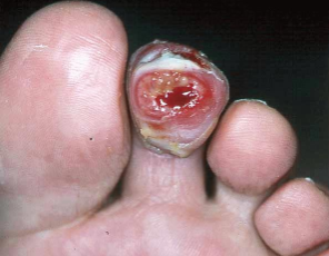
A 51-year-old man initially presented to the emergency department (ED) with a left second digital ulcer with osteomyelitis. Significant medical history included hypertension and deep vein thrombosis with an extensive history of alcohol abuse (with peripheral neuropathy) and smoking. He had previously undergone a left leg venous thrombectomy, which advanced his lower leg neuropathy. Upon his first encounter at our institution, active infection was found in his left second digit with purulence, edema, and erythema (Figure 1). Pedal pulses were nonpalpable along with claudication-type symptoms noted to both lower extremities. After treatment options were discussed with the patient, he elected to undergo a partial left second digital amputation for definitive treatment of underlying bone infection (Figure 2). Following the amputation, vascular noninvasive studies showed an ABI of 0.15 on the left and 0.40 on the right. The patient was discharged in stable condition and instructed to follow up with vascular surgery as an outpatient.
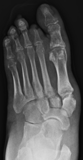
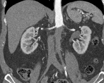
The patient failed to follow up with the vascular team regarding his poor circulation and presented to the ED 2 months later with a right hallux ulceration showing signs of underlying osteomyelitis. An angiogram was performed, demonstrating complete pararenal aortic occlusion extending to the iliacs (Figure 3), and the ABI had not changed from the previous testing. Given his young age, an aortobifemoral bypass was performed, restoring in-line flow. While recovering from the procedure in the hospital, the patient left against medical advice. He returned to the ED after 2 weeks with a left red, hot, swollen, and collapsed foot consistent with CN (Figure 4).
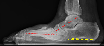
Faced with a potentially limb-threatening deformity and an unstable foot susceptible to re-ulcerations, the patient agreed to undergo CN reconstruction to create a plantigrade stable foot in which the long axis of the foot is perpendicular to the long axis of the lower leg without any significant osseous prominences. With the foot presenting in the acute active stage, the decision was made to first stabilize the deformity with external fixation (Figure 5).
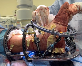
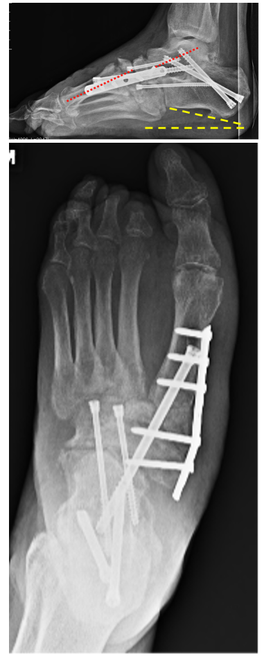
With non-weightbearing and immobilization, the CN deformity was gradually corrected using external fixation and then held statically to allow for transitioning into the coalescence phase radiographically. Definitive surgery was performed with a triple arthrodesis with medial midfoot column fusion (Figure 6). After successful revascularization, CN reconstruction created a plantigrade functional foot. With over a year of follow-up, the patient reported no complications and was able to ambulate with a custom ankle-foot orthosis (AFO) and a plantigrade foot. He will need to be monitored at least annually to ensure architecture is maintained without tissue breakdown. The patient will wear the AFO brace until 18 months after surgery to ensure sufficient osseous consolidation. Custom orthoses may be considered to provide additional support in medical shoegear once the AFO brace is no longer needed.
Discussion
In limb salvage scenarios, a multidisciplinary approach is essential to maximize the likelihood of success. Preventing both minor and major amputations is a critical objective that should not be underestimated. Five-year mortality rates with foot-related issues such as CN (29.0%), diabetic foot ulcer (30.5%), and minor (46.2%) and major (56.6%) amputations are comparable or higher than the pooled mortality rate for all reported cancers (31.0)%.16 These elevated mortality rates highlight the importance of collaborative care, particularly between vascular surgery and podiatric surgery, in achieving successful limb salvage outcomes.
Vascular management of CLTI requires timely revascularization to preserve limb viability, relieve pain, and improve survival. The tenet is for in-line reconstruction whenever possible. Both endovascular and open surgical options play essential roles, and treatment should be individualized based on anatomy, conduit availability, comorbidities, and life expectancy. Endovascular therapy—including angioplasty, stenting, atherectomy, and pedal interventions—offers a minimally invasive approach with lower perioperative morbidity, shorter hospital stay, and faster recovery. It is particularly attractive for older or high-risk patients and for focal or multilevel lesions amenable to catheter-based treatment. However, open surgery remains an important option for extensive occlusive disease, particularly aortic occlusions, long-segment femoropopliteal or tibial disease, failed prior endovascular therapy, or when durable long-term patency is prioritized. Evidence increasingly supports a “best therapy for the best patient” strategy rather than a one-size-fits-all model. Multidisciplinary limb salvage programs help optimize wound care, infection control, and revascularization selection.
Another important consideration in CN reconstruction is when to approach this in a single stage or a staged approach. When managing an ulcerated CN foot, there is a risk of underlying osteomyelitis. In this scenario, the use of internal hardware is typically contraindicated until infection has been ruled out where a staged approach is preferred. A retrospective study examined the effectiveness of a 2-stage surgical reconstruction for infected CN foot deformity in 22 patients, with a mean follow-up of 44 months.17 The first stage involved surgical debridement of all infected bone and tissue, application of antibiotic beads, stabilization with an external fixator, use of negative-pressure wound therapy, and targeted intravenous antibiotics based on intraoperative bone cultures. Once the infection was eradicated, the second stage focused on definitive treatment with internal hardware. After the 2-stage approach, limb salvage was successful in all but 1 patient, and at 1-year follow-up, all but 1 patient was fully weightbearing. The authors emphasized utilizing a multidisciplinary team approach to optimize successful limb salvage.
The timing of revascularization must also be considered after surgical infection control. Transcutaneous oxygen tension measurements were evaluated preoperatively and postoperatively.18 The authors noted transcutaneous oxygen tension levels significantly increased on the third postoperative day (P=.001) to optimize tissue perfusion and support healing. They concluded that tissue oxygenation can reach adequate levels after waiting at least 3 days following intervention.
In the case presented, a successful bypass procedure addressing CLTI allowed the patient to heal, significantly improving the chances of a successful midfoot fusion following multiple staged reconstructive surgeries to create a stable plantigrade foot. Ultimately, the multidisciplinary approach prevented lower-leg amputation and led to a successful limb salvage outcome.
Affiliations and Disclosures
Zeeshan S. Husain, DPM, FACFAS, Athanasios Garbis, DPM, and Cody Ingram, DPM, MPH are from McLaren Oakland Hospital, Pontiac, Michigan; Nicolas J. Mouawad, MD, MPH, MBA, RPVI, is from the Division of Vascular Surgery, McLaren Health, Bay City, Michigan.
The authors report no financial relationships or conflicts of interest regarding the content herein.
Manuscript accepted July 20, 2026.
Address for correspondence: Zeeshan S Husain, DPM, McLaren Oakland Hospital, 50 Perry St, Pontiac, MI 48342. Email: zeeshan.husain@mclaren.org
References
1. Mustapha JA, Saab FA, Martinsen BJ, et al. Digital subtraction angiography prior to an amputation for critical limb ischemia (CLI): an expert recommendation statement from the CLI Global Society to optimize limb salvage. J Endovasc Ther. 2020;27(4):540-546. doi:10.1177/1526602820928590
2. Caravaggi CMF, Sganzaroli AB, Galenda P, et al. Long-term follow-up of tibiocalcaneal arthrodesis in diabetic patients with early chronic Charcot osteoarthropathy. J Foot Ankle Surg. 2012;51(4):408-411. doi:10.1053/j.jfas.2012.04.007
3. Palena LM, Brocco E, Manzi M. Critical limb ischemia in association with Charcot neuroarthropathy: complex endovascular therapy for limb salvage. Cardiovasc Intervent Radiol. 2014;37(1):257-261. doi:10.1007/s00270-013-0642-y
4. Norgen L, Hiatt WR, Dormandy JA, Nehler MR, Harris KA, Fowkes FGR; TASC II Working Group. Inter-society consensus for the management of peripheral arterial disease (TASC II). J Vasc Surg. 2007;45 Suppl S:S5-67. doi:10.1016/j.jvs.2006.12.037
5. Wadehra V, Pupp G, Mustapha JA. Total ankle replacement (TAR): there is no patient too old or too sick. J Crit Limb Ischem. 2022;2(2):E55-E57.
6. Farber A, Menard MT, Conte MS, et al; BEST-CLI Investigators. Surgery or endovascular therapy for chronic limb-threatening ischemia. N Engl J Med. 2022;387(25):2305-2316. doi:10.1056/NEJMoa2207899
7. Gornik HL, Aronow HD, Goodney PP, et al; Peer Review Committee Members. 2024 ACC/AHA/AACVPR/APMA/ABC/SCAI/SVM/SVN/SVS/SIR/VESS guideline for the management of lower extremity peripheral artery disease: a report of the American College of Cardiology/American Heart Association joint committee on clinical practice guidelines. Circulation. 2024;149(24):e1313-e1410. doi:10.1161/CIR.0000000000001251
8. Bradbury AW, Adam DJ, Bell J, et al. Multicentre randomised controlled trial of the clinical and cost-effectiveness of a bypass-surgery-first versus a balloon-angioplasty-first revascularization strategy for severe limb ischaemia due to infrainguinal disease. The Bypass versus Angioplasty in Severe Ischaemia of the Leg (BASIL) trial. Health Technol Assess. 2010;14(14):1-210, iii-iv. doi:10.3310/hta14140
9. Bradbury AW, Moakes CA, Popplewell M, et al; BASIL-2 Investigators. A vein bypass first versus a best endovascular treatment first revascularisation strategy for patients with chronic limb threatening ischaemia who required an infra-popliteal, with or without an additional more proximal infra-inguinal revascularisation procedure to restore limb perfusion (BASIL-2): an open-label, randomised, multicentre, phase 3 trial. Lancet. 2023;401(10390):1798-1809. doi:10.1016/S0140-6736(23)00462-2
10. Zarrintan S, Hamouda M, Mills JL, Conte MS, Farber A, Malas MB. Bypass versus endovascular therapy for elective infrapopliteal interventions in chronic limb-threatening ischemia: propensity score-matched analyses of vascular quality initiative registry. Ann Surg. 2025;282(3):494-502. doi:10.1097/SLA.0000000000006827
11. Morisaki K, Matsuda D, Guntani A, et al. Infra-inguinal bypass surgery vs endovascular revascularization for chronic limb-threatening ischemia in average- and high-risk patients. J Vasc Surg. 2024;80(1):204-212.e3. doi:10.1016/j.jvs.2024.03.025
12. Giles KA, Farber A, Menard MT, et al. Surgery or endovascular therapy for patients with chronic limb-threatening ischemia requiring infrapopliteal interventions. J Vasc Surg. 2024;80(5):1515-1524. doi:10.1016/j.jvs.2024.05.049
13. Mumtaz A, Berlas MFT, Malik J, et al. Comparison of bypass surgery versus endovascular interventions for peripheral artery disease through systematic review and meta-analysis of randomized controlled trials. J Vasc Interv Radiol. 2025;36(6):933-949.e6. doi:10.1016/j.jvir.2025.02.005
14. Schon LC, Marks RM. The management of neuroarthropathy fracture-dislocations in the diabetic patient. Orthop Clin North Am. 1995;26(2):375-392. doi:10.1016/S0030-5898(20)31999-4
15. Armstrong DG, Todd WF, Lavery LA, Harkless LB, Bushman TR. The natural history of acute Charcot’s arthropathy in a diabetic foot specialty clinic. Diabet Med. 1997;14(5):357-363. doi:10.1002/(SICI)1096-9136(199705)14:5<357::AID-DIA341>3.0.CO;2-8
16. Armstrong DG, Swerdlow MA, Armstrong AA, Conte MS, Padula WV, Bus SA. Five year mortality and direct costs of care for people with diabetic foot complications are comparable to cancer. J Foot Ankle Res. 2020;13(1):16. doi:10.1186/s13047-020-00383-2
17. Kavarthapu V, Budair B. Two-stage reconstruction of infected Charcot foot using internal fixation: a promising functional limb salvage technique. Bone Joint J. 2021;103-B(10):1611-1618. doi:10.1302/0301-620X.103B10.BJJ-2021-0339.R2
18. Arroyo CI, Tritto VG, Buchbinder D, et al. Optimal waiting period for foot salvage surgery following limb revascularization. J Foot Ankle Surg. 2002;41(4):228-232. doi:10.1016/s1067-2516(02)80019-9