Diabetic Peripheral Neuropathy in the Macrocirculatory and Microcirculatory Chronic Limb-Threatening Ischemia Puzzle
Does It Act as an Additional Factor for Vascular Damage?
Does It Act as an Additional Factor for Vascular Damage?
J CRIT LIMB ISCHEM 2026:6(3):E107-E118. doi: 10.25270/jcli/CLIG-2600009
Key words: chronic limb-threatening ischemia, chronic hyperglycemia, diabetic microangiopathy, peripheral microcirculation, diabetic peripheral neuropathy, medial arterial calcification, diabetic neuroischemic foot, capillary disease, peripheral angioplasty, lower limb salvage
In 2019, approximately 9.3% of the global population was estimated to have type 1 or type 2 diabetes mellitus (DM), and about 4.2 million deaths were attributed to diabetes-related complications.1 Among these complications, sensorimotor diabetic peripheral neuropathy (DPN), frequently accompanied by autonomic involvement, represents the most common neural disorder affecting the lower limbs.2 Confirmed DPN prevalence ranges from 32% to 52%, depending on diabetes severity and duration, associated comorbidities, geographic variation, and diagnostic methodology.3 DPN substantially reduces quality of life and is associated with increased all-cause and cardiovascular mortality among patients with diabetes.3 Approximately 80% of patients with diabetic foot (DF) ulcers present varying degrees of DPN, while nearly 37% also demonstrate latent or clinically evident ischemic features.4,5
Historically, DPN has been closely associated with lower extremity arteriopathy5 and severe infection of the foot.4,6 This complex clinical entity was previously described as “multifactorial DF gangrene”,6 a concept initially proposed by Levy6 in the pioneering Textbook of Chiropody, published in 1914.6 The same work also introduced early recommendations for multidisciplinary management of neuroischemic wounds, an approach that has only gained widespread clinical validation in recent decades through improved outcomes and survival among patients with DM.7
Potential interactions8,9 between DPN and chronic limb-threatening ischemia (CLTI) were initially suggested by observations of more extensive infrapopliteal arterial calcification,7–9 aggressive below-the-knee (BTK) arterial occlusive disease,8,9 and delayed wound healing despite technically satisfactory angiographic outcomes. These observations have been increasingly reported over the past 4 decades,8 yet robust clinical evidence remains limited.8–11
The Global Vascular Guidelines (GVG)12 document on the management of CLTI emphasizes the importance of a systematic multidisciplinary approach in the treatment of complex morphologic and functional CLTI lesions, particularly those encountered in patients with neuroischemic DF syndrome.12
This review examines the current evidence on macrovascular and microvascular interactions between CLTI and DPN and evaluates their implications for the contemporary management of the neuroischemic DF, with the goal of improving tissue preservation and limb salvage rates.10,11 It is further hypothesized that progressive DPN may represent an additional risk factor in patients with diabetes who have peripheral arterial disease (PAD) or CLTI.4,12 Such interactions may contribute to greater perioperative technical complexity, increased incidence of major adverse limb events, 4,12 and poorer postinterventional reflow outcomes.2,4,8-10,12
Accordingly, as previously postulated by the Wound, Ischemia and foot Infection (WIfI) risk stratification system in CLTI,4 it appears important to systematically assess CLTI and parallel DPN neurovascular diagnostic interactions in routine DF wound care,2,4,12 with the goal of reducing tissue loss and major limb amputation according to current clinical knowledge.2,4,5,10-12
Materials and Methods
Publication screening and data selection
This scoping review examined relevant studies of DPN and CLTI in patients with DM and neuroischemic inferior limbs, with emphasis on publications from the past 2 decades. A PubMed search supplemented by an unrestricted online literature search was performed (Figure 1), focusing on specific morphologic and pathophysiologic aspects that may link DPN and CLTI, including potential bilateral interactions. Fourteen words and terms were searched in the database, including “diabetic neuroischemic foot”, “chronic hyperglycemia”, “chronic limb-threatening ischemia”, “diabetic peripheral neuropathy”, “diabetic autonomic neuropathy”, “diabetic microangiopathy”, “diabetic microcirculation”, “medial arterial calcification”, “medial arterial sclerosis”, “diabetic endothelial damage”, “lower limb salvage”, “below-the-knee arterial revascularization”, “diabetic foot ulceration”, and “systemic diabetic microangiopathy”.
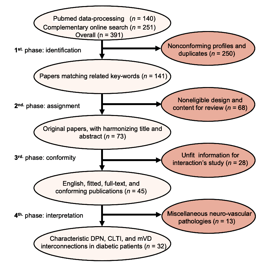
Abbreviations: CLTI, chronic limb-threatening ischemia; DPN, diabetic peripheral neuropathy; mVD, microvascular disease.
Design for data dispatching and analysis
Utilizing a 4-phase approach, the identification, assignment, conformity, and analytical processes are briefly illustrated in Figure 1.
Following data selection, distinct analysis of bilateral DPN/CLTI interplay in DF syndrome revealed little evidence-based information. Few publications matched the scope of this study and were further examined in a narrative review format.
Etiologic Aspects
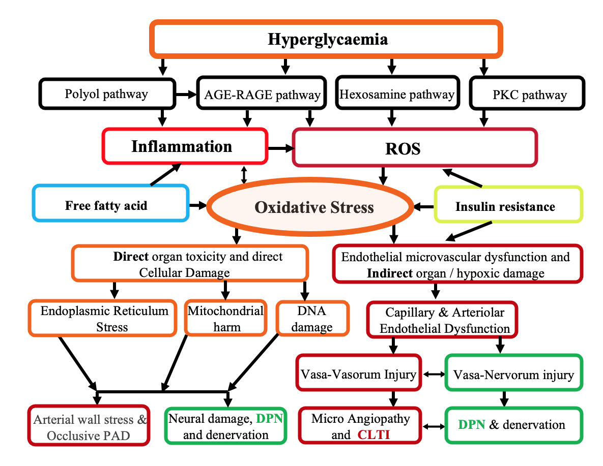
From an etiologic perspective, persistent hyperglycemia contributes to the development of DPN in approximately half of adults with DM.1-3 The pathogenesis of DPN is multifactorial and involves not only metabolic syndrome but also dyslipidemia, chronic inflammation, persistent hypoxia, and insulin resistance.1-3,13 Although the complete pathophysiologic sequence remains incompletely understood, several interconnected mechanisms have been identified (Figure 2) and are summarized below.
Direct hyperglycemic cellular damage
DPN arises through multiple molecular pathways that exert direct tissue toxicity during chronic hyperglycemia.14,15 Activation of the polyol (sorbitol) pathway and excessive formation of advanced glycation end products (AGEs) within neurons increase cellular oxidative stress and promote uncontrolled production of reactive oxygen species (ROS).14-16 These processes result in progressive mitochondrial harm, local tissue hypoxia, acidosis, and systemic cellular injury (Figure 2), including the endothelial cells in regional arterioles and capillaries that are critical to the viability of the neuroischemic limb.16,17
Indirect hyperglycemic damage to lower limb innervation
AGEs can simultaneously damage the microcirculatory system by disrupting both the morphology and function of endothelial structures,14,17 including the neural microvessels responsible for nerve perfusion, known as the vasa nervorum (VN).5,14,17 Initially confined to endothelial injury, AGE-mediated endothelial damage progressively involves the entire microvascular wall (Figure 2) and produces indirect cellular harmful effects through chronic tissue hypoxia.14,17
Consequently, the severely impaired perfusion caused by hypoxic microvascular disease (mVD) promotes regional microthrombosis and sclerotic tissue growth.16,17 This extensive and complex neurovascular microcirculatory impairment13,14,17 may drive the clinical evolution of neural tissue, which is highly susceptible to hypoxia.14,16
Lipid imbalance and insulin resistance
Recent studies demonstrate that dyslipidemia further accelerates the progression of DPN (Figure 2) and exerts neurotoxic effects,14,18,19 primarily mediated by free fatty acids and oxidized low-density lipoproteins.18,19 Notably, an adverse lipid profile characterized by hypercholesterolemia has been identified as an independent negative predictor of healing in patients with diabetic neuroischemic ulcers and is associated with increased risk of limb loss.18-20
Additional etiologic factors frequently associated with DPN include insulin resistance (Figure 2), vitamin D deficiency,21,22 and chronic inflammation.15 These factors involve multiple inflammatory pathways and collectively contribute to the initiation and progression of DPN. 2,15,23
Structural and Pathophysiologic Features
Morphologic changes in peripheral neural structures contribute to progressive sensory-motor and autonomic changes in lower limb peripheral nerves, revealing a bidirectional self-perpetuating neurovascular interaction. These interactions may operate during the early or subclinical stages of DPN.14,17,24,25
Progressive neural and perineural anatomical alterations
As in other tissues, the lower extremities contain reproducible neurovascular bundles that provide a consistent anatomic relationship between nerves and blood vessels.26 First described by Taylor et al26 in the early 1990s, this consistent topographic neurovascular arrangement is essential for normal tissue function and repair following injury.26 It enabled the authors to better define the complex role of these bundles as specialized vascular pathways that facilitate regional tissue monitoring.26 Pathologic remodeling originating from either the vascular or neural component of these bundles may result in harmful bilateral neurovascular interactions.24-26
However, these essential neurovascular bundles are not damaged solely by intrinsic neural or vascular dysfunction.25,26 Surrounding tissues that envelop these multifunctional hubs frequently undergo harmful extrinsic transformation, creating a rigid peri-neurovascular sclerotic environment with entrapment features. This diffuse perineural fibrosis appears to result from concomitant musculoskeletal diabetic microangiopathy and contributes to limited joint and fascial mobility, chronic ligamentous hypertension, bone deformities and lysis, and foot compartment syndromes. These conditions generate additional anatomical constraints that worsen the DPN clinical picture.15,17,24,27
Together, these intrinsic and extrinsic neurovascular structural alterations promote the creation of a hostile neurovascular environment in DF, facilitating early DPN and mVD development within the complex diabetic neuroischemic foot syndrome.4,12,13,15,24,25,27 Patients with diabetes experience combined multifactorial injury affecting arterial, venous, neural, and surrounding tissue structures.3,4,15,17
Conventionally, the peripheral arterial network is divided into macrocirculatory conduits, including arterial trunks and branches with diameters >100 μm and microcirculatory vessels with diameters <100 μm.25,27,28
Macrovascular arterial structural damage produces characteristic morphologic wall changes that lead to regional or global ischemia in DF tissues, including specific forms of axonal neuropathy observed in neuroischemic feet with CLTI.4,29 In addition, BTK and below-the-ankle (BTA) macrovascular occlusive disease affects arterial branches that may undergo medial arterial sclerosis (MAS) and medial arterial calcification (MAC).11,25 These macrovascular changes promote severe arterial chronic total occlusions (CTOs) with common continuous wall calcifications that often correspond to GVG/Global Limb Anatomic Staging System (GLASS)12 infrainguinal grades 3-4 severity lesions within the affected vessels.9,12,30,31
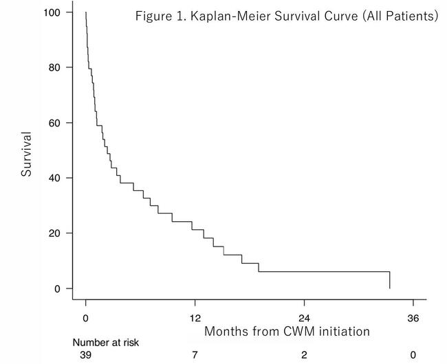
Although the mechanisms underlying MAS and MAC remain incompletely understood, Edmonds et al first reported a possible association between tibial and pedal artery calcification and autonomic DPN in 1982 (Figure 3).8 This association was subsequently confirmed by Gilbery et al9 and later investigations.10,17 In most DF patients with complex thrombocalcific occlusions (GVG/GLASS grades 3-4),12 tissue recovery attempts have been proposed through wound-oriented revascularization and regional foot reperfusion.26,32 These approaches have been collectively described as intentionally targeted revascularization30 and include the end-artery occlusive disease (EAOD) theory,31 the anatomical and functional angiosome model for CLTI foot perfusion,30 and the older concept of wound-related artery (inline flow),32 more recently called the woundosome concept.33 Despite encouraging wound-healing outcomes, robust evidence supporting their effectiveness remains limited,12,17,30,32 and consistent evidence of DPN recovery has not been established.25
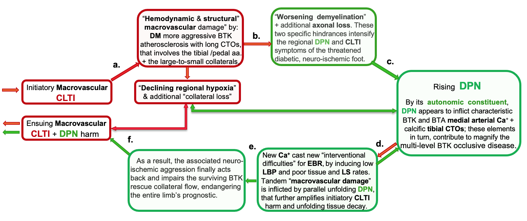
Abbreviations: DM, diabetes mellitus; CTO, chronic total occlusion.
Microvascular structural impairment simultaneously contributes to DF complications related to chronic hyperglycemia affecting foot tissue, including peripheral nerves. mVD involves irreversible proximal arteriolar damage associated with MAS and MAC9,11,17,28 and widespread capillary injury characterized by endothelial basement membrane thickening.24,25,27,28 These changes adversely affect neural structures, including the vasa vasorum and VN, leading to hypoxic depletion and impairment of axon-mediated vasomotor microcirculation.2,24,28
The disappearance of capillary networks and small nerve branches also results from inhibited arteriogenesis and angiogenesis.9,10,14,28 A characteristic bidirectional neurovascular ischemic injury occurs in limbs affected by DPN combined with CLTI.2,4,25,28 This injury damages multiple neural structures and leads to demyelination, distal-to-proximal neural dysfunction in the lower limbs, and eventually tissue denervation.28,29
Myelin plays a crucial structural role in normal axonal conduction and interneural signalling.14,19 Progressive demyelination contributes to DF regional denervation and indirectly exacerbates mVD.14,27
Functional amendments
Several pathophysiologic mechanisms associated with DPN contribute to progressive neural degeneration.14,24
- Macrovascular functional disturbances. Progressive stiffening of patent arterial branches leads to regional collateral alterations30-33 in which normal pulsatile peripheral collateral flow gradually transforms into abnormal laminar and slow flow in rigid containers.34,35 These hemodynamic changes, frequently observed in diabetic BTK arteries, generate higher stiffness and reduced wall shear stress, abnormal vascular remodeling, and diminished arteriogenesis. Collectively, these factors (Figure 3) increase the macrovascular propensity for regional vessel thrombosis.14,17,28,30
This hostile acidotic and prothrombotic environment may be intensified by local sepsis,4 tissue necrosis,4 scar tissue retraction, and inflammatory edema. These conditions mechanically distort macrovascular blood flow and progressively intensify regional hypoxia.5,14,17,25,28
Adjacent neural structures are particularly vulnerable to chronic hypoxia, and progressive oxygen deprivation contributes to additional DPN-related injury.17,27-29
- Microvascular functional disturbances. Similar to many pathophysiologic conditions, DPN arises from dysfunction involving both peripheral and central neural mechanisms. Myelin is present in most axons of lower limb nerves; it functions as a principal regulator of neural signal transmission, contributes to the formation of nodes of Ranvier, and ensures appropriate pacing of electrical conduction, thereby playing a crucial role in rapid nerve signal propagation.14,24,27
Schwann cells provide metabolic protection against cytotoxic agents such as AGEs and ROS.27,36,37 However, myelin is highly sensitive to hypoxic injury, and its depletion leads to slow, discontinuous axonal signal conduction.27,28
Long axons of the lower limbs are supplied by extensive microvascular networks through the VN.27,28 These axons are particularly sensitive to small hemodynamic variations and demonstrate increased vulnerability to multiple flow-dependent factors.14,25,27
Peripheral nerve injury frequently induces persistent structural and functional fiber disorders due to impaired autoregulation within the somatosensory system.29,37 Following such injury, the critical connection between peripheral nerves and central neural units becomes disrupted, ultimately resulting in neural loss.2,5,36,37 This theory aligns with current guidelines in DPN pathology and diagnostics.2,14 mVD associated with CLTI further reduces arteriolar and capillary perfusion28,30 and may compromise the VN (Figure 4). This process accelerates DPN progression and is directly linked to peripheral axonal degeneration.13,14,27-29
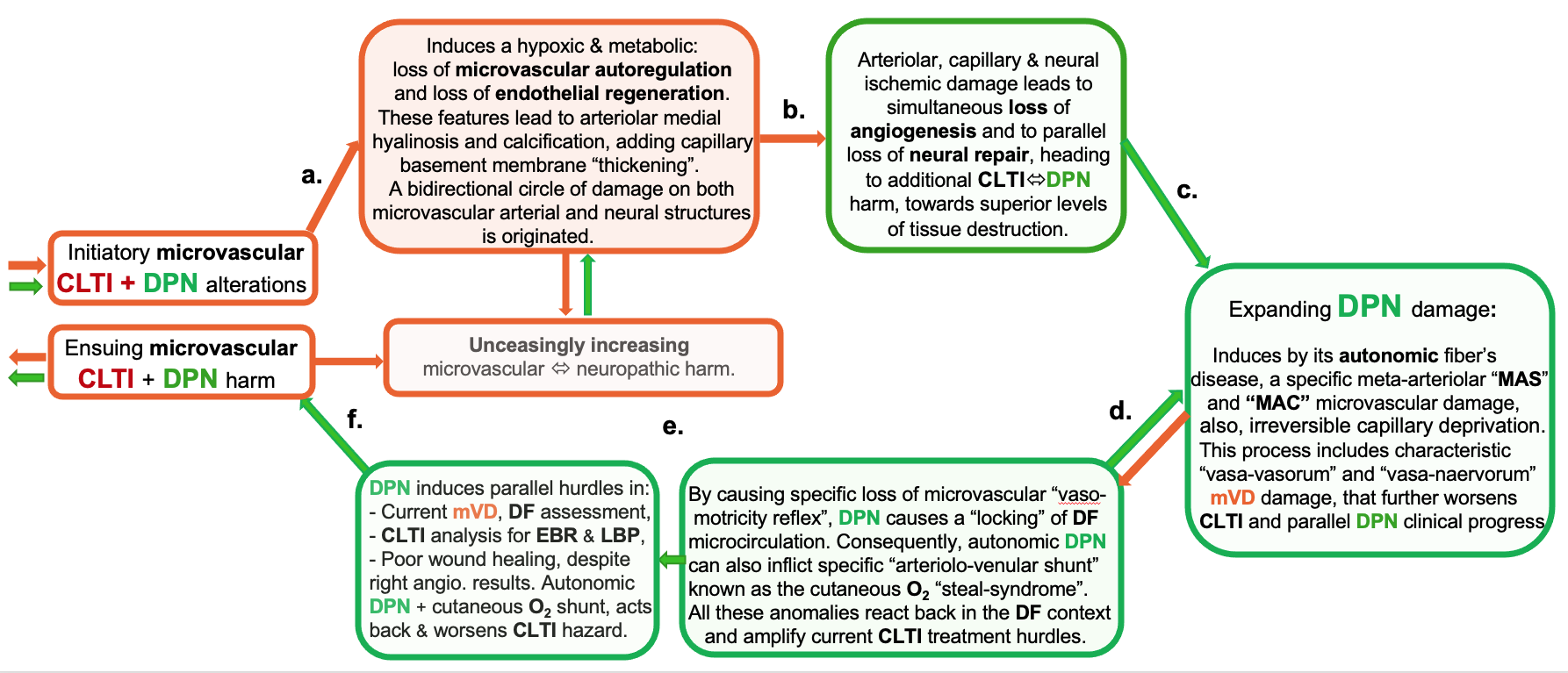
Peripheral nerve injury ultimately produces sympathetic denervation, increased local tissue hypoxia, and vasodilatation (Figure 4) along with microcirculatory arteriovenous shunting in cutaneous and muscular tissues.14,25,27,28
The DPN-related abnormal arteriovenous cutaneous steal phenomenon diverts oxygenated blood from the foot directly into the venous circulation, resulting in reduced microvascular perfusion of the skin, particularly in DPN-denervated territories.10,27 Consequently, the progressive deterioration of foot perfusion caused by autonomic DPN (Figure 4) further intensifies CLTI-induced hypoxic microvascular injury.24,25,27-30
Practical clinical point: The neuroischemic DF pathology presents a distinctive characteristic in which DPN and CLTI may alternately assume either a causal or resultant role at different stages of their bidirectional interaction. Although the precise mechanisms underlying the harmful interplay between DPN and CLTI remain incompletely defined, systematic assessment of mVD and DPN alongside contemporary vascular evaluation and treatment should be considered essential within an unremitting multidisciplinary DF approach.
Diagnostic Aspects
Synopsis of peripheral neurologic evaluation
Recent studies report from 35% to 63% symptomatic forms in patients with DM, including pDPN.4,42,49 pDPN represents a severe form of DPN that has been associated with a 5-year mortality rate of up to 43%.4,49 Several different types of DPN have been described, including distal symmetric bilateral polyneuropathy and sensory, motor, and autonomic neuropathies, as well as other forms of radiculoplexus neuropathies with or without associated pain.2,5,14,36,37,46 It should be emphasized that for most common DPN and pDPN clinical presentations, no current diagnostic test serves as a universally accepted gold standard for the diagnosis of DPN or pDPN.5,14,37,49 It is also important to note that for any neuropathic painful limb’presentation, a concomitant peripheral vascular examination is mandatory in order to exclude additional PAD.
For more clarity, several diagnostic evaluation levels2,5,14 could be implemented in assessing this group of patients:
The First Level of diagnostic evaluation should focus on detailed anamnesis, doubled by rigorous initial clinical neurological exam (parallel to the vascular investigation) of the threatened limb.
Over the past 2 decades, several scoring scales and “patient-reported symptoms” questionnaires have been proposed to identify and stratify the progression and severity of DPN or pDPN, including those described by Tesfaye et al,50 Scholz et al,51 Young et al,39 and Dyck et al.52 Current practice guidelines recommend a careful neurologic evaluation at both the systemic and affected limb levels, ideally via multidisciplinary participation.2,5,14,50
The Second Level of diagnostic evaluation consists of complementary noninvasive exams that provide objective assessment of DPN.
These quantitative measures have been described as helpful for DPN recognition and stratification, using quantitative neural analysis exams.14,37,49,50,51 Some of the current methods in use include:
- Electrophysiologic assessment.50-54 Applied to sensory nerves, it is a highly sensitive key examination for quantitative evaluation of DPN.50,53
- Point-of-care testing.47,51 Available devices include concomitant sural nerve conduction velocity analysis with plantar sweat analysis and assessment of sudomotor reflex function in the affected limbs.47,48,51
The Third Level of DPN assessment incorporates more performant, yet more expensive, diagnostic technologies that address targeted, complex neuropathic presentations, either in in isolation or in combination with CLTI.
Because concomitant systemic or peripheral mVD is common,2,47-50 it should be considered in all patients presenting with a neuroischemic foot.44,47-49 The use of these advanced diagnostic modalities should be individualized and guided by diabetic team consensus.51-53,55 Representative diagnostic techniques include:
- Skin biopsy. Evaluation of intraepidermal nerve fiber density offers valuable information on lower limb DPN and systemic mVD involvement.51-53
- Cornea confocal microscopy. This noninvasive technique evaluates corneal nerve fiber damage.55 Results require multidisciplinary team analysis.
- Eleven neurology-related serum proteins.56 These demonstrate a significant correlation between specific serum inflammatory proteins and pDPN severity. This new DPN monitoring method still needs to be validated by additional clinical research, including evaluation of forthcoming DPN biomarkers.56
Synopsis of associated vascular assessment
According to the same step-by-step approach to vascular assessment, 2 main levels2,5,14 can be acknowledged.
The First Level includes a comprehensive medical history and detailed examination of lower-extremity perfusion. Because most patients with diabetes and neurovascular disease have coexisting PAD or CLTI and varying degrees of DPN, concomitant screening for DPN is increasingly recommended in patients presenting with a neuroischemic foot.2-5,10,49 These patients may exhibit atypical hypoxic signs and symptoms, even in the early stages of neurovascular interactions. 5,25,41,49 Affected feet may appear warm2,5,41,49 with undistinguished cutaneous discoloration due to peripheral autonomic vasomotor reflex abolition.25,49 This dysfunction results in capillary vasodilatation with or without associated cutaneous oxygen steal syndrome.25,38
The Second Level of vascular assessment includes mandatory paraclinical exams that should include:
(a) Macrovascular atherosclerotic occlusive disease evaluation. Assessment should document the presence of MAS and MAC. Because of abnormal stiffness as a result of MAS and MAC, the segmental limb pressures and the ankle-brachial index (ABI) measurements often dwell without clinical pathological relevance.12,30,38,49
Contemporary approaches to standard macrovascular imaging were extensively reviewed in the last 3 decades concerning BTK arteriopathy in CLTI and are beyond the scope of this article.4,12,49 Most neurovascular interactions can be assessed at the macrovascular level using several standardized methods (starting with noninvasive, moving toward more invasive technology),12 including:
- Extravascular ultrasound (EVUS). A valuable noninvasive modality for evaluating MAS and MAC via direct visualization of the arterial wall,12 it also provides indirect analysis of the pulsatility index,34 pedal acceleration time,35 and distal foot collateral resistance.35,57
- Intravascular ultrasound (IVUS). Used to accurately evaluate the extent and morphology of MAS and MAC,58 its use is limited by the invasive nature of the procedure and reduced feasibility in occluded or severely stenotic arterial segments.12,40,58
- Conventional plane radiography. Particularly for MAC, it is recommended because it shows the characteristics and reproducible “railroad track” appearance.40,58,59
- Computed tomography (CT). It provides 70% sensitivity for MAC detection and correct quantitative arterial wall calcium analysis.58,59,60
(b) Current microvascular diagnostic methods. Targeting the arterial structures of <100-μm diameter (arterioles and capillaries), accurate mVD diagnosis can be done with several acknowledged methods in the absence of eventual arteriolovenular oxygen cutaneous shunting.10,25,41 These include:
- Transcutaneous pressure oxygen (TcPO2) monitoring. This can provide correct measurement of cutaneous oxygen levels in the foot.12,17,49
- Laser Doppler skin perfusion pressure. Combined with laser speckle flow imaging,61,62 it enables real-time analysis of regional microcirculatory flow in the foot.
- Near-infrared spectroscopy. Used to provide noninvasive assessment of tissue oxygen in specific regions of the foot microcirculation,63 but measurements may be affected by neuropathic influences.10,25,29,41
- Hyperspectral imaging. It provides reliable oxygen saturation data within targeted regions of the foot.64
- Cutaneous videocapillaroscopy. This method enables direct, high-resolution, noninvasive visualization of capillary morphology and function and functional skin capillary damange imaging.65
- Spatial frequency domain imaging. It combines assessment of cutaneous hemoglobin concentration with tissue oxygen saturation.66
- Nuclear imaging. Single-photon emission CT and positron emission tomography provide noninvasive quantification of intracellular mitochondrial ischemia.67 These imaging methods are not affected by neuropathic cutaneous shunting and may complement digital subtraction angiography analysis, particularly for vessels <500-μm diameter, where visual acuity is diminished.68
- Novel diagnostic techniques. Thse techniques focus on endovascular endothelial cell biopsy and provide accurate diagnosis and stratification of early-stage mVD.69 Independent from neuropathic interferences, this method has demonstrated a pooled technical success rate up to 91%.69
- Distal foot videocapillaroscopy. This method may aid differential diagnosis by distinguishing primary mVD (associated with CLTI) from secondary (DPN-induced) microvascular cutaneous arteriolovenular shunting.17,41,65,70
It is important to note that according to available guidelines, contemporary CLTI diagnostics should associate current macrovascular stratification (adding MAS/MAC evaluation)40, 41,58,59 to at least one microcirculatory exploration4,12,17,38,40,49,58 to rule out coupled mVD and its independently aggravating ischemic threat.
(c) Combined macrovascular and mVD diagnosis. This modern diagnostic technology can be successfully implemented (if it is available in reference DF centers) in association with other macro- and microvascular daily practice and above-mentioned exploration; It includes:
- Indocyanine green angiography. This technique provides accurate anatomic and functional imaging of macrocirculatory and microcirculatory foot perfusion.71 It promotes targeted topographic foot revascularization to optimize tissue healing.30,71
- Contrast-enhanced magnetic resonance imaging (MRI). Encompassing arterial spin labeling and blood oxygenation level-dependent techniques, it enables a direct flow (morpho-functional) assessment of macrovascular and microvascular perfusion in small arterial branches, MAC, arterioles, and capillaries affected by mVD.72
Treatment Insights
Current management of the diabetic neuroischemic foot in patients with CLTI requires rapid diagnosis of both macrovascular disease and mVD, combined with multidisciplinary evaluation that includes assessment for DPN, and timely macrovascular revascularization performed in conjunction with wound debridement.12,30,41,49
However, no standardized treatment algorithms or consensus recommendations specifically address the management of diabetic MAS, MAC, and mVD.17,40,41,49,59 Several studies have suggested that CLTI revascularization may provide clinical benefit, including potential improvement in DPN. However, the available evidence remains limited and inconsistent.12,24,74-76
Several studies have evaluated the effects of CLTI bypass revascularization on microvascular perfusion and DPN. In these investigations, TcPO2 and laser flowmetry were used to assess microvascular perfusion, while peroneal nerve conduction velocity was analyzed for DPN evaluation in patients undergoing CLTI revascularization.74-76 Some studies reported that reversal of limb hypoxia halted DPN progression,74 whereas others demonstrated significant improvement in limb perfusion accompanied by parallel improvement in neuropathic symptoms.75 In contrast, other investigators found no meaningful changes in DPN manifestations or nerve conduction parameters despite successful revascularization.10,76
Recently, Jujo et al77 reported findings from a prospective series of patients with neuroischemic CLTI. Three months after endovascular revascularization, both the current perception threshold (used to assess DPN) and ABI (used to evaluate CLTI) improved significantly.77 Patients who experienced improvement in DPN-related sensory symptoms also had a significantly higher 6-month survival rate.77
Based on the hypothesis that peripheral nerves in patients with diabetes are particularly vulnerable to compression at specific anatomical entrapment sites in the neuropathic foot, minimally invasive nerve decompression techniques have recently been proposed.49,78 These interventions have been associated with improved pain scores; however, consistent resolution of symptoms within 3 months has not been reported.78
Limitations
A major limitation of this scoping review is the limited quality and quantity of available evidence, as few studies directly address the central topic. Despite a rigorous literature search, relatively few publications adequately described the relevant mechanisms. Much of the available information focuses on relatively novel pathologic entities, such as DF mVD, with or without concomitant MAS and MAC. Evidence regarding the bidirectional interactions between CLTI and DPN is particularly limited and is derived largely from observational studies and clinical interpretations. Consequently, these findings do not provide a comprehensive etiopathogenetic classification or a complete mechanistic understanding of these processes.
Another important limitation is the lack of systematic DPN screening and severity stratification in most clinical studies of diabetic neuroischemic CLTI. Detailed neuropathic assessment is rarely incorporated into CLTI revascularization studies and, when reported, is often limited to descriptive or anecdotal observations.
Conclusion
Limbs affected by DPN may coexist with CLTI and appear to form closely interconnected pathologic processes that interact continuously at both the macrovascular and microvascular levels. These complex interactions may accelerate tissue injury and increase the risk of tissue damage and limb loss. Current evidence supports a comprehensive vascular assessment that includes evaluation of both macrovascular disease and mVD, together with systematic screening for DPN as part of multidisciplinary management. Additional research is needed to clarify the underlying mechanisms of these neurovascular interactions and establish evidence-based diagnostic and therapeutic strategies.
Affiliations and Disclosures
Vlad Adrian Alexandrescu, MD, PhD, Arnaud Kerzmann, MD, Evelyne Boesmans, MD, and Vincent Tchana-Sato, MD, PhD are from the Cardiovascular and Thoracic Surgery Department, CHU Sart-Tilman University Hospital, Liège, Belgium; Jos C. van den Berg, MD, PhD, is from the Centro Vascolare Ticino, Ospedale Regionale di Lugano, Switzerland.
The authors report no financial relationships or conflicts of interest regarding the content herein.
This research received no external funding.
Manuscript accepted June 24, 2026.
Address for correspondence: Vlad Adrian Alexandrescu, MD, PhD, CHU Sart Tilman University of Liège Hospital, Avenue de l’Hôpital 1, 4000 Liège, Belgium. Email: v.alex@skynet.be
References
1. Saeedi P, Petersohn I, Salpea P, et al; IDF Diabetes Atlas Committee. Global and regional diabetes prevalence estimates for 2019 and projections for 2030 and 2045: results from the International Diabetes Federation Diabetes Atlas, 9th edition. Diabetes Res Clin Pract. 2019;157:107843. doi:10.1016/j.diabres.2019.107843
2. Pop-Busui R, Boulton AJM, Feldman EL, et al. Diabetic neuropathy: a position statement by the American Diabetes Association. Diabetes Care. 2017;40(1):136-154. doi:10.2337/dc16-2042
3. Sun J, Wang Y, Zhang X, Zhu S, He H. Prevalence of peripheral neuropathy in patients with diabetes: a systematic review and meta-analysis. Prim Care Diabetes. 2020;14(5): 435-444. doi:10.1016/j.pcd.2019.12.005
4. Mills JL Sr, Conte MS, Armstrong DG, et al; Society for Vascular Surgery Lower Extremity Guidelines Committee. The Society for Vascular Surgery lower extremity threatened limb classification system: risk stratification based on wound, ischemia, and foot infection (WIfI). J Vasc Surg. 2014;59(1):220-234.e1-2. doi:10.1016/j.jvs.2013.08.003
5. Nukada H. Ischemia and diabetic neuropathy. Handb Clin Neurol. 2014;126:469-4487. doi:10.1016/B978-0-444-53480-4.00023-0
6. Lewi MJ, ed. The Textbook of Chiropody. The School of Chiropody of New York; 1914.
7. Sanders LJ, Robbins JM, Edmonds ME. History of the team approach to amputation prevention: pioneers and milestones. J Vasc Surg. 2010; 52(3 Suppl):3S-16S. doi:10.1016/j.jvs.2010.06.002
8. Edmonds ME, Morrison N, Laws JW, Watkins PJ. Medial arterial calcification and diabetic neuropathy. Br Med J (Clin Res Ed). 1982;284(6320):928-930. doi:10.1136/bmj.284.6320.928
9. Gilbey SG, Walters H, Edmonds ME, et al. Vascular calcification, autonomic neuropathy, and peripheral blood flow in patients with diabetic nephropathy. Diabet Med. 1989;6(1):37-42. doi:10.1111/j.1464-5491.1989.tb01136.x
10. Alexandrescu VA, Van Overmeire L, Makrygiannis G, et al. Clinical implications of diabetic peripheral neuropathy in primary infrapopliteal angioplasty approach for neuro-ischemic foot wounds. J Endovasc Ther. 2023;30(6):920-930. doi:10.1177/15266028221106312
11. Lanzer P, Boehm M, Sorribas V, et al. Medial vascular calcification revisited: review and perspectives. Eur Heart J. 2014;35(23):1515-1525. doi:10.1093/eurheartj/ehu163
12. Conte MS, Bradbury AW, Kolh P, et al; GVG Writing Group. Global vascular guidelines on the management of chronic limb-threatening ischemia. J Vasc Surg. 2019;69(6S):3S-125S.e40. doi:10.1016/j.jvs.2019.02.016
13. Powers AC. Type 1 diabetes mellitus: much progress, many opportunities. J Clin Invest. 2021;131(8):e142242. doi:10.1172/JCI142242
14. Chang MC, Yang S. Diabetic peripheral neuropathy essentials: a narrative review. Ann Paliat Med. 2023;12(2):390-398. doi:10.21037/apm-22-693
15. Cheng Y, Chen Y, Li K, et al. How inflammation dictates diabetic peripheral neuropathy: an enlightening review. CNS Neurosci Ther. 2024;30(4):e14477. doi:10.1111/cns.14477
16. Yang P, Feng J, Peng Q, Liu X, Fan Zhongcai. Advanced glycation end products: potential mechanism and therapeutic target in cardiovascular complications under diabetes. Oxid Med Cell Longev. 2019;2019:9570616. doi:10.1155/2019/9570616
17. Alexandrescu VA, Kerzmann A, Boesmans E, Tchana-Sato V. Microangiopathic disease in the diabetic neuro-ischemic feet: a threatening, often understated contributor, for tissue and limb loss. Medical Research Archives. 2025;13(7). doi:10.18103/mra.v13i7.6730
18. Iqbal Z, Bashir B, Ferdousi M, et al. Lipids and peripheral neuropathy. Curr Opin Lipidol. 2021;32(4):249-257. doi:10.1097/MOL.0000000000000770.
19. Wu S, Cao X, He R, Xiong K. Detrimental impact of hyperlipidemia on the peripheral nervous system: a novel target of medical epidemiological and fundamental research study. Neural Regen Res. 2012;7(5):392-399. doi:10.3969/j.issn.1673-5374.2012.05.011
20. Ahmadi SAY, Shirzadegan R, Mousavi N, Farokhi E, Soleimaninejad M, Jafarzadeh M. Designing a logistic regression model for a dataset to predict diabetic foot ulcer in diabetic patients: high-density lipoprotein (HDL) cholesterol was the negative predictor. J Diabetes Res. 2021;2021:5521493. doi:10.1155/2021/5521493
21. Yip J, Mattock MB, Morocutti A, Sethi M, Trevisan R, Viberti G. Insulin resistance in insulin-dependent diabetic patients with microalbuminuria. Lancet. 1993;342(8876):883-887. doi:10.1016/0140-6736(93)91943-g
22. Alam U, Petropoulos IN, Ponirakis G, et al. Vitamin D deficiency is associated with painful diabetic neuropathy. Diabetes Metab Res Rev. 2021;37(1):e3361. doi:10.1002/dmrr.3361
23. Jin HY, Park TS. Role of inflammatory biomarkers in diabetic peripheral neuropathy. J Diabetes Investig. 2018;9(5):1016-1018. doi:10.1111/jdi.12794
24. Horton WB, Barrett EJ. Microvascular dysfunction in diabetes mellitus and cardiometabolic disease. Endocr Rev. 2020;42(1):29-55. doi:10.1210/endrev/bnaa025
25. Alexandrescu VA, Kerzmann A, Tchana-Sato V. Diabetic neuro-ischaemic foot syndrome and the key underlying interactions between chronic arterial disease and peripheral neuropathy. Medical Research Archives. 2025;13(2). doi:10.18103/mra.v13i2.6364
26. Taylor GI, Gianoutsos MP, Morris SF. The neurovascular territories of the skin and muscles: anatomic study and clinical implications. Plast Reconstr Surg. 1994;94(1):1-36. doi:10.1097/00006534-199407000-00001
27. Mauricio D, Gratacòs M, Franch-Nadal J. Diabetic microvascular disease in non-classical beds: the hidden impact beyond the retina, the kidney, and the peripheral nerves. Cardiovasc Diabetol. 2023;22(1):314. doi:10.1186/s12933-023-02056-3
28. Mansour A, Mousa M, Abdelmannan D, Tay G, Hassoun A, Alsafar H. Microvascular and macrovascular complications of type 2 diabetes mellitus: exome wide association analyses. Front Endocrinol (Lausanne). 2023;14:1143067. doi:10.3389/fendo.2023.1143067
29. Weber S, Ziegler A. Axonal neuropathy in chronic peripheral arterial occlusive disease. Muscle Nerve. 2002; 26(4):471-476. doi:10.1002/mus.10235
30. Alexandrescu VA, Neville RF, Kerzmann A, et al. Applicability of intentional topographic revascularization following the angiosome model in patients with chronic limb-threatening ischemia: from theory, to current clinical challenges. Clin Surg. 2024; 9:3701. doi:10.25107/cis-v9-id3701
31. O’Neal LW. Surgical pathology of the foot and clinicopathologic correlations. The end-artery occlusive disease conception. In: Bowker JH, Pfeifer MA. Levin and O’Neal’s Diabetic Foot. 7th ed. 2007;Mosby Elsevier;367-401. doi:10.1016/B978-0-323-04145-4.50025-9
32. Iida O, Takahara M, Ohura N, et al; WARRIORS Investigators. Editor’s Choice - Impact of infrapopliteal revascularisation establishing in line flow to the wound in patients with chronic limb threatening ischaemia. Eur J Vasc Endovasc Surg. 2024;68(5):631-638. doi:10.1016/j.ejvs.2024.07.024
33. Patrone L, Pasqui E, Conte MS, et al. The “woundosome” concept and its impact on procedural outcomes in patients with chronic limb-threatening ischemia. J Endovasc Ther. 2025;32(6):1765-1776. doi:10.1177/15266028241231745
34. Mustapha JA, Diaz-Sandoval LJ, Saab F. Infrapopliteal calcification patterns in critical limb ischemia: diagnostic, pathologic and therapeutic implications in the search for the endovascular holy grail. J Cardiovasc Surg (Torino). 2017;58(3):383-401. doi:10.23736/S0021-9509.17.09878-0
35. Teso D, Sommerset J, Dally M, Feliciano B, Vea Y, Jones RK. Pedal acceleration time (PAT): a novel predictor of limb salvage. Ann Vasc Surg. 2021;75: 189-193. doi:10.1016/j.avsg.2021.02.038
36. Malik RA. Pathology of human diabetic neuropathy. Handb Clin Neurol. 2014;126:249-259. doi:10.1016/B978-0-444-53480-4.00016-3
37. Rosenberg DC, Blechschmidt V, Timmerman H, Wolff A, Treede R. Challenges of neuropathic pain: focus on diabetic neuropathy. J Neural Transm (Vienna). 2020;127(4):589-624. doi:10.1007/s00702-020-02145-7
38. Schraepen C, van der Laan L, Smet N, Meulenbroek A, Fourneau I. Chronic limb-threatening Ischemia does not enclose a homogeneous population: time for a more detailed classification. Int J Angiol. 2023;33(1):8-14. doi:10.1055/s-0043-1777414
39. Young MJ, Boulton AJ, Macleod AF, Williams DR, Sonksen PH. A multicentre study of the prevalence of diabetic peripheral neuropathy in the United Kingdom hospital clinic population. Diabetologia. 1993;36(2):150-154. doi:10.1007/BF00400697
40. Lanzer P, Ferraresi R. Medial sclerosis-epidemiology and clinical significance. Dtsch Arztebl Int. 2023;120(21):365-372. doi:10.3238/arztebl.m2023.0066
41. Alexandrescu VA, Deloose KR, Kerzmann A. From diabetic foot microvascular disease to medial arterial sclerosis and calcification in chronic limb-threatening ischemia: a potentially unified pathogenic and prognostic entity for tissue and limb loss. Clin Cardiol Cardiovasc Interv. 2026;9(2):1-10. doi:10.31579/2641-0419/547
42. Hicks CW, Selvin E. Epidemiology of peripheral neuropathy and lower extremity disease in diabetes. Curr Diab Rep. 2019;19(10):86. doi:10.1007/s11892-019-1212-8
43. Pop-Busui R, Lu J, Lopes N, Jones TLZ, BARI 2D Investigators. Prevalence of diabetic peripheral neuropathy and relation to glycemic control therapies at baseline in the BARI 2D cohort. J Peripher Nerv Syst. 2009;14(1):1-13. doi:10.1111/j.1529-8027.2009.00200.x
44. Behroozian A, Beckman JA. Microvascular disease increases amputation in patients with peripheral artery disease. Arterioscler Thromb Vasc Biol. 2020;40(3):534-540. doi:10.1161/ATVBAHA.119.312859
45. van den Berg JC. Angiosome perfusion of the foot: an old theory or a new issue? Semin Vasc Surg. 2018;31(2-4):56-65. doi:10.1053/j.semvascsurg.2018.12.002
46. Sharma S, Schaper N, Rayman G. Microangiopathy: is it relevant to wound healing in diabetic foot disease? Diabetes Metab Res Rev; 2020;36 Suppl 1:e3244. doi:10.1002/dmrr.3244
47. Petropoulos IN, Ponirakis G, Khan A, Almuhannadi H, Gad H, Malik RA. Diagnosing diabetic neuropathy: something old, something new. Diabetes Metab J. 2018;42(4):255-269. doi:10.4093/dmj.2018.0056
48. Yorec M, Malik RA, Calcutt NA, Vinik A, Yagihashi S. Diabetic neuropathy: new insights to early diagnosis and treatments. J Diabetes Res. 2018:5378439. doi:10.1155/2018/5378439
49. Hingorani A, LaMuraglia GM, Henke P, et al. The management of the diabetic foot: a clinical practice guideline by the Society for Vascular Surgery in collaboration with the American Podiatric Medical Association and the Society for Vascular Medicine. J Vasc Surg. 2016;63(2 Suppl):3S-21S. doi:10.1016/j.jvs.2015.10.003
50. Tesfaye S, Boulton AJM, Dyck PJ, et al; Toronto Diabetic Neuropathy Expert Group. Diabetes neuropathies: update on definitions, diagnostic criteria, estimation of severity, and treatments. Diabetes Care. 2010;33(10):2285-2293. doi:10.2337/ dc101303
51. Scholz J, Finnerup NB, Attal N, et al; Classification Committee of the Neuropathic Pain Special Interest Group (NeuPSIG). The IASP classification of chronic pain for ICD-11: chronic neuropathic pain. Pain. 2019;160(1):53-59. doi:10.1097/j.pain.0000000000001365
52. Dyck PJ, Albers JW, Andersen H, et al; Toronto Expert Panel on Diabetic Neuropathy. Diabetic polyneuropathies: update on research definition, diagnostic criteria and estimation of severity. Diabetes Metab Res Rev. 2011;27(7):620-628. doi:10.1002/dmrr.1226
53. England JD, Gronseth, GS, Franklin G, et al; American Academy of Neurology. Practice parameter: evaluation of distal symmetric polyneuropathy: role of autonomic testing, nerve biopsy, and skin biopsy (an evidence-based review) [RETIRED]. Report of the American Academy of Neurology, American Association of Neuromuscular and Electrodiagnostic Medicine, and American Academy of Physical Medicine and Rehabilitation. Neurology. 2009;72(2):177-184. doi:10.1212/01.wnl.0000336345.70511.0f
54. Dyck PJ, Overland CJ, Low PA, et al. Signs and symptoms versus nerve conduction studies to diagnose diabetic sensorimotor polyneuropathy: Cl vs. NPhys trial. Muscle Nerve. 2010;42(2):157-164. doi:10.1002/ mus.21661
55. Petropoulos IN, Ponirakis G, Ferdousi M, et al. Corneal confocal microscopy: a biomarker for diabetic peripheral neuropathy. Clin Ther. 2021;43(9):1457-1475. doi:10.1016/j.clinthera.2021.04.003
56. Bäckryd E, Themistocleous A, Larsson A, et al. Eleven neurology‐related proteins measured in serum are positively correlated to the severity of diabetic neuropathy. Sci Rep. 2024;14(1):17068. doi:10.1038/s41598-024-66471-6
57. Li M, Liang H, Chiang C, Wu T. Pulsatility-index adjusted doppler flow measurement of pedal arteries in peripheral artery disease patients. J Chin Med Assoc. 2023;86(1):98-104. doi:10.1097/JCMA.0000000000000843
58. Kim TI, Guzman RJ. Medial artery calcification in peripheral arterial disease. Front Cardiovasc Med. 2023;10:1093355. doi:10.3389/fcvm.2023.1093355
59. Chen L, Chen D, Gong H, et al. Pedal medial arterial calcification in diabetic foot ulcers: a significant risk factor of amputation and mortality. J Diabetes. 2024;16(4):e13527. doi:10.1111/1753-0407.13527
60. POSTER/ABSTRACT Kerzmann A, Valkenborgh C, Alexandrescu VA, et al. Calcium score for chronic limb-threatening ischemia: P-111. ESVS 39th Annual meeting in Istanbul, Türkiye, September 2025. Eur J Vasc Endovasc Surg; 2025. doi:10.1016/j.ejvs.2025.09.037
61. Utsunomiya M, Nakamura M, Nagashima Y, Sugi K. Predictive value of skin perfusion pressure after endovascular therapy for wound healing in critical limb ischemia. J Endovasc Ther. 2014;21(5):662-670. doi:10.1583/14-4675MR.1
62. Guven G, Dijkstra A, Kuijper TM, et al. Comparison of laser speckle contrast imaging with laser Doppler perfusion imaging for tissue perfusion measurement. Microcirculation. 2023;30(1):e12795. doi:10.1111/micc.12795
63. Boezeman RPE, Becx BP, van den Heuvel DAF, Ünlü Ç, Vos JA, de Vries JPPM. Monitoring of foot oxygenation with near-infrared spectroscopy in patients with critical limb ischemia undergoing percutaneous transluminal angioplasty: a pilot study. Eur J Vasc Endovasc Surg. 2016;52(5):650-656. doi:10.1016/j.ejvs.2016.07.020
64. Chiang N, Jain JK, Sleigh J, Vasudevan T. Evaluation of hyperspectral imaging technology in patients with peripheral vascular disease. J Vasc Surg. 2017;66(4):1192-1201. doi:10.1016/j.jvs.2017.02.047
65. Neubauer-Geryk J, Wielicka M, Hoffmann M, Myśliwiec M, Bieniaszewski L. The impact of disease duration on microcirculatory dysfunction in young patients with uncomplicated type 1 diabetes. Biomedicines. 2024;12(5):1020. doi:10.3390/biomedicines12051020
66. Jett S, Thompson MR, Awasthi S, et al. Stratification of microvascular severity in the foot using spatial frequency domain imaging. J Diabetes Sci Technol. 2023;17(1):25-34. doi:10.1177/19322968211024666
67. Stacy MR, Zhou W, Sinusas AJ. Radiotracer imaging of peripheral vascular disease. J Nucl Med Technol. 2015;43(3):185-192. doi:10.2967/jnumed.112.115105
68. Diehm N. Commentary: intra-arterial subtraction angiography: what you see is not always what you get. J Endovasc Ther. 2015;22(2):252-253. doi:10.1177/1526602815573231
69. Piercy C, Adam J, Harrison JN, Heiss C, Campagnolo P, Creagh-Brown B. Endovascular endothelial cell biopsy - a systematic review. Vasa. 2025;54(5):305-313. doi:10.1024/0301-1526/a001207
70. Baku T, Kotatsu B, Sayaka H, Hiroyuki T, Hideaki H. Observation and density estimation of a large number of skin capillaries using wide-field portable video capillaroscopy and semantic segmentation. J Biomed Opt. 2023; 28(10):106003. doi:10.1117/1.JBO.28.10.106003
71. Venermo M, Settembre N, Albäck A, et al. Pilot assessment of the repeatability of indocyanine green fluorescence imaging and correlation with traditional foot perfusion assessments. Eur J Vasc Endovasc Surg. 2016;52(4):527-533. doi:10.1016/j.ejvs.2016.06.018
72. Pantoja JL, Ali F, Baril DT, et al. Arterial spin labeling magnetic resonance imaging quantifies tissue perfusion around foot ulcers. J Vasc Surg Cases Innov Tech. 2022;8(4):817-824. doi:10.1016/j.jvscit.2022.09.015
73. Hunter L, Wiley A, Mckinney G, et al. Neuropathy screening for patients with peripheral vascular disease helps to identify those at an increased risk of amputation, revascularization, and death. Ann Vasc Surg. 2024;100:60-66. doi:10.1016/j.avsg.2023.10.021
74. Akbari CM, Gibbons GW, Habershaw GM, LoGerfo FW, Veves A. The effect of arterial reconstruction on the natural history of diabetic neuropathy. Arch Surg. 1997;132(2):148-152. doi:10.1001/archsurg.1997.01430260046008
75. Young MJ, Veves A, Smith JV, Walker MG, Boulton AJ. Restoring lower limb blood flow improves conduction velocity in diabetic patients. Diabetologia.1995;38(9):1051-1054. doi:10.1007/BF00402174
76. Veves A, Donaghue VM, Sarnow MR, Giurini JM, Campbell DR, LoGerfo FW. The impact of reversal of hypoxia by revascularization on the peripheral nerve function of diabetic patients. Diabetologia 1996;39(3):344-348. doi:10.1007/BF00418351
77. Jujo K, Saito K, Ishida I, et al. Pilot cohort study assessing the efficacy of endovascular revascularization in the restoration of peripheral sensory disturbance in patients with critical limb ischemia. Circ J. 2017;81(12):1919-1926. doi:10.1253/circj.CJ-17-0405
78. Chaudhry V, Russel J, Belzberg A. Decompressive surgery of lower limbs for symmetrical diabetic peripheral neuropathy. Cochrane Database Syst Rev. 2008;2008(3):CD006152. doi:10.1002/14651858.CD006152.pub2