Reverse Crossing During Transcatheter Arterialization of Deep Veins in No-Option Chronic Limb-Threatening Ischemia: A Case Report
Abstract
Deep vein arterialization (DVA) has emerged as a technique for limb salvage in patients with chronic limb-threatening ischemia who lack distal arterial targets for conventional revascularization. Multiple operative strategies to perform DVA exist, all involving creation of an arteriovenous (AV) connection between a tibial artery and its corresponding vein. While technical success rates range from 79% to 100%, procedural challenges remain, most notably the creation of the AV connection. We present a novel crossing strategy designed to facilitate AV connection formation during DVA using the LimFlow system (Stryker).
J CRIT LIMB ISCHEM 2026;6(3):E92-E95. doi: 10.25270/jcli/CLIG-2500011
Key words: deep vein arterialization, LimFlow, chronic limb-threatening ischemia, retrograde access, vein-to-artery, endovascular limb salvage
Chronic limb-threatening ischemia (CLTI) remains one of the most challenging pathologies in vascular surgery, particularly in patients with distal arterial occlusions and no suitable distal targets for bypass or endovascular revascularization. For these “no-option” patients, deep vein arterialization (DVA) has emerged as a viable limb salvage technique.1,2 Interventionalists have utilized a variety of different operative strategies to perform DVA; however, there is only one FDA-approved comprehensive system that is dedicated to treat this type of pathology.3 The technical success rate ranges from 79% to 100%.3-6 Despite this, all interventional strategies require the creation of an arteriovenous (AV) connection somewhere in the lower leg, and the ability to create this connection has served historically as a challenging component of any DVA procedure. We describe a modified transcatheter arterialization of the deep veins (TADV) technique to facilitate AV crossing in the setting of burdensome calcific inflow disease.
Case Report (Reverse Vein-to-Artery Technique)
Initial presentation
A 74-year-old man with a history of end-stage renal disease on hemodialysis, metastatic renal cell carcinoma, type 2 diabetes mellitus, hypertension, ischemic cardiomyopathy with implantable cardioverter defibrillator placement, and prior right below-the-knee (BTK) amputation presented at an outside hospital with a nonhealing left lower extremity foot wound. At admission, the patient had a Wound, Ischemia, and foot Infection score of 4. Diagnostic arteriography revealed no overt disease of his aortoiliac and femoropopliteal segments, with a patent anterior tibial artery and occlusions of his peroneal and posterior tibial arteries (PTAs). Below the ankle, he had no reconstitution of his plantar vessels, with patent but diminutive dorsalis pedis (DP) and lateral tarsal arteries.
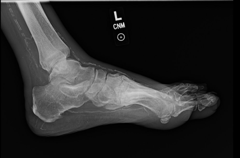
Given the lack of targets for either open bypass or retrograde pedal access, we elected to evaluate the patient for DVA. Preoperative evaluation included plain view x-rays of the foot, DVT ultrasound, and pedal vein mapping, which revealed the patient would be a suitable anatomic candidate for DVA based upon the PTA and posterior tibial vein (PTV). Additionally, medial arterial calcification score assessment of the patient was deemed to be 5, highlighting the extensive calcific burden of the tibial arteries and below-the-ankle (BTA) vessels (Figure 1).
Procedural overview
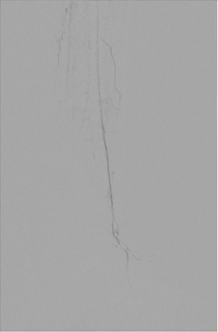
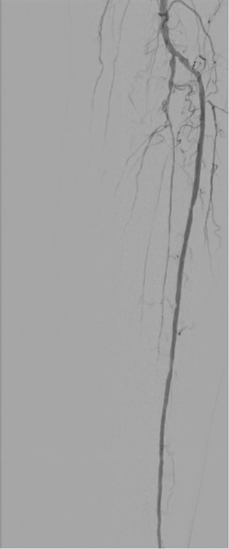
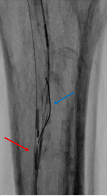
The patient was placed supine under general anesthesia. Ultrasound-guided access of the left superficial femoral artery was obtained with a 7F sheath and lateral plantar vein (LPV) using a 5F sheath. Prior diagnostic arteriography demonstrated patent proximal arteries (aorta to popliteal). BTK, the patient had a patent anterior tibial artery that continued BTA as an atretic DP and lateral tarsal artery with scant flow out to the mid-foot and toes, and both the peroneal artery and the PTA occluded in the proximal calf with no distal reconstitution (Figure 2). Of note, attempts at recanalization at both arteries had been unsuccessful (Figure 3). Based upon this anatomy, a PTA to PTV TADV was planned. We attempted to perform the TADV conventionally, first by crossing from the donor PTA into the PTV using the LimFlow ARC and V-Ceiver (Stryker). Despite our best efforts, the calcific burden of the distal popliteal artery and PTA made it challenging to deliver the ARC into the PTA. As a result, we elected to “reverse” our crossing strategy. We subsequently upsized our pedal venous sheath from a 5F sheath to a 6/7F slender sheath to facilitate the delivery of the ARC device from our venous sheath. Following this, we were subsequently able to deliver the ARC device to the proximal PTV and the V-Ceiver to the proximal PTA (Figure 4). Following this, we were able to successfully cross from the PTV into the PTA and obtain through-and-through wire access. The remainder of the case was performed conventionally.
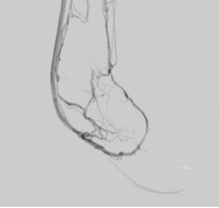
Then, we proceeded to balloon the PTV distal to our anastomosis with a 5 x 220-mm balloon. Following this, we used the Vector (Stryker), the forward-cutting valvulotome, and a 5 x 40-mm Serranator balloon (Cagent Vascular) to disrupt the distal venous valves at the level of the malleolus and proximal LPV. Following vein preparation, we sequentially deployed two 5.5 x 150-mm LimFlow stents from the level of the base of the calcaneus to 2 centimeters proximal to our AV crossing. We then deployed the 3.5 -5.5 mm x 60-mm LimFlow crossing stent. The procedure was successful based upon brisk flow through the venous stents into the LPV as well as the dorsal superficial and deep venous systems of the foot (Figure 5).
Postoperatively, the patient remained hemodynamically stable, with improved foot perfusion and Doppler signals noted along the plantar venous arch. On postoperative day 1, DVA ultrasound was obtained and demonstrated a flow volume of 145 mL/min in the LPV.
Follow-up and outcomes
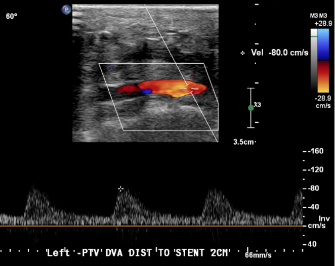
Over the course of the following 6 months, the patient’s clinical course has been uncomplicated. His TADV remains patent (Figure 6), he has undergone guillotine transmetatarsal amputation and subsequent grafting, and continues to follow up.
Discussion
DVA provides a vital solution for patients with advanced BTK and BTA disease, particularly those who cannot be treated using conventional endovascular or open strategies.1,2 The initial description of a LimFlow procedure reported by Kum et al7 utilized a dedicated system consisting of 4 main components: an arterial catheter with a needle, a venous catheter, a covered nitinol stent in a 7F delivery system, and an ultrasound alignment system with a laptop computer. The primary mechanism to facilitate crossing from artery to vein was based upon an ultrasound-emitting and ultrasound-receiving probe on the tips of the arterial and venous catheters, respectively. Since this publication, there have been technical and procedural refinements of the LimFlow system, most notably, the utilization of a needle-based crossing catheter (ARC) and dedicated catheter-based snare (V-Ceiver). This iterative change, which eliminated the use of ultrasound-based crossing to the procedure, yielded consistent technical success as demonstrated in the PROMISE I and II trials.3,8 The standard LimFlow procedure, also known as TADV, is performed utilizing fluoroscopic-guided artery-to-vein alignment between a tibial artery and one of its corresponding paired veins. An AV crossing is created using the LimFlow catheters, and covered stents are deployed to establish continuous flow from the artery into the venous system.1,2 This configuration allows for perfusion of the foot through the venous network. The success of the LimFlow system, both domestically and internationally, in the treatment of “no-option” CLTI patients led to FDA approval of the system in 2023.3,9 Despite this, prior to commercial approval, many operators routinely performed DVA procedures in an “off-the-shelf” fashion, using currently available products. Given that, the mechanistic strategy in performing AV crossing has been varied and includes the following techniques: venous arterialization simplified technique (VAST), Pioneer Peschiera Revascularization technique (PiPeR), GUNSIGHT, and Gandini technique.5,10-12 These strategies generally rely on some combination of the following off-the-shelf tools: re-entry catheters, balloon catheters, snares, needles, and wires.
Despite the availability of a variety of strategies to facilitate AV crossing, in this case, the technical issue that led to our decision to “reverse” our crossing strategy was our inability to deliver the ARC device into the ideal location due to eccentric luminal calcium. The ARC device was simply too large in comparison to the lumen gained following angioplasty. Recognizing this as the primary factor in our inability to perform AV crossing, we hypothesized that the smaller, more malleable V-Ceiver would be deliverable from our arterial access, and the ARC would be deliverable through a patent, accommodating tibial vein. While previously mentioned alternative AV crossing strategies may have led to successful AV crossing, each would have required opening additional products and contributing to increased case cost, and very likely, would have resulted in a longer case duration. Ultimately, our decision proved to be successful and demonstrates the feasibility and safety of this “reverse” crossing strategy when performing TADV. Insights gained from this case may allow for crossing when doing TADV to be performed in more distal, diseased locations due to the characteristics of the V-Ceiver and its ease of deliverability compared to the ARC device.
The evidence for the VAST technique reported no major complications, with 75% complete wound healing, rest pain resolution, and arterial venous fistula patency after 20 weeks of follow-up9 while the PROMISE II trial demonstrated a 76% limb salvage rate at 6 months using this approach, with 66% amputation-free survival at 1 year.3 However, these studies exclusively utilized arterial-to-venous crossings, the device was designed for antegrade use relative to arterial inflow, and no published data describe routine deployment from the venous side. In this case, conventional arterial access failed due to the heavy calcific burden of the PTA. Thus, the system was deployed in a retrograde direction from the vein, creating a vein-to-artery crossing. This reversal of direction required careful imaging alignment to avoid subintimal dissection or vessel misalignment. To our knowledge, this is the first report of reverse LimFlow deployment from the venous access site. Experimental or hybrid techniques involving surgical cutdowns and manual graft tunneling have been reported, but these differ significantly from the percutaneous approach performed for the procedure. In this case, we were able to successfully execute a technique that expands options for operators to achieve optimal AV formation with the use of an off-the-shelf device in the setting of adverse anatomy.
Conclusions
Our crossing technique represents an innovative and technically feasible alternative when standard crossing using the LimFlow system is unsuccessful. While this patient’s clinical course is ongoing, he continues to improve. This case demonstrates procedural technical success utilizing the ARC and V-Ceiver in an unconventional fashion in a no-option CLTI patient. Additional studies are needed to better understand the application of this technique in a larger population of no-option CLTI patients.
Affiliations and Disclosures
Antonio Solano, MD, Carla Scott, MD, Suyue M. Zhang, MD, Kathryn Naumann, MS, MBA, Jarrett Hubbard, BSA, Madison Baehner, BS, Gerardo Gonzalez-Guardiola, MD, Khalil Chamseddin, MD, Aaron Wagner, MD, Michael Shih, MD, M. Shadman Baig, MD, Melissa L Kirkwood, MD, and Michael Siah, MD are from the Division of Vascular Surgery, UT Southwestern Medical Center, Dallas, Texas, USA; Abdulaziz Shammaa, BS, is from the University of the Incarnate Word School of Osteopathic Medicine, San Antonio, Texas, USA.
Dr Baig has received research support from Cook Medical Inc and W.L. Gore & Associates; Dr Kirkwood is a consultant for W.L. Gore & Associates, Mölnlycke Health Care, and Egg Medical; and Dr Siah is a consultant for Stryker. The other editors and reviewers of this article have no relevant financial relationships to disclose. No funding was provided.
Manuscript accepted June 10, 2026.
Corresponding author: Michael C. Siah, MD, UT Southwestern Medical Center, Professional Office Building 1, Ste. 620, 5959 Harry Hines Blvd, Dallas, TX 75390-9157. Email: michael.siah@utsouthwestern.edu
References
1. Ho VT, Gologorsky R, Kibrik P, et al. Open, percutaneous, and hybrid deep venous arterialization technique for no-option foot salvage. J Vasc Surg. 2020;71(6):2152-2160. doi:10.1016/j.jvs.2019.10.085
2. Montero-Baker M, Sommerset J, Miranda JA. Hybrid superficial venous arterialization and endovascular deep venous arterialization. J Vasc Surg Cases Innov Tech. 2023;9(3):101160. doi:10.1016/j.jvscit.2023.101160
3. Shishehbor MH, Powell RJ, Montero-Baker MF, et al; PROMISE II Investigators. Transcatheter arterialization of deep veins in chronic limb-threatening ischemia. N Engl J Med. 2023;388(13):1171-1180. doi:10.1056/NEJMoa2212754
4. Saab FA, Mustapha JA, Ansari M, et al. Percutaneous deep venous arterialization: treatment of patients with end-stage plantar disease. J Soc Cardiovasc Angiogr Interv. 2022;1(6):100437. doi:10.1016/j.jscai.2022.100437
5. Ysa A, Lobato M, Mikelarena E, et al. Homemade device to facilitate percutaneous venous arterialization in patients with no-option critical limb ischemia. J Endovasc Ther. 2019;26(2):213-218. doi:10.1177/1526602819830983
6. Siah M, Reynolds K, Mashhood Ali Bokhari SM, et al. Real-world multicenter retrospective study of outcomes following transcatheter arterialization of the deep veins for no-option chronic limb-threatening ischemia. J Vasc Surg. 2026:S0741-5214(26)01172-9. doi:10.1016/j.jvs.2026.05.029
7. Kum S, Tan YK, Schreve MA, et al. Midterm outcomes from a pilot study of percutaneous deep vein arterialization for the treatment of no-option critical limb ischemia. J Endovasc Ther. 2017;24(5):619-626. doi:10.1177/1526602817719283
8. Clair DG, Mustapha JA, Shishehbor MH, et al. PROMISE I: early feasibility study of the LimFlow System for percutaneous deep vein arterialization in no-option chronic limb-threatening ischemia: 12-month results. J Vasc Surg. 2021;74(5):1626-1635. doi:10.1016/j.jvs.2021.04.057
9. Schmidt A, Schreve MA, Huizing E, et al. Midterm outcomes of percutaneous deep venous arterialization with a dedicated system for patients with no-option chronic limb-threatening ischemia: the ALPS multicenter study. J Endovasc Ther. 2020;27(4):658-665. doi:10.1177/1526602820922179
10. Naga A, Hamo N, Lakin C, Thulasidasan N, Zayed H. Off-the-shelf percutaneous deep vein arterialization (pDVA). Vasa. 2026;55(3):190-195. doi:10.1024/0301-1526/a001229
11. Hasanlli A, Rundback JH, Herman K, Mendoza E. Deep vein arterialization for acute limb ischemia: initial experience in three patients. J Crit Limb Ischem. 2026:6(1)E10-E15. doi:10.25270/jcli/CLIG-2500008
12. Gandini R, Merolla S, Scaggiante J, et al. Endovascular distal plantar vein arterialization in dialysis patients with no-option critical limb ischemia and posterior tibial artery occlusion: a technique for limb salvage in a challenging patient subset. J Endovasc Ther. 2018;25(1):127-132. doi:10.1177/1526602817750211