






No-Option Chronic Limb-Threatening Ischemia: Percutaneous Deep Vein Arterialization for Limb Salvage in Buerger Disease
Abstract
Thromboangiitis obliterans (TAO) is a rare nonatherosclerotic vasculopathy that may progress to chronic limb-threatening ischemia without revascularization options. This case report describes a 27-year-old man with biopsy-confirmed TAO and refractory chronic limb-threatening ischemia who underwent percutaneous deep vein arterialization after failure of medical, endovascular, and surgical therapies. Despite technical success, he developed hyperperfusion syndrome, limb edema, and persistent ischemia. Repeated interventions failed to improve perfusion, resulting in below-the-knee amputation. The patient subsequently achieved good functional recovery with a prosthesis. This case highlights the uncertain efficacy of percutaneous deep vein arterialization in TAO and the need for further investigation and careful patient selection.
J CRIT LIMB ISCHEM 2026:6(4):E126-E132. doi: 10.25270/jcli/CLIG-2600008
Key words: thromboangiitis obliterans, percutaneous deep vein arterialization, endovascular therapy
Introduction
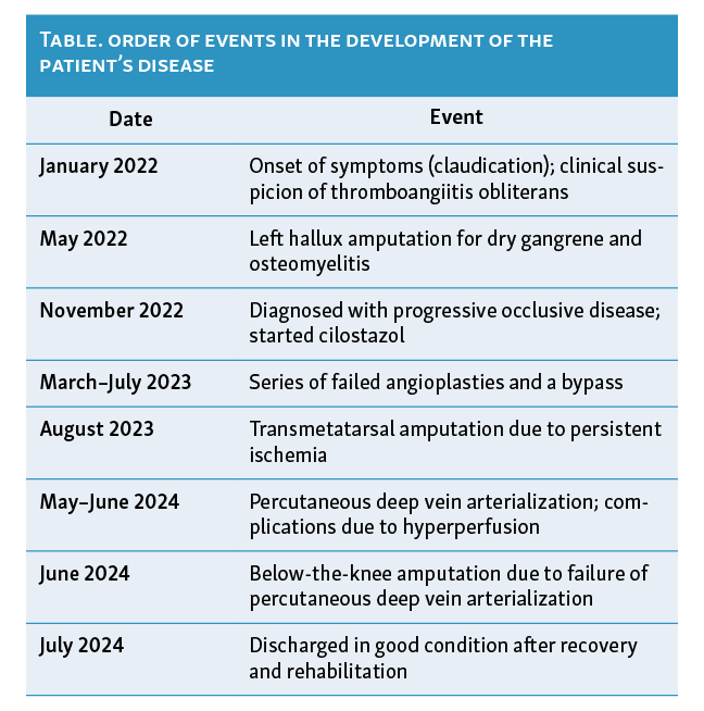
Buerger disease, also known as thromboangiitis obliterans (TAO), is a nonatherosclerotic, inflammatory occlusive disease of small- and medium-sized vessels that spares the arterial media but causes segmental thrombosis and distal microcirculatory failure. While tobacco exposure is a known prerequisite, the precise immune, genetic, and endothelial mechanisms of its pathogenesis remain undefined. Chronic limb-threatening ischemia (CLTI) in patients without conventional revascularization options remains a major therapeutic challenge. Percutaneous deep vein arterialization (pDVA) has emerged as a novel salvage approach in this setting, although its use has been primarily studied in atherosclerotic or diabetic populations. We present a case of advanced TAO managed with pDVA, highlighting the procedural feasibility, clinical challenges, and outcome in a nonatherosclerotic patient. A summarized timeline of events can be seen in the Table. This case report was written in accordance with the CARE guidelines.1
Patient Information
A 27-year-old man from Buenos Aires, Argentina, working in a family-owned cleaning supplies business, presented with progressive lower extremity ischemia. He had no history of hypertension, diabetes, dyslipidemia, autoimmune disease, or cardiovascular disease. The patient reported a 6.5 pack-year smoking history, having quit in June 2022. There was no history of substance use or significant family medical history. He was independent in daily activities and psychosocially stable.
Clinical Findings
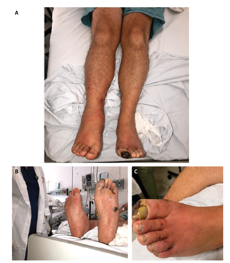
Initial symptoms in January 2022 included intermittent claudication at 100 meters. This progressed over months to severe rest pain, exacerbated in the supine position and partially relieved with leg dependency. By mid-2022, he developed a nonhealing blister on the left hallux, which progressed to dry gangrene. Key physical findings included bilateral absence of pedal and posterior tibial pulses, distal pallor and rubor with cold extremities consistent with Raynaud phenomenon, positive Allen and Buerger tests, a necrotic ischemic ulcer over the left hallux, and no sensory deficits, joint inflammation, or superficial phlebitis. The right foot was clinically asymptomatic throughout, with no ischemic symptoms reported at any point during follow-up. Clinical photographs obtained at initial evaluation are shown in Figure 1.
Diagnostic Assessment and Diagnosis
Following the identification of bilateral distal ischemia with digital ulceration and rest pain, diagnostic evaluation focused on characterizing the pattern of vascular involvement and excluding other etiologies. The case was initially assessed in a multidisciplinary setting by the rheumatology and vascular surgery departments, who coordinated the diagnostic workup.
Doppler ultrasound and angiographic imaging demonstrated bilateral infrapopliteal arterial occlusions involving all 3 tibial vessels—the anterior tibial, posterior tibial, and peroneal arteries—bilaterally, without distal continuity. The popliteal arteries were patent bilaterally. On the right side, vascular assessment was performed by Doppler ultrasound only, as the patient was clinically asymptomatic on that limb and formal arteriography was not indicated. Preserved proximal arterial flow and absence of calcifications or atherosclerotic plaques were confirmed; there were no hemodynamically significant proximal stenoses. Laboratory testing, including complete blood count, renal and liver function tests, inflammatory markers, thrombophilia screening, and autoimmune serologies (antinuclear antibodies, antineutrophil cytoplasmic antibodies, antiphospholipid antibodies), showed no abnormalities. Blood cultures were negative. A transoesophageal echocardiogram did not identify cardiac sources of emboli.
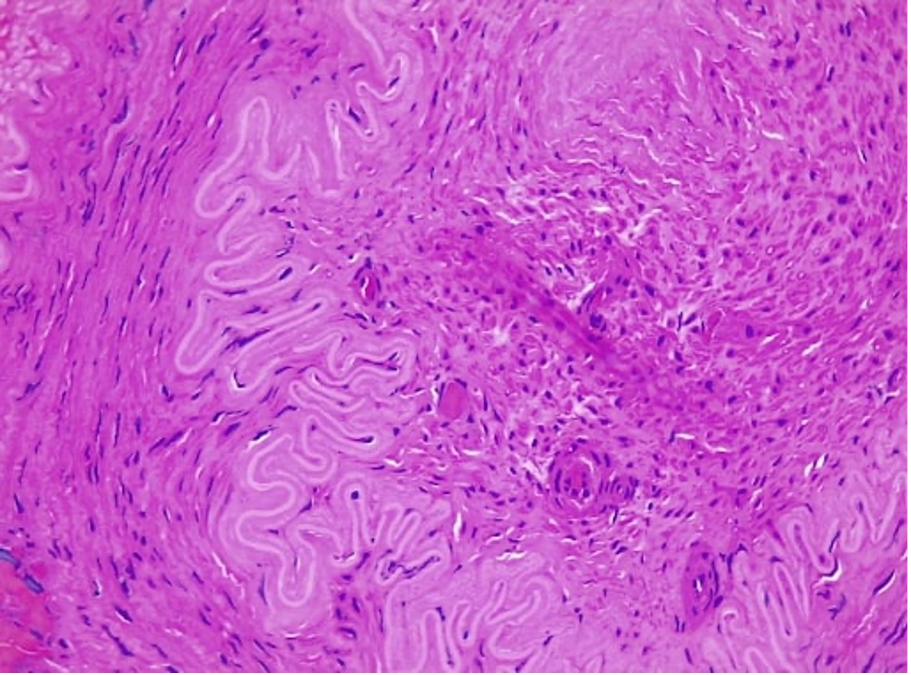
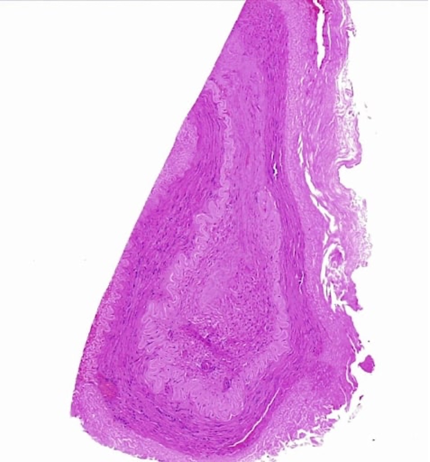
In June 2023, histopathologic examination of a distal arterial segment obtained during surgery showed features of acute vasculitis of small vessels, including transmural inflammatory infiltrates and occlusive thrombosis, without evidence of atherosclerosis or systemic vasculitis.
The diagnosis of TAO was established based on patient age, smoking history, distal nonatherosclerotic arterial involvement, absence of systemic disease, and confirmatory histological findings as shown in Figure 2 and Figure 3.
Therapeutic Interventions
Initial management focused on strict tobacco cessation, which the patient achieved and maintained from June 2022 onward. Despite this, the symptoms progressed. Pharmacologic therapy was initiated with cilostazol 100 mg bid in November 2022; the regimen was reassessed due to persistent claudication, and nifedipine 10 mg tid was added to address Raynaud phenomenon. Bosentan 62.5 mg bid, an endothelin receptor antagonist (Actelion Pharmaceuticals), was later introduced to manage ischemic pain but was discontinued due to a severe dermatologic adverse reaction. None of the medical treatments provided significant clinical improvement.
Revascularization Attempts
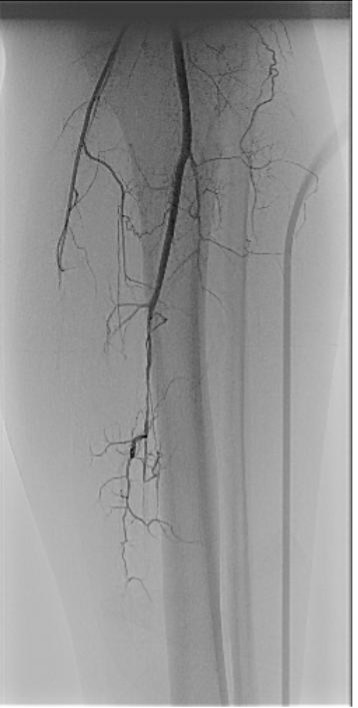
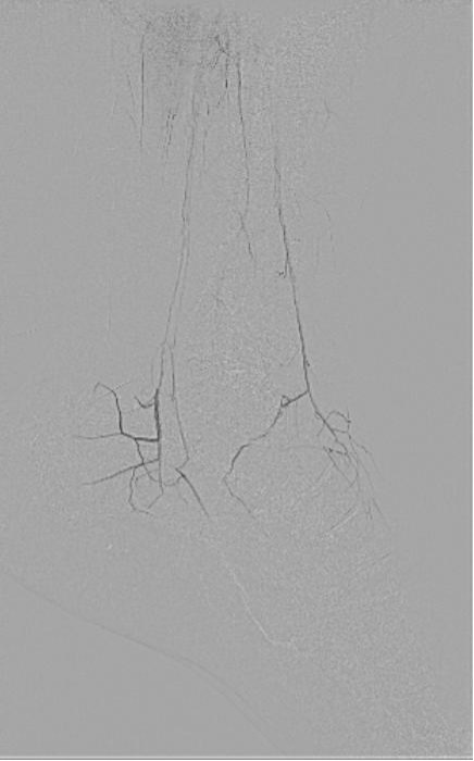
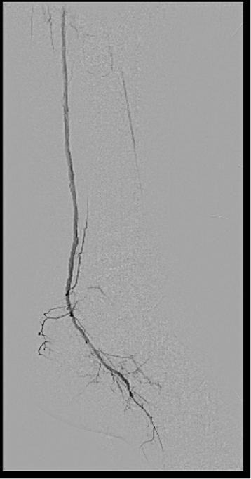
Between March and July 2023, the patient underwent multiple attempts at endovascular revascularization, including balloon angioplasties of the posterior tibial and plantar arteries, as well as stent placement. These interventions failed to achieve sustained perfusion or symptom relief. Pre- and post-procedural angiographic images are shown in Figures 4, 5, and 6.
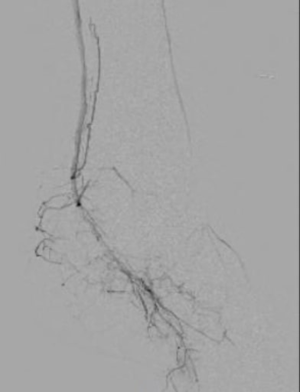
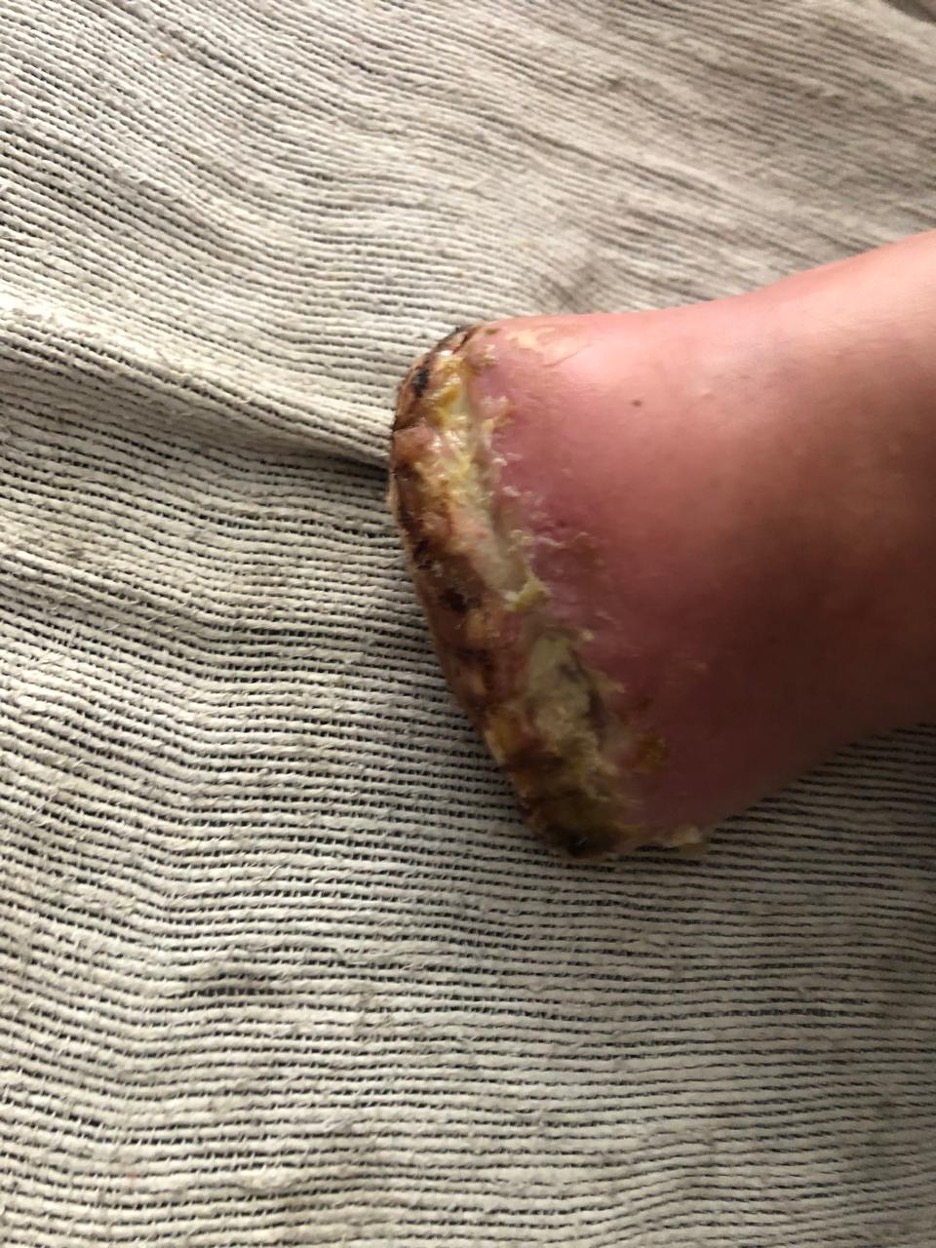
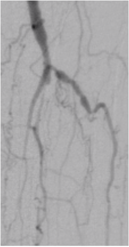
A surgical bypass using the internal saphenous vein from the popliteal artery to the distal third of the anterior tibial artery was performed in July 2023 but did not result in meaningful revascularization due to the extremely small caliber of the target vessels, with immediate postoperative occlusion of the graft. Figure 7 shows an angiogram obtained during this period, demonstrating that the anterior tibial artery reaches the foot via pedal vessels of very small caliber; the bypass itself is not identifiable on this image due to immediate occlusion. Following progressive trophic changes and critical ischemia, a hallux amputation was performed in May 2022 in the context of persistent rest pain refractory to conservative management, and a transmetatarsal amputation was required in August 2023 following failure of revascularization attempts. The clinical status of the left foot following transmetatarsal amputation is shown in Figure 8.
Percutaneous Deep Vein Arterialization
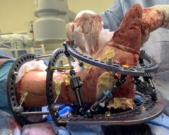
Given the absence of conventional surgical or endovascular options, and in the context of no-option CLTI, a pDVA procedure was performed in May 2024 as a limb salvage attempt. The procedure was performed under fluoroscopic guidance using the VAST system. Percutaneous access of the left posterior tibial artery at the proximal level was obtained and an arteriovenous fistula (AVF) was created. A Viabahn covered stent (W.L. Gore & Associates) was deployed from the popliteal artery (P3 segment) as the proximal landing zone to the retromaleolar posterior tibial vein as the distal landing zone, directing arterial blood retrograde through the posterior tibial vein into the plantar venous system. The venous loop was managed using a Wolverine cutting balloon (5 x 20 mm; Boston Scientific) followed by a Mustang percutaneous transluminal angioplasty balloon (5 x 80 mm; Boston Scientific). Intraoperative Doppler confirmed fistulous flow measuring 1269 mL/min at the plantar level.
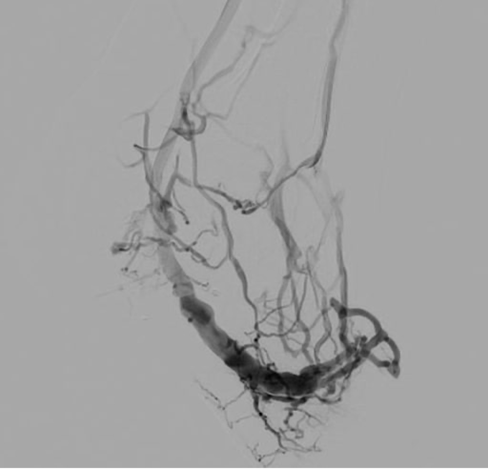
Although technically successful, the procedure was complicated by a high-flow arteriovenous syndrome resulting in significant limb edema and pain. In the immediate postprocedural period, elastic bandage compression was applied and limb elevation maintained; no pharmacological adjustments were made at this stage. Given the persistence of hyperperfusion syndrome, embolization of plantar venous branches was performed 10 days after the procedure (May 30), followed by surgical ligation of the marginal vein on June 12. Flow-limiting banding was also attempted prior to the decision to proceed with amputation. Despite these efforts, perfusion remained inadequate and critical ischemia persisted. Figure 7 shows the residual antegrade arterial flow pre-procedure, and Figure 9 the retrograde venous flow post-procedure.
Follow-up and Outcomes
A control angiogram performed following the banding procedure demonstrated occlusion of the AVF. Given the persistence of critical ischemia and exhaustion of further endovascular options, the patient underwent a below-the-knee (BTK; infrapatellar) amputation in June 2024. The postoperative course was favorable, with good wound healing, effective pain control, and no further complications. The patient was discharged after a 29-day hospital stay and began a structured rehabilitation program. At follow-up, the patient demonstrated excellent recovery and full adaptation to a lower limb prosthesis. He resumed his professional and social activities, including sports participation, and remained tobacco-free. At the most recent follow-up, the patient reported no ischemic symptoms, maintained full autonomy, and exhibited stable psychosocial wellbeing. The right lower limb, which had demonstrated reduced flow on Doppler ultrasound consistent with bilateral TAO involvement but had remained clinically asymptomatic throughout, showed no deterioration and required no intervention.
Patient Perspective
The patient approached his condition with determination and accepted smoking cessation as essential to treatment. He viewed the BTK amputation not as a setback but as a necessary step that finally alleviated his refractory ischemic pain. At final follow-up, he had completed rehabilitation, resumed his daily and professional activities, and expressed a positive outlook, feeling physically well and optimistic about his future.
Discussion
TAO, or Buerger disease, is a rare, nonatherosclerotic, segmental inflammatory vasculopathy that predominantly affects young male smokers and small- and medium-caliber arteries and veins of the distal extremities.2 The only intervention known to halt disease progression is absolute smoking cessation.3 However, in advanced cases presenting as CLTI, patients often progress to critical distal tissue loss with no revascularization options. Unlike patients with atherosclerotic peripheral arterial disease, where endovascular or surgical bypass techniques may be feasible, the distal distribution, lack of suitable target vessels, and segmental occlusions in TAO make conventional revascularization frequently unsuccessful.4-6
pDVA has emerged as a limb salvage technique for patients with no-option CLTI. Initially developed in animal models, this method establishes an arteriovenous connection to deliver arterial blood retrograde into the venous system, perfusing ischemic foot tissue via the venous plexus.7 Early trials in atherosclerotic and diabetic CLTI populations have demonstrated success rates above 90%, with 6-month amputation-free survival rates around 70% to 75% in PROMISE I and II studies, as well as ALPS registry data.8,9
Although endovascular interventions have been applied in TAO with variable short-term success, the role of pDVA in this cohort is scarcely documented. No large series or controlled data exist for pDVA in TAO. Unlike diabetic or atherosclerotic disease, TAO may compromise not only the arterial inflow but also the venous outflow and microcirculation, which are critical for pDVA efficacy. Moreover, venous fibrosis and inflammation may predispose to complications. A limited but informative body of case reports documents the use of DVA in patients with TAO, each offering distinct insights into the technical feasibility and clinical unpredictability of this approach. Ryu et al reported the first case of pDVA in a patient with TAO, a 38-year-old man with BTK triple-vessel occlusion and digital gangrene.10 Using an intravascular ultrasound-guided technique with Viabahn stents and a balloon-mounted covered stent for the arteriovenous anastomosis at the tibioperoneal trunk, they achieved technically successful retrograde arterialization of the deep venous arch. The patient subsequently required transmetatarsal amputation due to forefoot osteomyelitis but achieved wound granulation and fistula patency at follow-up with successful skin grafting, constituting a major limb salvage outcome. Smith et al described pDVA using commercially available off-the-shelf devices in a 55-year-old man with TAO and prior contralateral BTK amputation, presenting with a “desert foot” pattern on angiography.11 Technically successful arterialization was achieved via the lateral plantar vein, with Viabahn stents bridging the tibioperoneal trunk into the plantar venous system. Early detection of siphoning tributaries and timely coil embolization at 2 weeks were critical to optimizing foot perfusion. The patient underwent transmetatarsal amputation at 2.5 months with complete healing and remained ambulatory at 13-month follow-up, representing a successful limb salvage outcome. Moriarty et al reported pDVA in a 30-year-old man with TAO and a nonhealing hallux wound refractory to prior angioplasty.12 A posterior tibial artery-to-vein DVA with a 5-mm x 10-cm Viabahn stent resulted in initial clinical improvement, including wound granulation and restored Doppler signals at 1 month. A proximal stent stenosis at 3 months required reintervention with drug-coated balloon angioplasty and stent extension, with restoration of adequate perfusion. The patient ultimately required a transmetatarsal amputation, which healed completely, and remained ambulatory, representing successful prevention of major amputation. In contrast, Wu et al reported an open surgical DVA—a superficial femoral artery-to-superficial femoral vein anastomosis with distal valve disruption—in a patient with TAO that yielded no clinical improvement in ankle-brachial index or claudication distance. Ten years later, the patient developed a ruptured venous aneurysm secondary to inflammatory destruction of the venous wall, requiring emergent surgical repair.13 Notably, ligation of the shunt produced no further deterioration in distal perfusion, suggesting the DVA had contributed minimally to tissue perfusion—an outcome the authors attributed in part to the overly proximal anastomotic site and the underlying venous wall compromise inherent to TAO.
Taken together, these reports reveal a heterogeneous landscape. Three cases achieved major limb salvage with distal-level pDVA, while 1 open surgical DVA with a proximal anastomosis yielded no benefit and resulted in a serious, delayed complication. The cases of successful limb salvage share several features: predominantly arterial disease with relatively preserved venous architecture, technically distal fistula creation, and a disease pattern that, though severe, did not appear to prominently involve the venous outflow. These distinctions are relevant to understanding the failure observed in our patient.
In our patient, pDVA was pursued after failure of all conventional options, including multiple angioplasties and a distal bypass. Preprocedural criteria appeared favorable: the superficial venous system was patent and proximal inflow was intact. Nevertheless, despite technically successful fistula creation, clinical improvement did not ensue. The intraoperative Doppler-confirmed flow of 1269 mL/min at the plantar level exceeded the range typically observed in pDVA for atherosclerotic disease and may reflect underlying venous noncompliance secondary to inflammatory TAO involvement. This high-flow state, rather than restoring tissue perfusion, translated into hyperperfusion syndrome with edema and congestion that could not be controlled despite embolization and venous ligation. In retrospect, the magnitude of initial flow should have prompted immediate intraoperative flow reduction strategies, such as flow-limiting stent sizing, partial embolization of outflow branches prior to completing the procedure, or staged intervention, before concluding the case. No validated flow thresholds to guide intraoperative decision-making in pDVA currently exist in the literature, representing an important area for future investigation.
A further consideration is the role of the posterior tibial artery in preprocedural perfusion. As demonstrated in Figure 7, the posterior tibial artery represented the dominant vessel with extensive antegrade flow to the foot at the time of the pDVA. The placement of the covered stent to create the AVF effectively abolished this residual antegrade arterial supply. This may have significantly contributed to the acute ischemic complications observed postoperatively, as the microcirculation—already rendered dysfunctional by active vasculitis—was deprived of its remaining arterial input while simultaneously subjected to high-pressure retrograde venous flow. Whether a hybrid approach preserving some degree of antegrade flow or a flow-limiting technique might have altered the outcome remains speculative, but it warrants prospective consideration in future cases with similar anatomy.
It is also worth noting that the surgical bypass performed in July 2023 may have contributed to disease progression. In TAO, surgical trauma to already-inflamed distal tissues can act as a trigger for local disease exacerbation, given the underlying active vasculitis. The pedal incisions required for bypass access in a setting of microvascular inflammatory disease may have accelerated trophic deterioration, ultimately contributing to the need for transmetatarsal amputation prior to the pDVA attempt. While this does not alter the indication for revascularization at the time, it underscores the vulnerability of patients with TAO to procedural tissue injury and the importance of minimizing surgical trauma in this population.
Regarding the initial management of the left hallux gangrene, watchful waiting was considered and initially adopted. However, progressive rest pain refractory to conservative management ultimately prompted the decision to intervene, precluding a purely expectant approach. While dry gangrene in the absence of systemic infection may sometimes be managed expectantly with the goal of autoamputation, the severity of ischemic symptoms in this patient made continued observation untenable.
Though pDVA holds promise as a limb salvage technique in no-option CLTI, its application in TAO remains investigational. The limited published experience means that efficacy, safety, and long-term outcomes in TAO-specific populations remain unknown. Systematic investigations or prospective registries are needed to establish anatomical and clinical predictors of success, refine patient selection criteria, define intraoperative flow management thresholds, and evaluate complication rates specific to this nonatherosclerotic population.
Conclusion
pDVA may represent a potential salvage option for patients with Buerger disease who lack traditional revascularization opportunities. However, due to the diffuse inflammatory nature of TAO, venous involvement, and risk of serious complications, pDVA should be approached cautiously and reserved for carefully selected patients, specifically those with patent venous outflow, adequate proximal arterial inflow, absence of active venous inflammatory involvement, and realistic expectations regarding limb salvage rates, within a multidisciplinary care framework. The high intraoperative flow rates observed in this case further suggest that proximal anastomotic level and the absence of intraoperative flow titration may represent modifiable risk factors for hyperperfusion in this population. Further research is essential before broader adoption in TAO can be advocated.
Affiliations and Disclosures
Oliver Antonio Gómez-Gutiérrez, MD, Oscar A. De la Torre, MD, and Mario Alejandro Fabiani, MD, are from the Tecnológico de Monterrey Escuela de Medicina y Ciencias de la Salud Monterrey, N.L., Mexico; Franco Imbrognio, MD, and Ignacio Pepe, MD, are from the Instituto Universitario CEMIC Escuela de Medicina Buenos Aires, Buenos Aires, Argentina; Rocío Gau, MD, and Diego Herrera-Vegas, MD, are from the Instituto Universitario CEMIC Escuela de Medicina, Cirugía Vascular Periférica y Trasplante Renal Buenos Aires, Buenos Aires, Argentina.
The authors report no financial relationships or conflicts of interest regarding the content herein.
Patient written informed consent was obtained prior to submission for information and images used in this article. Institutional Review Board approval was not required for the elaboration of this article per institutional policies.
Manuscript accepted August 4, 2026.
Corresponding author: Diego Herrera-Vegas, MD, Avellaneda 3940 1ro B, Olivos, Buenos Aires 1636, Argentina. Email: drherreravegas@gmail.com
References
1. Gagnier JJ, Kienle G, Altman DG, Moher D, Sox H, Riley D; CARE Group. The CARE guidelines: consensus-based clinical case reporting guideline development. Glob Adv Health Med. 2013;2(5):38-43. doi:10.7453/gahmj.2013.008
2. Olin JW. Thromboangiitis obliterans (Buerger’s disease). N Engl J Med. 2000;343(12):864-869. doi:10.1056/NEJM200009213431207
3. Qaja E, Muco E, Hashmi MF. Buerger Disease. In: StatPearls. StatPearls Publishing; 2025. Accessed November 22, 2025. http://www.ncbi.nlm.nih.gov/books/NBK430858
4. Carneiro FCF, Almeida BM, Cacione DG. Endovascular treatment for thromboangiitis obliterans (Buerger’s disease). Cochrane Database Syst Rev. 2023;2023(1):CD014886. doi:10.1002/14651858.CD014886
5. Watanabe Y, Sekar N, Sahu T, et al. Short review of diagnosis and revascularization for thromboangiitis obliterans. JVS-Vascular Insights. 2025;3:100162. doi:10.1016/j.jvsvi.2024.100162
6. Shekouhi R, Mumtaz M, Naqvi H, et al. Treatment options for Buerger disease: a systematic review and meta-analysis of outcomes. J Surg Res. 2025;306:371-381. doi:10.1016/j.jss.2024.12.034
7. Migliara B, Cappellari TF, Mirandola M, et al. Percutaneous saphenous vein arterialization. J Endovasc Ther. 2021;28(6):823-827. doi:10.1177/15266028211030530
8. Clair DG, Mustapha JA, Shishehbor MH, et al. PROMISE I: early feasibility study of the LimFlow System for percutaneous deep vein arterialization in no-option chronic limb-threatening ischemia: 12-month results. J Vasc Surg. 2021;74(5):1626-1635. doi:10.1016/j.jvs.2021.04.057
9. Schmidt A, Schreve MA, Huizing E, et al. Midterm outcomes of percutaneous deep venous arterialization with a dedicated system for patients with no-option chronic limb-threatening ischemia: the ALPS multicenter study. J Endovasc Ther. 2020;27(4):658-665. doi:10.1177/1526602820922179
10. Ryu SW, Tan GWL, Pua U. Percutaneous deep vein arterialization in Buerger’s disease. J Vasc Interv Radiol. 2020;31(11):1897-1900.e1. doi:10.1016/j.jvir.2020.01.024
11. Smith JA, Kandala J, Turner JT, Cho JS, Shishehbor MH. Off-the-shelf percutaneous deep vein arterialization for no-option chronic limb-threatening ischemia related to Buerger disease. J Vasc Surg Cases Innov Tech. 2023;9(3):101211. doi:10.1016/j.jvscit.2023.101211
12. Moriarty KT, Rundback JH, Shackles C. Percutaneous deep venous arterialization for the treatment of Buerger’s disease. J Vasc Med Surg. 2023;1(3):103. doi:10.47363/JVMS/2023(1)103
13. Wu W, Liu C, Liu B, Ye W, Chen Y, Jiang X. Successful repair for a ruptured venous aneurysm: an unusual complication of venous arterialization in Buerger’s disease. Ann Vasc Surg. 2010;24(5):693.e5-e7. doi:10.1016/j.avsg.2009.10.015