






Prevalence and Geometric Analysis of Tibial Bifurcation Disease in the Office-Based Lab
Abstract
Objectives: Infrapopliteal peripheral arterial disease (PAD) involving tibial bifurcations presents challenges in chronic limb-threatening ischemia (CLTI). Above-knee endovascular success has not translated reliably to below-knee disease due to elastic recoil, plaque shift, and limited device options. The prevalence and anatomic features of tibial bifurcation disease are not well described. This study aimed to determine the prevalence, lesion characteristics, and branching geometry of tibial bifurcation disease. Methods: A retrospective review was performed of 194 consecutive patients undergoing lower extremity angiography for symptomatic PAD or CLTI from January to April 2023. Popliteal and tibioperoneal trunk (TPT) bifurcations were evaluated. Significant disease was defined as greater than 50% stenosis or flow-limiting dissection within 5 mm of the bifurcation. Lesions were classified using the Medina system. A true bifurcation is defined as Medina classification 1,1,1; 1,0,1; or 0,1,1. In a subset of disease-free bifurcations, branching geometry was assessed using the ratio of mother-vessel diameter to the sum of daughter-vessel diameter. Results: After excluding 21 patients for inadequate imaging, 173 were analyzed. Significant tibial bifurcation disease was present in 83% (144/173) of patients, and true bifurcation lesions were identified in 45% (78/173). Among patients with significant bifurcation disease, true bifurcation lesions accounted for 54% (78/144). Disease involved the TPT alone in 14%, popliteal alone in 17%, and both in 69%. Mean geometric ratios were 0.718 ± 0.090 for popliteal and 0.716 ± 0.106 for TPT bifurcations. Conclusions: Tibial bifurcation disease is highly prevalent in symptomatic PAD and CLTI. Prospective studies are needed to better characterize this vascular territory and guide the development of dedicated treatment approaches.
J CRIT LIMB ISCHEM 2026:6(4):E145-E149. doi: 10.25270/jcli/CLIG-2600002
Key words: tibial bifurcation, chronic limb-threatening ischemia, Medina classification
Introduction
Despite continued advancements in endovascular techniques and devices for the treatment of peripheral arterial disease (PAD), patients with chronic limb-threatening ischemia (CLTI) remain particularly challenging to treat due to the frequency of infrapopliteal disease characterized by long occlusive lesions, vascular calcification, and elastic recoil.1,2 Favorable technical and clinical outcomes achieved with contemporary therapies in above-knee vascular territories have not consistently translated to below-knee interventions.
Infrapopliteal disease involving tibial bifurcations presents additional challenges beyond those encountered in straight vessel segments. In addition to elastic recoil, intervention at bifurcations carries the risk of plaque shift and carina shift, potentially compromising flow into adjacent branch vessels. Carina shift refers to the displacement of the bifurcation apex separating the daughter vessels during treatment, resulting in narrowing of the untreated branch. Furthermore, plain balloon angioplasty, the most commonly employed treatment modality in this vascular territory, is associated with residual stenosis, flow-limiting dissection (FLD), and the potential need for stent implantation with inadvertent jailing of adjacent tibial vessels.3
Despite the complexity of tibial bifurcations, their disease prevalence and anatomical characteristics remain poorly described. Dedicated devices and treatment strategies for tibial bifurcation disease are lacking, and contemporary approaches are frequently extrapolated from the coronary literature, often involving off-label use of coronary devices because of similarities in vessel caliber and branching architecture. However, important anatomical and physiological differences between the coronary and tibial circulations may limit the applicability of coronary-derived concepts in this setting.
Improved characterization of tibial bifurcation disease may inform procedural planning and facilitate the development of dedicated techniques and device platforms tailored to this challenging vascular territory. Therefore, the objective of this study was to determine the prevalence, lesion characteristics, and branching geometry of tibial bifurcation disease in patients undergoing angiography for symptomatic PAD and CLTI in the office-based lab (OBL) setting.
Methods
The records of 194 consecutive patients undergoing lower extremity angiography for symptomatic PAD or CLTI at multiple sites of an OBL (Palm Vascular Centers, FL) between January and April 2023 were retrospectively reviewed to assess the prevalence of significant tibial bifurcation disease in either lower extremity. Both the popliteal and tibioperoneal trunk (TPT) bifurcations were analyzed.
Significant tibial bifurcation disease was defined as (1) significant (>50%) stenosis involving the main branch or side branch (SB) within 5 mm of the vessel ostium or (2) FLD within 5 mm of either tibial bifurcation. A 5-mm extension from the bifurcation ostium was used to capture disease anatomically associated with the bifurcation while limiting inclusion of more remote vessel disease, consistent with established bifurcation lesion definitions and quantitative angiographic methodology.4,5 FLD was defined as an angiographically evident dissection associated with impaired antegrade flow requiring additional treatment. Patients were excluded when angiographic assessment of tibial bifurcation anatomy was not feasible or when lesion characterization could not be performed reliably because of inadequate angiographic technique, absence of dedicated tibial imaging, limited field of view, variant anatomy, or severe occlusive disease.
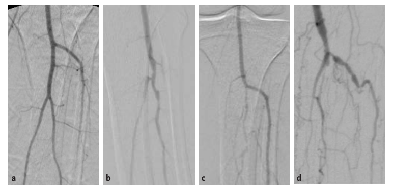
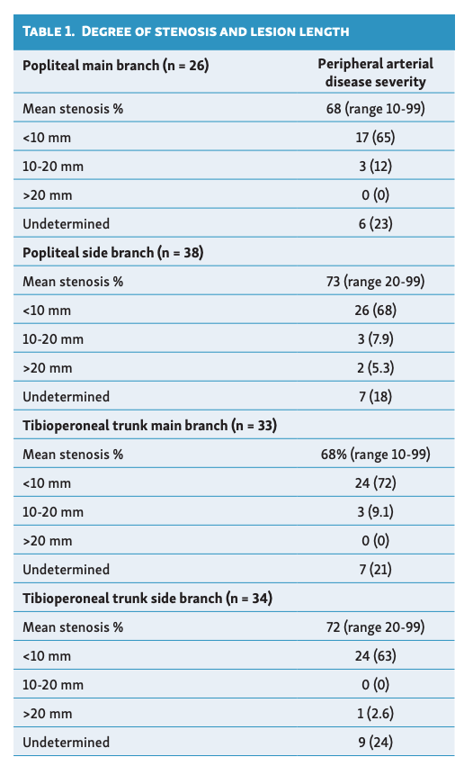
The Medina classification was used to categorize bifurcation lesions according to the presence or absence of significant disease in the proximal main vessel, distal main vessel, and SB.5 A true bifurcation lesion was defined as Medina classification 1,1,1; 1,0,1; or 0,1,1. Representative angiographic images of the tibial bifurcations (Figure 1), operative reports, and demographic data were exported into a relational database program for analysis. The degree of stenosis and lesion length were determined by visual estimation of angiographic images by 2 interventional radiologists, one with over 20 years of experience (Table 1).
For the purposes of this analysis, the main branch was defined as the larger-caliber continuation of the parent vessel distal to the bifurcation, while the SB represented the smaller daughter vessel arising from the bifurcation. In a subset of patients with disease-free tibial vessels, the branching geometry of the popliteal and TPT bifurcations was characterized by calculating the ratio of mother-vessel diameter (DM) to the sum of the 2 daughter-vessel diameters (DD1 and DD2) similar to the method described by Finet et al (see also Figure 2):6

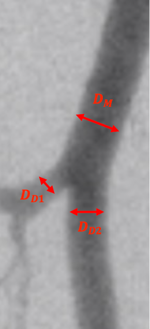
Disease-free bifurcations were selected to characterize native tibial branching geometry independent of plaque-related alteration in vessel caliber. Thirty-two bifurcation sites with angiographic visualization available in at least 2 projections were randomly selected for geometric analysis.
Descriptive statistics were used to summarize demographic, anatomical, and procedural characteristics. Continuous variables are presented as mean ± standard deviation and ranges where applicable. Categorical variables are reported as counts and percentages.
Results
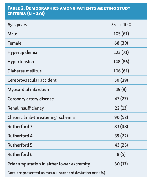
Twenty-one patients were excluded because angiographic assessment of tibial bifurcation anatomy was not feasible or lesion characterization could not be performed reliably due to inadequate angiographic technique, absence of dedicated tibial imaging, limited field of view, variant tibial anatomy, or severe occlusive disease. The prevalence of significant tibial bifurcation disease in the cohort was 83% (144/173), with an average age of 75.1 ± 10.0 years (M:F 1.5). Additional demographic and patient data are provided in Table 2.
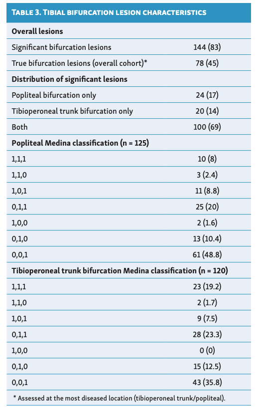
True bifurcation lesions, defined as Medina classifications 1,1,1; 1,0,1; or 0,1,1, were present in 45% (78/173) of the overall cohort and accounted for 54% (78/144) of patients with significant tibial bifurcation disease. The location of significant bifurcation lesions was 14% (20/144) involving the TPT bifurcation only, 17% (24/144) involving the popliteal bifurcation only, and 69% (100/144) involving both bifurcations; 17% (29/173) of patients had disease-free tibial bifurcations. The distribution of Medina classifications for popliteal and TPT bifurcation lesions is shown in Table 3.
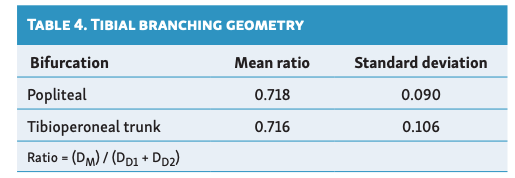
Thirty-two disease-free tibial bifurcation sites from 32 patients were analyzed to characterize native tibial branching geometry. The average ratio of DM to the sum of DD1 and DD2 was 0.718 ± 0.090 for popliteal bifurcations and 0.716 ± 0.106 for TPT bifurcations (Table 4).
Discussion
To our knowledge, this is the first study to quantify the prevalence and characterize the anatomical features of tibial bifurcation disease in a contemporary cohort of patients undergoing angiography for symptomatic PAD and CLTI in the OBL setting. Several important findings emerged.
First, significant tibial bifurcation disease was highly prevalent, affecting 83% of the study population. Second, nearly half of all patients (45%) had true bifurcation lesions, which represented 54% of patients with significant bifurcation disease. Finally, tibial bifurcation geometry appeared remarkably consistent between the popliteal and TPT bifurcations, demonstrating a mother-to-daughter vessel ratio approximating 0.72.
The high prevalence of tibial bifurcation disease observed in this study highlights an important and often underrecognized aspect of infrapopliteal PAD and CLTI. Although endovascular therapies for below-the-knee disease continue to evolve, most available devices and treatment algorithms have been developed for straight vessel segments rather than bifurcation anatomy. The finding that over half of patients with significant tibial bifurcation disease exhibited true bifurcation involvement suggests that compromise of adjacent branch vessels during intervention may be more common than previously appreciated. As a result, bifurcation anatomy warrants specific consideration during procedural planning and device development.
The geometric analysis revealed a mean tibial bifurcation ratio of approximately 0.72, modestly higher than the coronary fractal ratio of 0.678 described by Finet et al.6 While both vascular beds exhibit branching characteristics consistent with fractal principles, differences in anatomy and physiology may account for this observation. Unlike the coronary circulation, which undergoes repeated branching to supply metabolically active myocardium, tibial arteries distribute blood flow across longer vascular segments with relatively few major branch points before reaching the foot. Differences in distal resistance, vessel remodeling, and developmental adaptation may therefore contribute to subtle deviations from coronary bifurcation geometry.
Although the clinical implications of this difference remain uncertain, fractal relationships have informed coronary bifurcation vessel sizing strategies, procedural algorithms, and dedicated device development.7 The identification of a consistent tibial bifurcation ratio of approximately 0.72 may therefore provide an anatomical framework for future studies evaluating tibial-specific sizing approaches, bifurcation treatment techniques, and the design of dedicated tibial bifurcation devices. However, outcomes-based validation of these geometric relationships remains necessary before specific procedural recommendations can be made.
From a clinical perspective, the high prevalence of true bifurcation lesions observed in this study underscores the importance of recognizing bifurcation anatomy when treating patients with advanced limb ischemia. Current treatment strategies are frequently extrapolated from coronary interventions because of similarities in vessel diameter; however, dedicated devices and evidence-based treatment approaches for tibial bifurcation disease remain lacking. Improved understanding of bifurcation prevalence and geometry may ultimately facilitate the development of treatment algorithms and device platforms specifically designed for this vascular territory.
Several limitations warrant consideration. This study was retrospective and included patients treated within a single practice, potentially limiting generalizability. Angiographic interpretation relied upon visual estimation rather than independent core laboratory adjudication. Patients in whom lesion characterization could not be performed reliably due to severe occlusive disease were excluded from analysis; therefore, the prevalence of tibial bifurcation disease reported herein may underestimate the true burden of disease among patients with advanced PAD and CLTI. In addition, geometric analyses were intentionally restricted to disease-free bifurcations to characterize native tibial branching anatomy independent of plaque-related alterations in vessel caliber. Finally, no angiographic or clinical follow-up outcomes were assessed, precluding conclusions regarding the impact of bifurcation anatomy on long-term treatment success.
Conclusion
In conclusion, tibial bifurcation disease is highly prevalent among patients undergoing angiography for symptomatic PAD and CLTI and frequently involves true bifurcation lesions. Tibial branching geometry appears to follow predictable patterns that differ modestly from those reported in the coronary circulation. These findings provide a rationale for greater recognition of tibial bifurcation disease in clinical practice and support future prospective studies, multicenter registries, and the development of dedicated devices and treatment strategies tailored to this challenging vascular territory.
Affiliations and Disclosures
Daniel A. Braga, MD, if from the Mallinckrodt Institute of Radiology, Washington University School of Medicine, St. Louis, Missouri; Anne Beasley, BS, Jennifer Gimeno, MS, and Robert E. Beasley, MD, FSIR, FSCAI, are from Palm Vascular Centers, Miami Beach and Coral Gables, Florida.
The authors report no financial relationships or conflicts of interest regarding the content herein.
Manuscript accepted October 9, 2026.
Address for correspondence: Robert E. Beasley, MD, FSIR, FSCAI, Palm Vascular Centers, 2601 SW 37th Ave #704, Miami, FL 33133. Email: bbeaz@aol.com
References
1. Graziani L, Silvestro A, Bertone V, et al. Vascular involvement in diabetic subjects with ischemic foot ulcer: a new morphologic categorization of disease severity. Eur J Vasc Endovasc Surg. 2007;33(4):453-460. doi:10.1016/j.ejvs.2006.11.022
2. Conte MS, Bradbury AW, Kolh P, et al; GVG Writing Group. Global vascular guidelines on the management of chronic limb-threatening ischemia. J Vasc Surg. 2019;69(6S):3S-125S.e40. doi:10.1016/j.jvs.2019.02.016
3. Zeller T, Baumgartner I, Scheinert D, et al; IN.PACT DEEP Trial Investigators. Drug-eluting balloon versus standard balloon angioplasty for infrapopliteal arterial revascularization in critical limb ischemia: 12-month results from the IN.PACT DEEP randomized trial. J Am Coll Cardiol. 2014;64(15):1568-1576. doi:10.1016/j.jacc.2014.06.1198
4. Ramcharitar S, Onuma Y, Aben J, et al. A novel dedicated quantitative coronary analysis methodology for bifurcation lesions. EuroIntervention. 2008;3(5):553-557. doi:10.4244/eijv3i5a100
5. Louvard Y, Medina A. Definitions and classifications of bifurcation lesions and treatment. EuroIntervention. 2015;11 Suppl V:V23-26. doi:10.4244/EIJV11SVA5
6. Finet G, Gilard M, Perrenot B, et al. Fractal geometry of arterial coronary bifurcations: a quantitative coronary angiography and intravascular ultrasound analysis. EuroIntervention. 2008;3(4):490-498. doi:10.4244/eijv3i4a87
7. Burzotta F, Lassen JF, Lefèvre T, et al. Percutaneous coronary intervention for bifurcation coronary lesions: the 15th consensus document from the European Bifurcation Club. EuroIntervention. 2021;16(16):1307-1317. doi:10.4244/EIJ-D-20-00169