


Chronic Tibialis Anterior Tendon Ruptures: Considering A Key Surgical Option
Tibialis anterior tendon ruptures are quite rare. Anzel reported approximately one percent of 1014 tendon injuries of the foot and ankle involved ruptures of tibialis anterior tendon.1 Although a clinical rarity, rupture of this tendon is a debilitating injury, and if left untreated may result in significant disability. This particular injury may result from traumatic or atraumatic mechanisms. Traumatic ruptures typically involve direct injury (acute laceration), an eccentric load against a plantarflexed ankle2 or a forceful contraction (concentric load) producing an overload beyond the tensile strength of the tendon. Atraumatic, spontaneous, low-energy ruptures may occur in the presence of underlying factors that predispose the tendon to rupture. Systemic factors (inflammatory conditions, autoimmune disorders, diabetes mellitus, exposure to fluoroquinolones and systemic steroids) and local factors (local steroid injections, collagen abnormalities and repetitive microtauma) are examples of such predisposing factors in spontaneous rupture.3
Atraumatic ruptures may go unnoticed and there is often a delay in diagnosis. Patients may not seek immediate care thinking they suffered “just a sprain.” They’ll often exhibit an abnormal gait but may have intact dorsiflexion due to secondary function of the extensor hallucis longus (EHL) and extensor digitorum longus muscles (EDL).2 It may be weeks or months before the patient receives a referral or decides to seek treatment. Loss of function over time is a typical motivating factor that causes the patient to present for treatment.4
Acute tendon ruptures that are a result of blunt trauma or laceration are typically painful injuries. Patients may recall hearing or feeling a “pop” at the time of injury. They will present with pain and swelling around the ankle region. In contrast, with spontaneous atraumatic injuries, patients may present with vague pain and swelling and if there is a delay in treatment, as often is the case, patients may actually be pain-free at the time of evaluation.
On initial clinical presentation of both acute and chronic injuries, patients have alterations in gait and may complain of weakness compared to the contralateral limb. Patients report difficulty clearing the affected foot while walking. A gait exam will often reveal a steppage gait pattern with foot slap or foot drop, and increased hip flexion (to prevent the toes from hitting the ground in swing phase).2
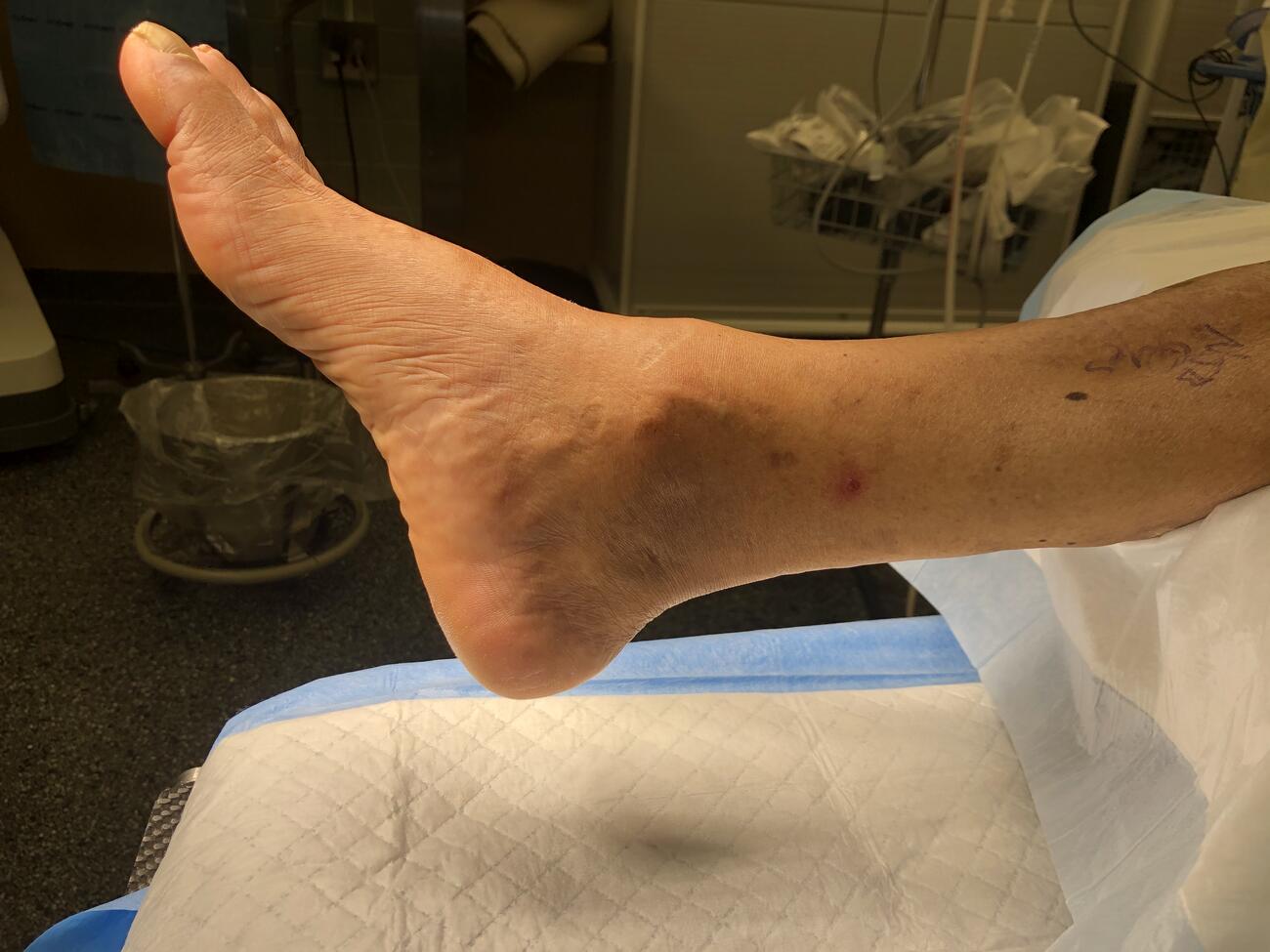
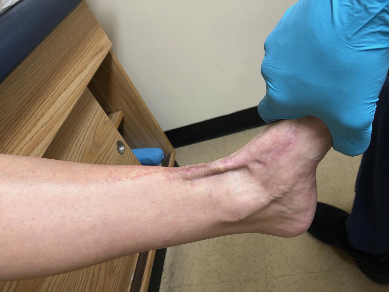
On clinical examination, one will note the loss of contour of the tibialis anterior tendon over the ankle and the tendon is not visible or palpable during dorsiflexion resistance. Patients may present with a painless mass, or pseudotumor, along the anterior medial aspect of the ankle joint (see first photo above). This mass represents the distal aspect of the tendon, retracted proximally. There is weakened dorsiflexory strength as compared to the contralateral limb. One may see the extensor substitution (EDL and EHL contracting) for active dorsiflexion of the foot.2
Physical exam findings are fairly consistent, and in my experience often all one needs to diagnose acute, traumatic ruptures. However in cases of a chronic, spontaneous rupture, magnetic resonance imaging (MRI) is helpful in confirming the diagnosis and determining the extent of tendon injury and size of the defect. MRI provides valuable information for surgical planning and allows for a more detailed evaluation of the tendinous structures.
After establishing a diagnosis, it is prudent to educate patients on available treatment options. Conservative treatment should only be considered in the elderly, inactive, or patients who are poor surgical candidates. Surgery should be considered for active individuals, with the goal to re-establish the function of the tibialis anterior tendon.
Acute ruptures are treated with a direct repair if amendable, with end-to-end repair of the ruptured tendon ends. If the tendon avulsed off the bone, or if the distal fibers are nonviable and require debridement, the surgeon can directly attach the tendon to the medial cuneiform and fixate using an interference screw or suture anchor.2
Chronic or neglected injuries require a more complex reconstruction. A rupture is considered chronic when the delay in treatment is longer than four weeks. One may find tendon retraction and poor-quality tissue with chronic ruptures, resulting in large defects that make end-to-end repair impossible.5
There are numerous described techniques to overcome the deficit caused by tendon retraction and aid in overall strength of the tendon, including slide lengthening, tendon turndown, interposition or tendon grafting.2
This article will present a surgical technique for chronic ruptures with large segmental defects using a tendon allograft, transferring the tendon into the medial cuneiform and fixating with a cortical button and interference screw. This technique restores dorsiflexory strength and improves patients’ functional status without the donor site morbidity associated with autograft harvest or tendon transfer.
Understanding A Surgical Option For Chronic Anterior Tibial Tendon Ruptures
For this procedure, patient placement is supine and under general anesthesia with an option for a regional nerve block to assist in peri-operative pain control. A well-padded thigh tourniquet assists in hemostasis and prepping and draping takes place in the usual fashion.
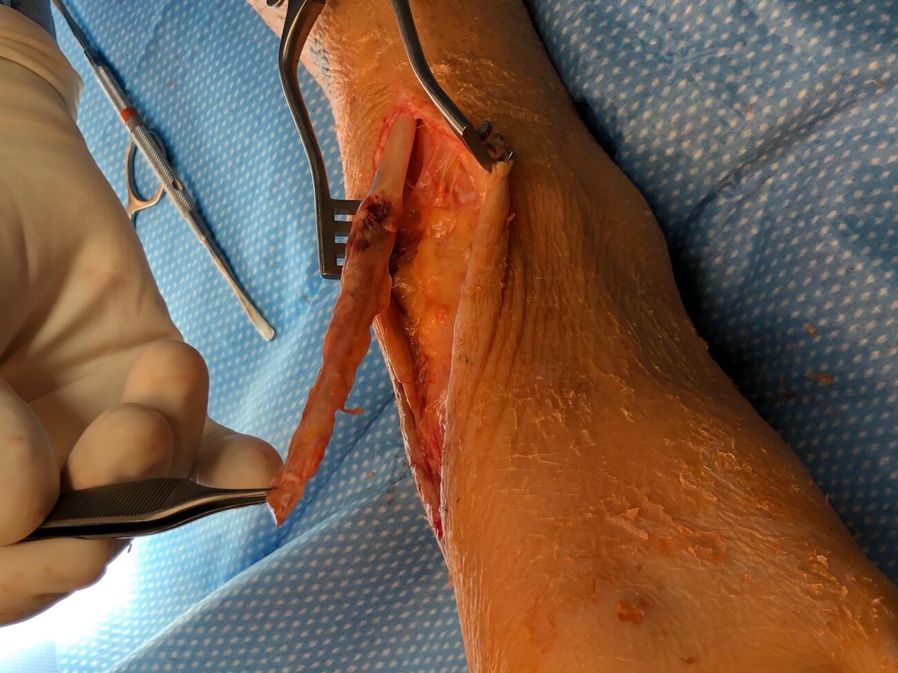
The surgeon makes the initial incision over the anterior medial aspect of the ankle at the level of the proximal tendon stump and extends distally over the course of the tibialis anterior tendon to the medial cuneiform. Careful dissection down through the subcutaneous tissues is next, being careful to isolate and protect neurovascular structures. One then identifies and proximally frees the tibialis anterior tendon stump (see second photo above). The distal tendon segment is then identified and is often poor quality, which warrants debridement as well. One should note that the debridement will increase the size of the defect, and when applying tension to the proximal segment it becomes clear that direct attachment is not possible.
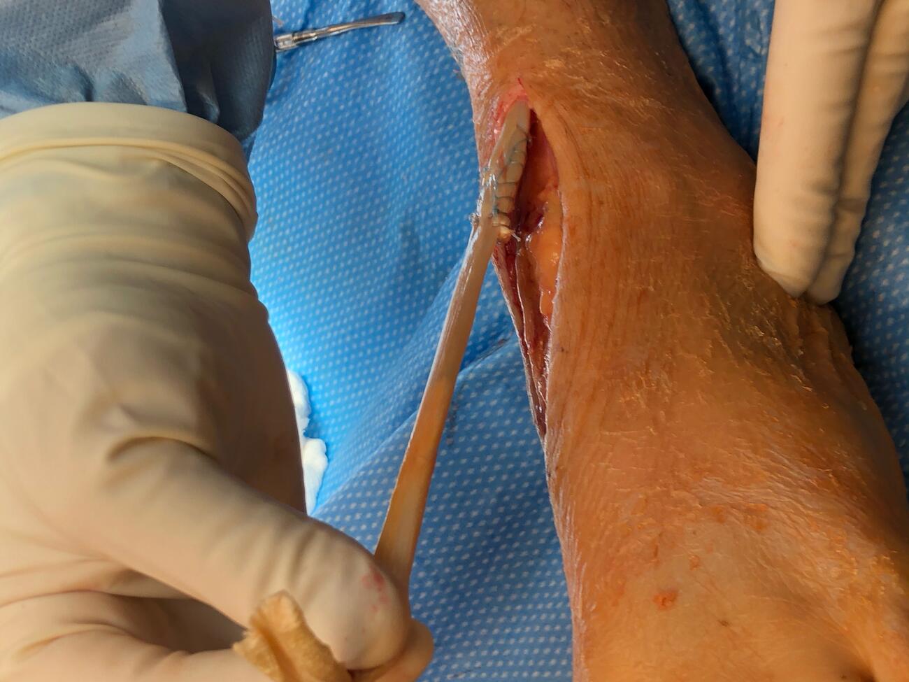
Accordingly, one prepares an appropriately sized tendon allograft (donor) and attaches it to the proximal tendon segment (recipient) using one’s preferred attachment technique (see third photo above). Placement of a traction stitch takes place on the distal end of the tendon and a tendon sizer measures the diameter to determine the appropriate size implant and drill.
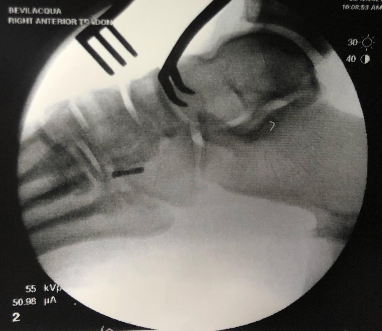
Next, the surgeon identifies the dorsal aspect of the medial cuneiform and centralizes a guide wire on the bone using intraoperative fluoroscopy. The guide wire placement is perpendicular to the dorsal cortex and advanced bicortically. One directly visualizes and confirms via fluoroscopy the position and orientation of the guide pin, exercising caution to prevent penetrating the proximal or distal joints.
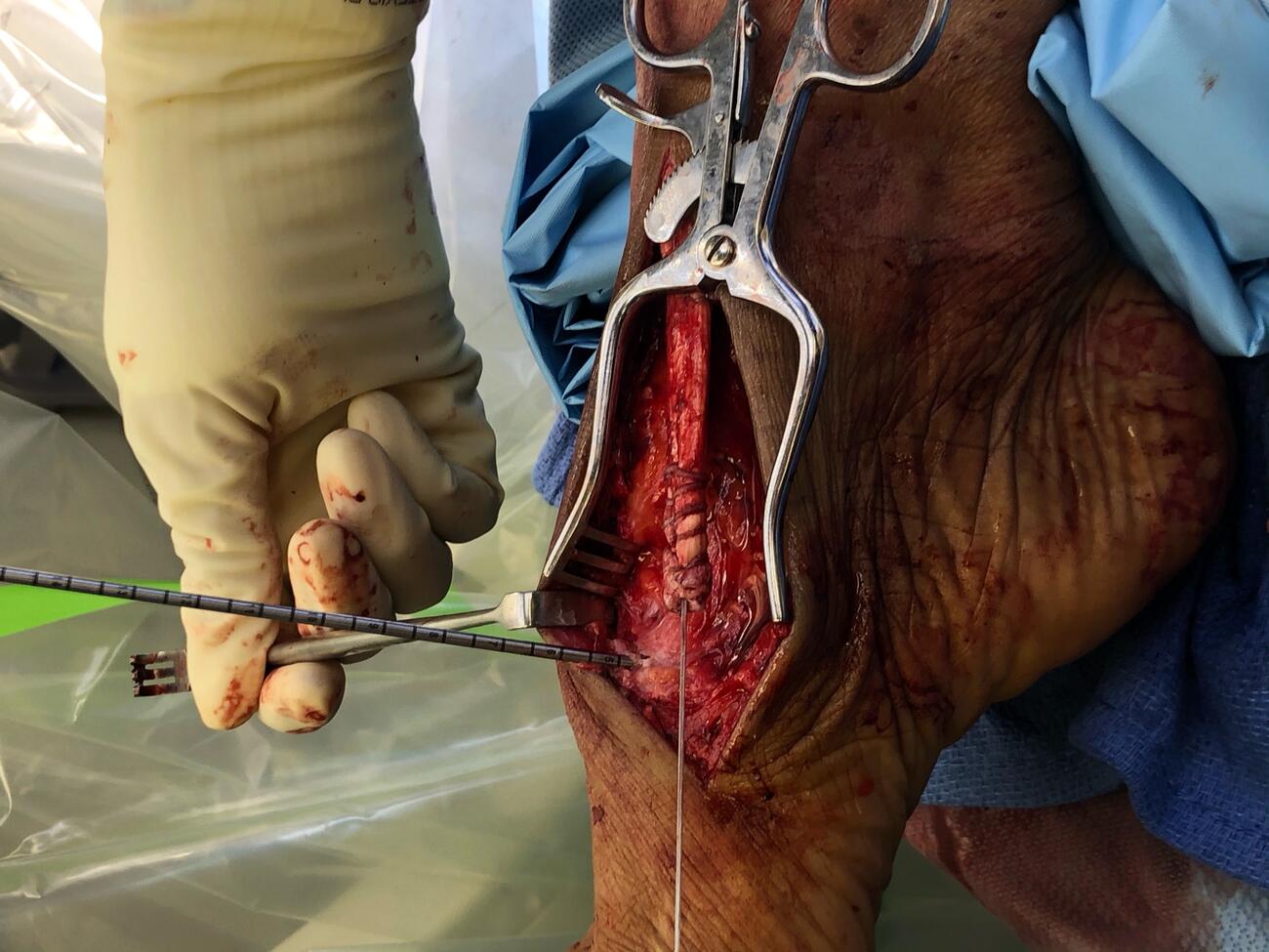
Next the surgeon places the reconstructed tendon under physiologic tension, bringing it to the medial cuneiform and guide pin interface and cutting to the appropriate length. The desired length of tendon should reach the plantar aspect of the medial cuneiform with the foot in dorsiflexion and inversion. Resection of excess tendon helps avoid redundancy that would compromise tensioning and future function.6 One then places a whip stitch on the distal end of the tendon (see fourth photo above).
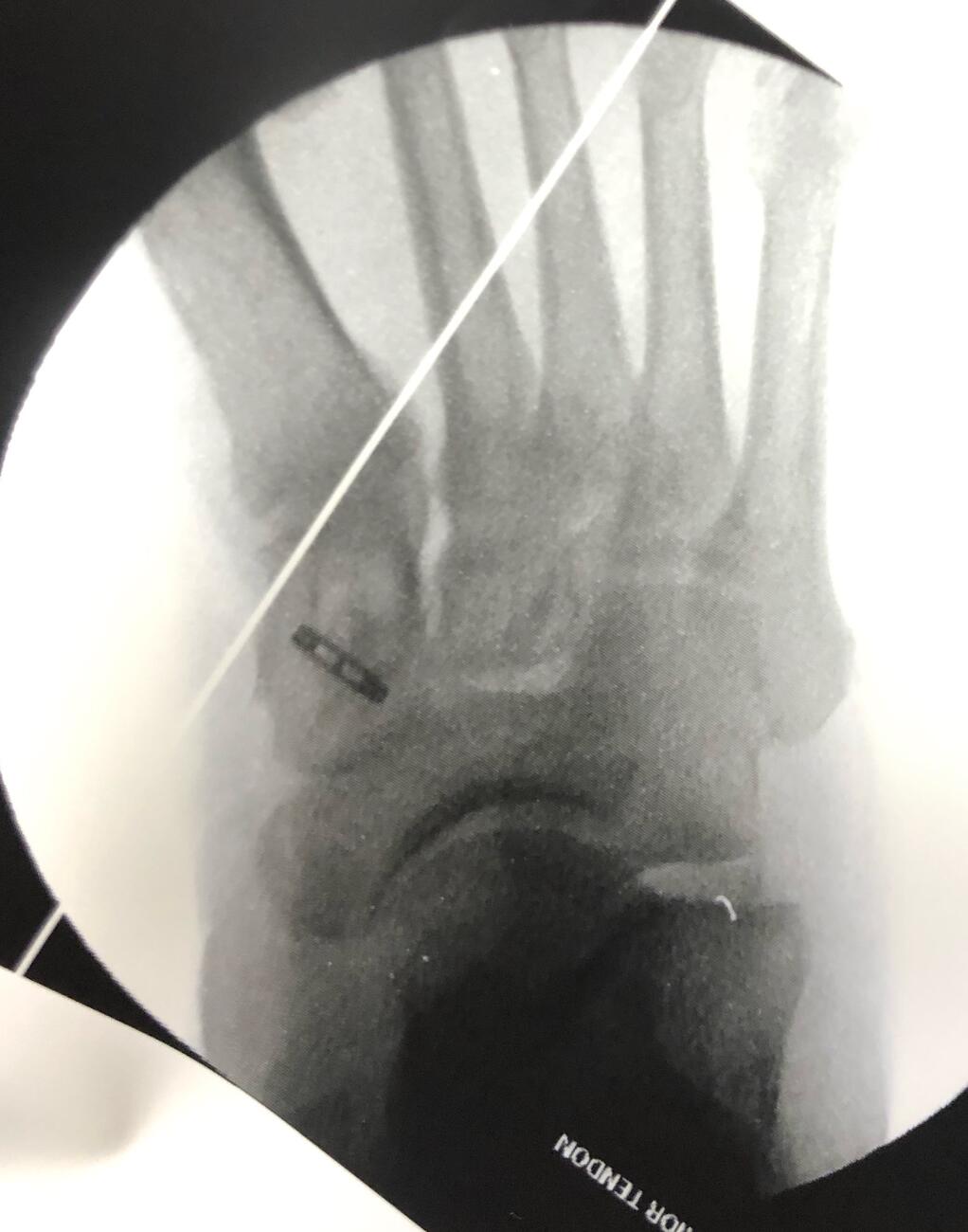
Drilling of an appropriately sized bone tunnel then takes place over the guide wire in the medial cuneiform from dorsal to plantar, being careful not to penetrate the plantar cortex. One then threads the suture limbs (from the previously placed whip stitch) through the cortical button, and the button inserter advances the button through the medial cuneiform from dorsal to plantar. Fluoroscopy confirms the button has flipped and is in proper position, sitting flush against the plantar cortex (see fifth and sixth photos above). The tendon is then carefully brought into the tunnel and advanced by pulling the sutures with controlled tension. Once the tendon is in satisfactory position, with the appropriate tension, insertion of a tenodesis screw further secures the tendon in place.
The procedure concludes with careful layered wound closure and application of a well-padded posterior splint in dorsiflexion, which helps keep tendon stretch to a minimum during the healing process.6 Patients are kept non-weightbearing for six weeks and then transitioned to weight bearing as tolerated (initially using a CAM walking boot). After wound healing, active dorsiflexion and plantarflexion can begin as permitted. Physical therapy focuses on active and passive range of motion, strength, endurance and proprioception exercises and patients gradually wean out of the boot and return to normal activity at eight weeks. If there is concern about fixation at any time during the post-operative period, radiographs of the foot can confirm the suture button position.
What Impact Might This Surgical Option Have On Patient Care?
Tibialis anterior tendon ruptures are a rare clinical entity and low energy spontaneous injuries can often result in a delay in diagnosis due to the inconspicuous nature of the injury. Continued functional impairment and alterations in gait cause patients to eventually seek treatment. Detailed history and physical exam are key factors in establishing a diagnosis and initiating a treatment plan. Historically, patients with a chronic tibialis anterior tendon rupture received conservative treatment. Conservative treatment options include ankle-foot orthoses, braces and modification of activity, which one may consider in the elderly or inactive patient.3 Conservative management results in the loss of function of tibialis anterior tendon and the resultant muscle imbalance will ultimately lead to long-term biomechanical problems.
In a letter to the editor published in the Journal of the American Podiatric Medical Association (JAPMA) in 1999, Cohen and Gordon detailed the long-term effects of an untreated tibialis anterior tendon rupture.7 The authors noted that the EDL and EHL tendons compensate for the lost dorsiflexion strength and over time, become stronger and adapt to better perform this task. However, as these muscles develop, there is a disruption in the delicate balance between the muscles, resulting in foot deformities. The authors’ observations and treatment of these late complications suggest that early intervention and repair of the tendon may result in a more satisfactory outcome.7
More recent data shows that good outcomes are possible with delayed surgical intervention.2 This paper presents a surgical technique for the successful management of chronic tibialis anterior tendon ruptures with large segmental defects. Tendon allograft reconstructs the tendon and bridges the defect without the donor site morbidity associated with autograft harvest or tendon transfer. The combination of cortical button fixation and tenodesis screw allows for a precise and strong anatomic fixation construct to permit early postoperative activation and mobilization of the reconstructed tendon for improved outcomes (see seventh photo above).
Dr. Bevilacqua is a fellowship-trained foot and ankle surgeon with North Jersey Orthopaedic Specialists in Teaneck and Englewood, NJ. He is board-certified by the American Board of Foot and Ankle Surgeons, and is a Fellow of the American College of Foot and Ankle Surgeons.
References
1. Anzel JR. Tendon injuries about the ankle. Orthop Clin North Am. 1980;11:801–811.
2. Patel R, Fallat L. Surgical techniques for repair of atraumatic tibialis anterior tendon ruptures: a report of two cases. J Foot Ankle Surg. 2017;56(6):1343-1349.
3. DiDomenico LA, Williams K. Petrolla AF. Spontaneous rupture of the anterior tibial tendon in a diabetic patient: results of operative treatment. J Foot Ankle Surg. 2008;47(5):463-467.
4. Burton A, Aydogan U. Repair of chronic tibialis anterior tendon rupture with a major defect using gracilis allograft. Foot Ankle Spec. 2016;9(4):345-350.
5. Huh J, Boyette DM, Parekh SG, Nunley JA 2nd. Allograft reconstruction of chronic tibialis anterior tendon ruptures. Foot Ankle Int. 2015;36(10):1180-1189
6. Rhee C, Burgesson B, Orlik B, Logan K. Suture button technique for tibialis anterior tendon transfer for the treatment of residual clubfoot. Foot Ankle Orthop. 2020;5(2). Available at: https://journals.sagepub.com/doi/full/10.1177/2473011420923591 . Published May 14, 2020. Accessed July 20, 2021.
7. Cohen DA, Gordon DH. The long-term effects of an untreated tibialis anterior tendon rupture. J Am Podiatr Med Assoc. 1999;89(3):149-152.

