What Caused This Hyperpigmentation?
Case Report
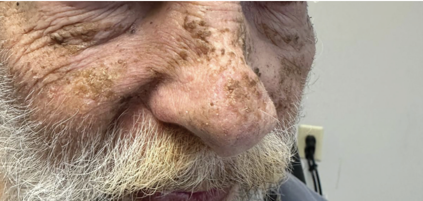
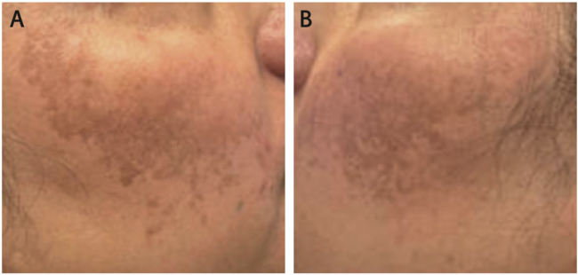
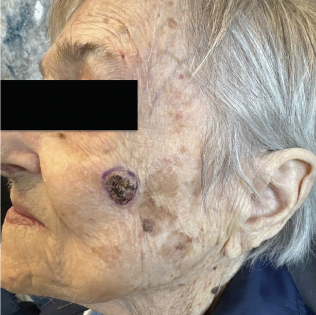
A 75-year-old male veteran, living in a trailer, sought dermatologic evaluation for new skin lesions appearing on his face and neck(Figure). The patient had a prior history of advanced basal cell carcinoma on the right ear but denied any current systemic symptoms.On examination, several brownish, hyperpigmented patches with a rough and wrinkled texture were observed, predominantly on the face and extending down the neck.
What’s Your Diagnosis?
Scroll below to find out!
Diagnosis: Terra Firma-Forme Dermatosis
Terra firma-forme dermatosis (TFFD), also referred to as Duncan’s dirty dermatosis, is a benign skin condition characterized by the presence of hyperpigmented, rough patches that resemble dried, cracked earth.1,2 The term terra firma, derived from the Latin meaning dry land, refers to the characteristic “dirt-like” hyperpigmented patches or plaques seen in this condition, whichwas first described and named by Duncan in 1987.1,3,4
TFFD is considered underreported and is more prevalent in children and adolescents. Most case series and retrospective reviews indicate a predominance in pediatric populations, with mean ages reported around 10.4 years and a female predominance in some cohorts.1,5 The most commonly affected sites are the face, neck, trunk, ankles, and navel, with symmetrical distribution in many cases.5-9
Interestingly, TFFD and dermatosis neglecta (DN) were once widely considered to be the same clinical entity.4 However, some authors have since proposed a distinction based on clinical presentations and age groups. While TFFD is predominantly seen in older children and adolescents with characteristic hygiene habits, DN can affect patients of all ages, especially those with inadequate hygiene in specific areas.4,10 This differentiation is supported by studies suggesting that TFFD occurs more commonly in pediatric populations, whereas DN is not limited by age.4,11
Clinical Presentation
TFFD is characterized by brown to gray-black patches that resemble dirt.1,9 These lesions often have a surface that appears verrucous or papillomatous, with a reticulated pattern and islands of normal skin interspersed throughout.1 The condition typically presents as asymptomatic, with patients reporting no discomfort.1 Patients also typically report that routine washing with soap and water does not remove the discoloration.9,10 Awareness of this characteristic presentation is crucial to prevent unnecessary investigations and treatments.
Pathophysiology and Etiology
The underlying pathophysiologic mechanism of TFFD is thought to be abnormal retention of keratinized corneocytes and melanin within the stratum corneum, leading to the formation of hyperpigmented, dirt-like plaques that are resistant to routine washing but removable with 70% isopropyl alcohol.1,7,8,10,12 The etiology remains unknown, but contributing factors may include less rigorous hygiene, inadequate pressure during cleansing, individual variations in skin barrier function, an atopic background, or possibly local factors such as occlusion or friction.1,7,12 Although inadequate cleansing may contribute, TFFD can also occur in individuals with normal or even vigorous hygiene, suggesting that factors beyond hygiene, such as local friction or barrier variability, play a role.9,10
Histopathologic features of TFFD demonstrate prominent lamellar hyperkeratosis with focal whorled orthokeratosis and keratin globules within the stratum corneum.13-15 Mild acanthosis and papillomatosis may be present, accompanied by focal perivascular lymphocytic infiltration in the papillary dermis.13,14 Additionally, there is often increased melanin pigment within retained keratinocytes and occasionally in the basal layer of the epidermis, with minimal overall dermal inflammation.16,17
Dermoscopy
Dermoscopy has been reported as a useful noninvasive tool in the diagnosis of TFFD. Lesions of TFFD characteristically show large polygonal, plate-like brown scales arranged in a mosaic pattern under polarized light examination.18 These findings reflect the underlying histopathologic features of compact orthokeratosis and papillomatosis, providing a prompt and discreet means of supporting the clinical diagnosis.18
Differential Diagnosis
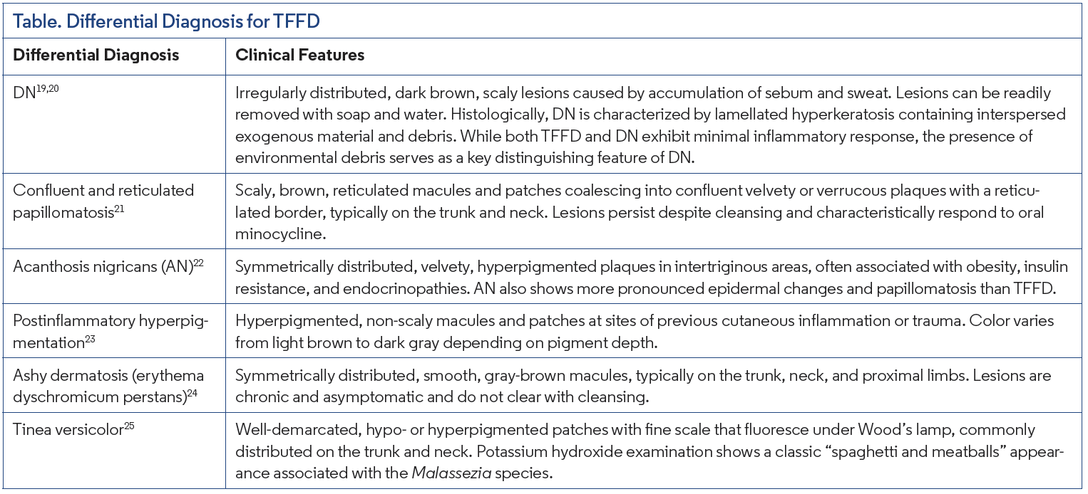
Due to its appearance as hyperpigmented, dirt-like patches, TFFD can resemble a variety of pigmentary and keratinization disorders. The distinguishing characteristics of these conditions are summarized in the Table.
Management
The primary treatment for TFFD involves gently swabbing the affected areas with 70% isopropyl alcohol, a method that both confirms the diagnosis and effectively clears the lesions.1,5,9,10,16 Routine washing with soap and water does not address the discoloration, and unnecessary treatments, such as topical corticosteroids or urea, should be avoided once the diagnosis is established through alcohol swabbing.9,16 In rare instances where alcohol use is contraindicated or poorly tolerated, an alternative option is the application of 5% salicylic acid in petrolatum ointment.3 Educating patients on proper cleansing methods and reassuring them about the benign nature of the condition are essential steps to prevent recurrence and alleviate any anxiety or embarrassment.7,12
Our Patient
After gently rubbing the lesions with 70% isopropyl alcohol, the brownish discoloration cleared completely, revealing normal underlying skin. The patient expressed surprise and relief at the immediate improvement. He was counseled on the benign nature of the condition and advised on gentle but thorough skin cleansing methods.
Conclusion
This case highlights a patient who presents with classic findings of TFFD, offering insight into the unique features and diagnostic approach for this often-overlooked dermatologic condition.
TFFD is distinctive but can easily be mistaken for other dermatologic disorders. Its diagnosis relies on the unique response to isopropyl alcohol, which differentiates it from similar conditions. Recognizing TFFD early can help prevent unnecessary investigations and reduce patient anxiety.
Reference
1. Sechi A, Patrizi A, Savoia F, Leuzzi M, Guglielmo A, Neri I. Terra firma-forme dermatosis: a systematic review. Int J Dermatol. 2021;60(8):933-943. doi:10.1111/ijd.15301
2. Campos MS, Fukuma LO, Delgado JC, Criado PR, Prigenzi KCK, e Dinato SLM. Exuberant terra firma-forme dermatosis in an elderly patient. An Bras Dermatol. 2025;100(4):501131. doi:10.1016/j.abd.2025.501131
3. Vakirlis E, Theodosiou G, Lallas A, Apalla Z, Sotiriou E. Terra firma-forme dermatosis: differential diagnosis and response to salicylic acid therapy. Pediatr Dermatol. 2019;36(4):501-504. doi:10.1111/pde.13807
4. Martín-Gorgojo A, Alonso-Usero V, Gavrilova M, Jordá-Cuevas E. Dermatosis neglecta or terra firma-forme dermatosis. Actas Dermo-Sifiliográficas.2012;103(10):932-934. doi:10.1016/j.adengl.2011.12.006
5. Aslan NÇ, Güler Ş, Demirci K, Isiyel E. Features of terra firma-forme dermatosis. Ann Fam Med. 2018;16(1):52-54. doi:10.1370/afm.2175
6. Browning J, Rosen T. Terra firma-forme dermatosis revisited. Dermatol Online J. 2005;11(2):15.
7. Ashique KT, Kaliyadan F, Goyal T. Terra firma-forme dermatosis: report of a series of 11 cases and a brief review of the literature. Int J Dermatol. 2016;55(7):769-774. doi:10.1111/ijd.12974
8. Guarneri C, Guarneri F, Cannavò SP. Terra firma-forme dermatosis. Int J Dermatol. 2008;47(5):482-484. doi:10.1111/j.1365-4632.2008.03516.x
9. Berk DR. Terra firma-forme dermatosis: a retrospective review of 31 patients. Pediatr Dermatol. 2012;29(3):297-300. doi:10.1111/j.1525-1470.2011.01422.x
10. Berk DR, Bruckner AL. Terra firma-forme dermatosis in a 4-month-old girl. Pediatr Dermatol. 2011;28(1):79-81. doi:10.1111/j.1525-1470.2010.01220.x
11. Lucas JL, Brodell RT, Feldman SR. Dermatosis neglecta: a series of case reports and review of other dirty-appearing dermatoses. Dermatol Online J. 2006;12(7):5.
12. Sechi A, Patrizi A, Savoia F, Neri I. Terra firma-forme dermatosis. Clin Dermatol. 2021;39(2):202-205. doi:10.1016/j.clindermatol.2020.10.019
13. Erkek E, Sahin S, Çetin ED, Sezer E. Terra firma-forme dermatosis. Indian J Dermatol Venereol Leprol. 2012;78(3):358-360. doi:10.4103/0378-6323.95455
14. Das A, Bhattacharya S, Kumar P, et al. Unilateral nevoid acanthosis nigricans: uncommon variant of a common disease. Indian Dermatol Online J. 2014;5(Suppl 1):S40-S43. doi:10.4103/2229-5178.144529
15. Berk DR, Mutizwa MM. Comment regarding the histopathology of terra firmaforme dermatosis. J Cutan Pathol. 2012;39(2):300-301; author reply 302-303. doi:10.1111/j.1600-0560.2011.01818.x
16. Demirci GT, Mansur AT, Demiralay E. Terra firma-forme dermatosis misdiagnosed as nevoid acanthosis nigricans. Am J Dermatopathol. 2017;39(10):782-784. doi:10.1097/DAD.0000000000000885
17. Zhou Y, Zhang J, Zheng L, Yang F. Penile terra firma-forme dermatosis in children. Pediatr Dermatol. 2023;40(5):894-896. doi:10.1111/pde.15277
18. Errichetti E, Stinco G. Dermoscopy in terra firma-forme dermatosis and dermatosis neglecta. Int J Dermatol. 2017;56(12):1481-1483. doi:10.1111/ijd.13686
19. Greywal T, Cohen PR. Terra firma-forme dermatosis: a report of ten individuals with Duncan’s dirty dermatosis and literature review. Dermatol Pract Concept. 2015;5(3):29-33. doi:10.5826/dpc.0503a08
20. Palaniappan V, Sadhasivamohan A, Karthikeyan K. Dermatosis neglecta. Clin Exp Dermatol. 2022;47(7):1265-1274. doi:10.1111/ced.15184
21. Davis MDP, Weenig RH, Camilleri MJ. Confluent and reticulate papillomatosis (Gougerot-Carteaud syndrome): a minocycline-responsive dermatosis without evidence for yeast in pathogenesis. A study of 39 patients and a proposal of diagnostic criteria. Br J Dermatol. 2006;154(2):287-293. doi:10.1111/j.1365- 2133.2005.06955.x