PATIENT PRESENTATION
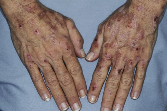
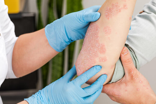
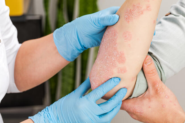
A 63-year-old man presented for evaluation of newly appearing, diffusely distributed, pruritic skin lesions. The patient’s medical history was significant for essential thrombocytosis initially diagnosed in 2007 that was unresponsive to several treatments, including hydroxyurea and anagrelide. He was admitted to the hospital, where he was seen in consultation for evaluation of recently developed anemia and thrombocytopenia; a bone marrow biopsy also showed 80% blasts. Induction chemotherapy with bendamustine per study protocol was initiated for newly diagnosed acute myelogenous leukemia. Physical examination revealed erythematous papules, nodules and plaques on the scalp, face, chest, back and upper extremities (Figures 1 and 2, above, left to right). Examination of the oral cavity demonstrated a 1-cm ulcer on the buccal mucosa and a small stellate fissure on the distal tip of the tongue. Punch biopsies of representative skin lesions on the right chest and left cheek were obtained.
WHAT IS YOUR DIAGNOSIS?
DIAGNOSIS:LEUKEMIA CUTIS
Skin biopsies revealed extensive perivascular dermal infiltrates of medium-sized mononuclear cells with irregularly shaped nuclei and an open chromatin pattern. Immunohistochemical staining of the neoplastic cells demonstrated positivity for CD4, CD33, CD43 and CD68. The histology was consistent with acute myelogenous leukemia involving the skin (leukemia cutis).
Leukemia cutis is a term used to describe the cutaneous manifestations of leukemia, characterized by the infiltration of malignant neoplastic leukocytes or their precursors into the epidermis, dermis or subcutis.1-5 Leukemia cutis is more commonly observed in association with myeloid leukemias6 and occurs most frequently in patients with acute myelogenous leukemia, with an estimated prevalence of 10% to 15%.1
The frequency of leukemia cutis is higher in children than in adults.1 For example, among children with congenital acute myelogenous leukemia in which the diagnosis is established at birth or in the first month of life, leukemia cutis is present in about 25% to 30% of patients.7
The symptoms of leukemia cutis are variable; hence, it is a challenge to diagnose this condition. However, since its presence tends to herald a poor prognosis, making an early diagnosis is vital to successful treatment.1,3,5
Clinical Presentation
Leukemia cutis can present as various lesion morphologies, such as erythematous or purpuric papules, nodules, plaques and hemorrhagic ulcers.2,6 However, erythematous papules and nodules are the most common clinical presentation.1 Skin lesions usually involve the legs, head, neck and trunk.1,2,6 Different leukemia subtypes often produce cutaneous lesions of remarkable uniformity, yet variation of the lesion morphology over time is possible even within the same patient.1,5 Approximately 90% of patients with leukemia cutis have involvement of additional extra-medullary sites, particularly the meninges.1,5,8
Atypical presentations of leukemia cutis include marked thickening of the gums, oral petechiae, leonine faces, eczematous lesions, penile and scrotal ulcers and panniculitis resembling erythema nodosum.1 Our patient exhibited multiple maculopapular hemorrhagic skin lesions on the face, anterior neck and chest, but no lesions on his lower abdomen or legs.
Leukemia cutis may also localize preferentially to sites of recent trauma.1 One patient, a 62-year-old man with plasma cell leukemia, presented with skin lesions limited to recent sites of needle and catheter insertion.9 Another patient, a 65-year-old woman diagnosed with acute myelomonocytic leukemia, also noted new infiltrated nodules consistent with leukemia cutis at the site of a recent Hickman catheter placement.10 In general, recent surgeries, trauma, burns, herpes zoster and intramuscular injections are highly likely to be sites for cutaneous leukemic infiltration.1,3,5,10
Pathology
The diagnosis of leukemia cutis depends on recognizing the morphologic pattern of skin infiltration, the cytologic features and the immunophenotype of the tumor cells.1 In most types of leukemia cutis, involvement shows either a perivascular and/or periadnexal pattern or a dense diffuse or nodular infiltrate involving the dermis and subcutis, but with a Grenz zone that spares the epidermis.1,3,5 Stromal fibrosis is also commonly seen.1
The diagnosis of a specific type of leukemia cutis, based only on the findings using hematoxylin and eosin stains, is often insufficient.6,11 Therefore, the use of immunohistochemical stains and immunophenotyping are recommended. Depending on the positive reactions, the correct subtype of leukemia may be identified.1,5,6,11
In Acute Myeloid (myelogenous, granulocytic) Leukemia, leukemia cutis is characterized histologically by the general infiltration of myeloid cells into the dermis and subcutis, as well as granulocytic maturation in the tumors and mitotic figures.3,5,6 Lysozyme and CD68 are the most sensitive immunohistochemical stains for all subtypes of myeloid leukemia cutis.3,5,6
In Acute Monocytic Leukemia, mucocutaneous leukemia cutis is present in 10% to 33% of patients. The histological features include leukemic infiltration of the entire dermis and superficial panniculus.3,5 Tumor cells are monomorphous and have large, kidney-shaped nuclei.3,5 Once again, the CD68 and lysozyme stain positively, although CD15, CD43 and CD45 also stain immunohistochemically.3,5
Acute Myelomonocytic Leukemia patients have been reported to develop leukemia cutis in 13% to 27% of cases. Acute myelomonocytic leukemia is very similar to acute monocytic leukemia in terms of histology and immunohistochemistry due to their common monocytic lineage. However, acute myelomonocytic leukemia also displays chloracetate esterase in some leukemia cells because of the myeloid component3 and demonstrates a distinct grenz zone.5
Leukemia cutis is more common in Chronic Lymphocytic Leukemia than its acute version, with an occurrence rate of about 8%. Histology reveals lymphocytes with dense nuclear chromatin in the dermis and subcutaneous tissue.3,5 Immunohistochemically, chronic lymphocytic leukemia originates almost exclusively from B-cells, staining negatively for CD3 and CD45 RO.3,5 Monoclonality of immunoglobulin light chain restriction is pathognomonic for B-cell malignancy and the predominant subtypes seen in chronic lymphocytic leukemia are IgM or IgM and IgD.3
Leukemia cutis has been reported in 40% to 70% of cases of Acute T-cell Leukemia. Histologically, the dermis and the area around blood vessels show a dense infiltrate of lymphoid cells with convoluted nuclei and mitotic figures.3 In addition, the presence of clonal T-cell receptor β gene rearrangement and integration of HTLV-1 confirms the diagnosis of acute T-cell leukemia.5 Immunohistochemically, CD3, CD4, CD5 and CD25 stain strongly positive.3,5
Leukemia cutis only occurs rarely in patients with Hairy Cell Leukemia. Histologically, there is a dense, uniform, malignant mononuclear infiltration in the dermis and subcutis that spares the papillary dermis. Immunohistochemically, tartrate-resistant acid phosphatase stains positively.3,5
Differential Diagnosis
Leukemia cutis is notoriously difficult to diagnose due to its varied and nonspecific clinical morphology.6,11,12 Many conditions present with lesions similar or identical to those of leukemia cutis, including benign lymphocytoma cutis, cutaneous metastatic eccrine carcinoma, drug eruptions, drug-induced gingival hyperplasia, erythema nodosum, hypereosinophilic syndrome, neutrophilic eccrine hidradenitis, pyoderma gangrenosum, sarcoidosis and Sweet’s syndrome.2
An accurate patient medical history is essential for diagnosis, as leukemia cutis usually presents in association with a systemic leukemia.1,2 Very rarely, aleukemic leukemia cutis arises, characterized by the invasion of the skin by leukemic cells before their appearance in the peripheral blood.5,8,12-14
The lesions of Sweet’s syndrome may resemble those of leukemia cutis, and Sweet’s syndrome may be seen in the context of an underlying hematologic malignancy, making differentiation between diseases difficult.15,16 However, it is important to remember that the infiltrate of Sweet’s syndrome consists of mature neutrophils without vasculitis. In contrast, biopsy specimens of leukemia cutis exhibit malignant, immature myeloblasts.10,12
Pathogenesis
The mechanism for migration of the leukemic cells to the skin is not known. Some authors suggest that the etiology for the migration of neutrophils to the skin in leukemia cutis may be similar to that observed in Sweet’s Syndrome.16 In the latter condition, skin-homing mechanisms have been postulated to be controlled by a combination of chemokines and adhesion molecules.15,16
In patients with T-cell dyscrasias, the skin-homing mechanism of T cells is thought to be regulated by co-expression of cutaneous lymphocyte antigen (CLA) with chemokine receptors. The CLA interacts with E-selectin, thus initiating rolling and tethering of T cells on the endothelial cells. Chemokine binds to T cell receptors, activating lymphocyte function-associated antigen-1. This, in turn, interacts with the intercellular adhesion molecule, which may cause the transmigration of T cells into the dermis.1
Treatment/Prognosis
The treatment of leukemia cutis is targeted to the management of the underlying leukemia.1,2,5 Long-term success is best achieved by combination chemotherapy and targeted local therapy of bone marrow, skin and other sites of extramedullary infiltration.1,5 In some individuals, total skin electron irradiation has not only been found to be effective in managing leukemia cutis (including some of the patients who have aleukemic leukemia cutis),2,5,13 but has also been shown to be effective in preventing skin relapse.8 It has been suggested that aggressive chemotherapy should continue even after remission to prevent further relapse.2
The prognosis of leukemia cutis tends to be grave, with 88% of patients dying within one year of diagnosis.1,3,5 Another study cited an average survival of 5 months after diagnosis.7 Patients with chronic lymphocytic leukemia have the best prognosis, surviving an average of 16 months after the onset of leukemia cutis.5 However, the prognosis of patients with congenital leukemia is not affected by the onset of leukemia cutis.1
Our patient was diagnosed with leukemia cutis occurring in conjunction with his underlying acute myelogenous leukemia. He began induction chemotherapy, but died approximately one month later due to complications related to multi-organ dysfunction, including failure of the bone marrow, heart, liver and kidneys, as well as an opportunistic infection with the mold species Fusarium.
Conclusion
A timely and accurate diagnosis of leukemia cutis is dependent upon investigating newly appearing skin lesions in a patient with a hematologic malignancy. Routine histology and confirmatory immunostaining of a lesional biopsy can establish the diagnosis. Since leukemia cutis typically heralds a very poor prognosis, aggressive treatment should be directed at the underlying leukemia.
Mr. Jin is a student at the St. Mark’s School of Texas, Dallas, TX.
Dr. Martinelli is a dermatologist and practices dermatologic surgery at Texas Skin Surgery Center, P.A., Plano, TX; Department of Dermatology, University of Texas-Southwestern Medical School, Dallas, TX.
Dr. Cohen is a dermatologist at the University of Houston Health Center, University of Houston, Houston, TX; Department of Dermatology, MD Anderson Cancer Center, Houston, TX; Department of Dermatology, University of Texas-Houston Medical School, Houston, TX.
Disclosure: The authors have no conflicts of interest or financial disclosures to report.

 PATIENT PRESENTATION
A 63-year-old man presented for evaluation of newly appearing, diffusely distributed, pruritic skin lesions. The patient’s medical history was significant for essential thrombocytosis initially diagnosed in 2007 that was unresponsive to several treatments, including hydroxyurea and anagrelide. He was admitted to the hospital, where he was seen in consultation for evaluation of recently developed anemia and thrombocytopenia; a bone marrow biopsy also showed 80% blasts. Induction chemotherapy with bendamustine per study protocol was initiated for newly diagnosed acute myelogenous leukemia. Physical examination revealed erythematous papules, nodules and plaques on the scalp, face, chest, back and upper extremities (Figures 1 and 2, above, left to right). Examination of the oral cavity demonstrated a 1-cm ulcer on the buccal mucosa and a small stellate fissure on the distal tip of the tongue. Punch biopsies of representative skin lesions on the right chest and left cheek were obtained.
WHAT IS YOUR DIAGNOSIS?
DIAGNOSIS:LEUKEMIA CUTIS
Skin biopsies revealed extensive perivascular dermal infiltrates of medium-sized mononuclear cells with irregularly shaped nuclei and an open chromatin pattern. Immunohistochemical staining of the neoplastic cells demonstrated positivity for CD4, CD33, CD43 and CD68. The histology was consistent with acute myelogenous leukemia involving the skin (leukemia cutis).
Leukemia cutis is a term used to describe the cutaneous manifestations of leukemia, characterized by the infiltration of malignant neoplastic leukocytes or their precursors into the epidermis, dermis or subcutis.1-5 Leukemia cutis is more commonly observed in association with myeloid leukemias6 and occurs most frequently in patients with acute myelogenous leukemia, with an estimated prevalence of 10% to 15%.1
The frequency of leukemia cutis is higher in children than in adults.1 For example, among children with congenital acute myelogenous leukemia in which the diagnosis is established at birth or in the first month of life, leukemia cutis is present in about 25% to 30% of patients.7
The symptoms of leukemia cutis are variable; hence, it is a challenge to diagnose this condition. However, since its presence tends to herald a poor prognosis, making an early diagnosis is vital to successful treatment.1,3,5
Clinical Presentation
Leukemia cutis can present as various lesion morphologies, such as erythematous or purpuric papules, nodules, plaques and hemorrhagic ulcers.2,6 However, erythematous papules and nodules are the most common clinical presentation.1 Skin lesions usually involve the legs, head, neck and trunk.1,2,6 Different leukemia subtypes often produce cutaneous lesions of remarkable uniformity, yet variation of the lesion morphology over time is possible even within the same patient.1,5 Approximately 90% of patients with leukemia cutis have involvement of additional extra-medullary sites, particularly the meninges.1,5,8
Atypical presentations of leukemia cutis include marked thickening of the gums, oral petechiae, leonine faces, eczematous lesions, penile and scrotal ulcers and panniculitis resembling erythema nodosum.1 Our patient exhibited multiple maculopapular hemorrhagic skin lesions on the face, anterior neck and chest, but no lesions on his lower abdomen or legs.
Leukemia cutis may also localize preferentially to sites of recent trauma.1 One patient, a 62-year-old man with plasma cell leukemia, presented with skin lesions limited to recent sites of needle and catheter insertion.9 Another patient, a 65-year-old woman diagnosed with acute myelomonocytic leukemia, also noted new infiltrated nodules consistent with leukemia cutis at the site of a recent Hickman catheter placement.10 In general, recent surgeries, trauma, burns, herpes zoster and intramuscular injections are highly likely to be sites for cutaneous leukemic infiltration.1,3,5,10
Pathology
The diagnosis of leukemia cutis depends on recognizing the morphologic pattern of skin infiltration, the cytologic features and the immunophenotype of the tumor cells.1 In most types of leukemia cutis, involvement shows either a perivascular and/or periadnexal pattern or a dense diffuse or nodular infiltrate involving the dermis and subcutis, but with a Grenz zone that spares the epidermis.1,3,5 Stromal fibrosis is also commonly seen.1
The diagnosis of a specific type of leukemia cutis, based only on the findings using hematoxylin and eosin stains, is often insufficient.6,11 Therefore, the use of immunohistochemical stains and immunophenotyping are recommended. Depending on the positive reactions, the correct subtype of leukemia may be identified.1,5,6,11
In Acute Myeloid (myelogenous, granulocytic) Leukemia, leukemia cutis is characterized histologically by the general infiltration of myeloid cells into the dermis and subcutis, as well as granulocytic maturation in the tumors and mitotic figures.3,5,6 Lysozyme and CD68 are the most sensitive immunohistochemical stains for all subtypes of myeloid leukemia cutis.3,5,6
In Acute Monocytic Leukemia, mucocutaneous leukemia cutis is present in 10% to 33% of patients. The histological features include leukemic infiltration of the entire dermis and superficial panniculus.3,5 Tumor cells are monomorphous and have large, kidney-shaped nuclei.3,5 Once again, the CD68 and lysozyme stain positively, although CD15, CD43 and CD45 also stain immunohistochemically.3,5
Acute Myelomonocytic Leukemia patients have been reported to develop leukemia cutis in 13% to 27% of cases. Acute myelomonocytic leukemia is very similar to acute monocytic leukemia in terms of histology and immunohistochemistry due to their common monocytic lineage. However, acute myelomonocytic leukemia also displays chloracetate esterase in some leukemia cells because of the myeloid component3 and demonstrates a distinct grenz zone.5
Leukemia cutis is more common in Chronic Lymphocytic Leukemia than its acute version, with an occurrence rate of about 8%. Histology reveals lymphocytes with dense nuclear chromatin in the dermis and subcutaneous tissue.3,5 Immunohistochemically, chronic lymphocytic leukemia originates almost exclusively from B-cells, staining negatively for CD3 and CD45 RO.3,5 Monoclonality of immunoglobulin light chain restriction is pathognomonic for B-cell malignancy and the predominant subtypes seen in chronic lymphocytic leukemia are IgM or IgM and IgD.3
Leukemia cutis has been reported in 40% to 70% of cases of Acute T-cell Leukemia. Histologically, the dermis and the area around blood vessels show a dense infiltrate of lymphoid cells with convoluted nuclei and mitotic figures.3 In addition, the presence of clonal T-cell receptor β gene rearrangement and integration of HTLV-1 confirms the diagnosis of acute T-cell leukemia.5 Immunohistochemically, CD3, CD4, CD5 and CD25 stain strongly positive.3,5
Leukemia cutis only occurs rarely in patients with Hairy Cell Leukemia. Histologically, there is a dense, uniform, malignant mononuclear infiltration in the dermis and subcutis that spares the papillary dermis. Immunohistochemically, tartrate-resistant acid phosphatase stains positively.3,5
Differential Diagnosis
Leukemia cutis is notoriously difficult to diagnose due to its varied and nonspecific clinical morphology.6,11,12 Many conditions present with lesions similar or identical to those of leukemia cutis, including benign lymphocytoma cutis, cutaneous metastatic eccrine carcinoma, drug eruptions, drug-induced gingival hyperplasia, erythema nodosum, hypereosinophilic syndrome, neutrophilic eccrine hidradenitis, pyoderma gangrenosum, sarcoidosis and Sweet’s syndrome.2
An accurate patient medical history is essential for diagnosis, as leukemia cutis usually presents in association with a systemic leukemia.1,2 Very rarely, aleukemic leukemia cutis arises, characterized by the invasion of the skin by leukemic cells before their appearance in the peripheral blood.5,8,12-14
The lesions of Sweet’s syndrome may resemble those of leukemia cutis, and Sweet’s syndrome may be seen in the context of an underlying hematologic malignancy, making differentiation between diseases difficult.15,16 However, it is important to remember that the infiltrate of Sweet’s syndrome consists of mature neutrophils without vasculitis. In contrast, biopsy specimens of leukemia cutis exhibit malignant, immature myeloblasts.10,12
Pathogenesis
The mechanism for migration of the leukemic cells to the skin is not known. Some authors suggest that the etiology for the migration of neutrophils to the skin in leukemia cutis may be similar to that observed in Sweet’s Syndrome.16 In the latter condition, skin-homing mechanisms have been postulated to be controlled by a combination of chemokines and adhesion molecules.15,16
In patients with T-cell dyscrasias, the skin-homing mechanism of T cells is thought to be regulated by co-expression of cutaneous lymphocyte antigen (CLA) with chemokine receptors. The CLA interacts with E-selectin, thus initiating rolling and tethering of T cells on the endothelial cells. Chemokine binds to T cell receptors, activating lymphocyte function-associated antigen-1. This, in turn, interacts with the intercellular adhesion molecule, which may cause the transmigration of T cells into the dermis.1
Treatment/Prognosis
The treatment of leukemia cutis is targeted to the management of the underlying leukemia.1,2,5 Long-term success is best achieved by combination chemotherapy and targeted local therapy of bone marrow, skin and other sites of extramedullary infiltration.1,5 In some individuals, total skin electron irradiation has not only been found to be effective in managing leukemia cutis (including some of the patients who have aleukemic leukemia cutis),2,5,13 but has also been shown to be effective in preventing skin relapse.8 It has been suggested that aggressive chemotherapy should continue even after remission to prevent further relapse.2
The prognosis of leukemia cutis tends to be grave, with 88% of patients dying within one year of diagnosis.1,3,5 Another study cited an average survival of 5 months after diagnosis.7 Patients with chronic lymphocytic leukemia have the best prognosis, surviving an average of 16 months after the onset of leukemia cutis.5 However, the prognosis of patients with congenital leukemia is not affected by the onset of leukemia cutis.1
Our patient was diagnosed with leukemia cutis occurring in conjunction with his underlying acute myelogenous leukemia. He began induction chemotherapy, but died approximately one month later due to complications related to multi-organ dysfunction, including failure of the bone marrow, heart, liver and kidneys, as well as an opportunistic infection with the mold species Fusarium.
Conclusion
A timely and accurate diagnosis of leukemia cutis is dependent upon investigating newly appearing skin lesions in a patient with a hematologic malignancy. Routine histology and confirmatory immunostaining of a lesional biopsy can establish the diagnosis. Since leukemia cutis typically heralds a very poor prognosis, aggressive treatment should be directed at the underlying leukemia.
Mr. Jin is a student at the St. Mark’s School of Texas, Dallas, TX.
Dr. Martinelli is a dermatologist and practices dermatologic surgery at Texas Skin Surgery Center, P.A., Plano, TX; Department of Dermatology, University of Texas-Southwestern Medical School, Dallas, TX.
Dr. Cohen is a dermatologist at the University of Houston Health Center, University of Houston, Houston, TX; Department of Dermatology, MD Anderson Cancer Center, Houston, TX; Department of Dermatology, University of Texas-Houston Medical School, Houston, TX.
Disclosure: The authors have no conflicts of interest or financial disclosures to report.
PATIENT PRESENTATION
A 63-year-old man presented for evaluation of newly appearing, diffusely distributed, pruritic skin lesions. The patient’s medical history was significant for essential thrombocytosis initially diagnosed in 2007 that was unresponsive to several treatments, including hydroxyurea and anagrelide. He was admitted to the hospital, where he was seen in consultation for evaluation of recently developed anemia and thrombocytopenia; a bone marrow biopsy also showed 80% blasts. Induction chemotherapy with bendamustine per study protocol was initiated for newly diagnosed acute myelogenous leukemia. Physical examination revealed erythematous papules, nodules and plaques on the scalp, face, chest, back and upper extremities (Figures 1 and 2, above, left to right). Examination of the oral cavity demonstrated a 1-cm ulcer on the buccal mucosa and a small stellate fissure on the distal tip of the tongue. Punch biopsies of representative skin lesions on the right chest and left cheek were obtained.
WHAT IS YOUR DIAGNOSIS?
DIAGNOSIS:LEUKEMIA CUTIS
Skin biopsies revealed extensive perivascular dermal infiltrates of medium-sized mononuclear cells with irregularly shaped nuclei and an open chromatin pattern. Immunohistochemical staining of the neoplastic cells demonstrated positivity for CD4, CD33, CD43 and CD68. The histology was consistent with acute myelogenous leukemia involving the skin (leukemia cutis).
Leukemia cutis is a term used to describe the cutaneous manifestations of leukemia, characterized by the infiltration of malignant neoplastic leukocytes or their precursors into the epidermis, dermis or subcutis.1-5 Leukemia cutis is more commonly observed in association with myeloid leukemias6 and occurs most frequently in patients with acute myelogenous leukemia, with an estimated prevalence of 10% to 15%.1
The frequency of leukemia cutis is higher in children than in adults.1 For example, among children with congenital acute myelogenous leukemia in which the diagnosis is established at birth or in the first month of life, leukemia cutis is present in about 25% to 30% of patients.7
The symptoms of leukemia cutis are variable; hence, it is a challenge to diagnose this condition. However, since its presence tends to herald a poor prognosis, making an early diagnosis is vital to successful treatment.1,3,5
Clinical Presentation
Leukemia cutis can present as various lesion morphologies, such as erythematous or purpuric papules, nodules, plaques and hemorrhagic ulcers.2,6 However, erythematous papules and nodules are the most common clinical presentation.1 Skin lesions usually involve the legs, head, neck and trunk.1,2,6 Different leukemia subtypes often produce cutaneous lesions of remarkable uniformity, yet variation of the lesion morphology over time is possible even within the same patient.1,5 Approximately 90% of patients with leukemia cutis have involvement of additional extra-medullary sites, particularly the meninges.1,5,8
Atypical presentations of leukemia cutis include marked thickening of the gums, oral petechiae, leonine faces, eczematous lesions, penile and scrotal ulcers and panniculitis resembling erythema nodosum.1 Our patient exhibited multiple maculopapular hemorrhagic skin lesions on the face, anterior neck and chest, but no lesions on his lower abdomen or legs.
Leukemia cutis may also localize preferentially to sites of recent trauma.1 One patient, a 62-year-old man with plasma cell leukemia, presented with skin lesions limited to recent sites of needle and catheter insertion.9 Another patient, a 65-year-old woman diagnosed with acute myelomonocytic leukemia, also noted new infiltrated nodules consistent with leukemia cutis at the site of a recent Hickman catheter placement.10 In general, recent surgeries, trauma, burns, herpes zoster and intramuscular injections are highly likely to be sites for cutaneous leukemic infiltration.1,3,5,10
Pathology
The diagnosis of leukemia cutis depends on recognizing the morphologic pattern of skin infiltration, the cytologic features and the immunophenotype of the tumor cells.1 In most types of leukemia cutis, involvement shows either a perivascular and/or periadnexal pattern or a dense diffuse or nodular infiltrate involving the dermis and subcutis, but with a Grenz zone that spares the epidermis.1,3,5 Stromal fibrosis is also commonly seen.1
The diagnosis of a specific type of leukemia cutis, based only on the findings using hematoxylin and eosin stains, is often insufficient.6,11 Therefore, the use of immunohistochemical stains and immunophenotyping are recommended. Depending on the positive reactions, the correct subtype of leukemia may be identified.1,5,6,11
In Acute Myeloid (myelogenous, granulocytic) Leukemia, leukemia cutis is characterized histologically by the general infiltration of myeloid cells into the dermis and subcutis, as well as granulocytic maturation in the tumors and mitotic figures.3,5,6 Lysozyme and CD68 are the most sensitive immunohistochemical stains for all subtypes of myeloid leukemia cutis.3,5,6
In Acute Monocytic Leukemia, mucocutaneous leukemia cutis is present in 10% to 33% of patients. The histological features include leukemic infiltration of the entire dermis and superficial panniculus.3,5 Tumor cells are monomorphous and have large, kidney-shaped nuclei.3,5 Once again, the CD68 and lysozyme stain positively, although CD15, CD43 and CD45 also stain immunohistochemically.3,5
Acute Myelomonocytic Leukemia patients have been reported to develop leukemia cutis in 13% to 27% of cases. Acute myelomonocytic leukemia is very similar to acute monocytic leukemia in terms of histology and immunohistochemistry due to their common monocytic lineage. However, acute myelomonocytic leukemia also displays chloracetate esterase in some leukemia cells because of the myeloid component3 and demonstrates a distinct grenz zone.5
Leukemia cutis is more common in Chronic Lymphocytic Leukemia than its acute version, with an occurrence rate of about 8%. Histology reveals lymphocytes with dense nuclear chromatin in the dermis and subcutaneous tissue.3,5 Immunohistochemically, chronic lymphocytic leukemia originates almost exclusively from B-cells, staining negatively for CD3 and CD45 RO.3,5 Monoclonality of immunoglobulin light chain restriction is pathognomonic for B-cell malignancy and the predominant subtypes seen in chronic lymphocytic leukemia are IgM or IgM and IgD.3
Leukemia cutis has been reported in 40% to 70% of cases of Acute T-cell Leukemia. Histologically, the dermis and the area around blood vessels show a dense infiltrate of lymphoid cells with convoluted nuclei and mitotic figures.3 In addition, the presence of clonal T-cell receptor β gene rearrangement and integration of HTLV-1 confirms the diagnosis of acute T-cell leukemia.5 Immunohistochemically, CD3, CD4, CD5 and CD25 stain strongly positive.3,5
Leukemia cutis only occurs rarely in patients with Hairy Cell Leukemia. Histologically, there is a dense, uniform, malignant mononuclear infiltration in the dermis and subcutis that spares the papillary dermis. Immunohistochemically, tartrate-resistant acid phosphatase stains positively.3,5
Differential Diagnosis
Leukemia cutis is notoriously difficult to diagnose due to its varied and nonspecific clinical morphology.6,11,12 Many conditions present with lesions similar or identical to those of leukemia cutis, including benign lymphocytoma cutis, cutaneous metastatic eccrine carcinoma, drug eruptions, drug-induced gingival hyperplasia, erythema nodosum, hypereosinophilic syndrome, neutrophilic eccrine hidradenitis, pyoderma gangrenosum, sarcoidosis and Sweet’s syndrome.2
An accurate patient medical history is essential for diagnosis, as leukemia cutis usually presents in association with a systemic leukemia.1,2 Very rarely, aleukemic leukemia cutis arises, characterized by the invasion of the skin by leukemic cells before their appearance in the peripheral blood.5,8,12-14
The lesions of Sweet’s syndrome may resemble those of leukemia cutis, and Sweet’s syndrome may be seen in the context of an underlying hematologic malignancy, making differentiation between diseases difficult.15,16 However, it is important to remember that the infiltrate of Sweet’s syndrome consists of mature neutrophils without vasculitis. In contrast, biopsy specimens of leukemia cutis exhibit malignant, immature myeloblasts.10,12
Pathogenesis
The mechanism for migration of the leukemic cells to the skin is not known. Some authors suggest that the etiology for the migration of neutrophils to the skin in leukemia cutis may be similar to that observed in Sweet’s Syndrome.16 In the latter condition, skin-homing mechanisms have been postulated to be controlled by a combination of chemokines and adhesion molecules.15,16
In patients with T-cell dyscrasias, the skin-homing mechanism of T cells is thought to be regulated by co-expression of cutaneous lymphocyte antigen (CLA) with chemokine receptors. The CLA interacts with E-selectin, thus initiating rolling and tethering of T cells on the endothelial cells. Chemokine binds to T cell receptors, activating lymphocyte function-associated antigen-1. This, in turn, interacts with the intercellular adhesion molecule, which may cause the transmigration of T cells into the dermis.1
Treatment/Prognosis
The treatment of leukemia cutis is targeted to the management of the underlying leukemia.1,2,5 Long-term success is best achieved by combination chemotherapy and targeted local therapy of bone marrow, skin and other sites of extramedullary infiltration.1,5 In some individuals, total skin electron irradiation has not only been found to be effective in managing leukemia cutis (including some of the patients who have aleukemic leukemia cutis),2,5,13 but has also been shown to be effective in preventing skin relapse.8 It has been suggested that aggressive chemotherapy should continue even after remission to prevent further relapse.2
The prognosis of leukemia cutis tends to be grave, with 88% of patients dying within one year of diagnosis.1,3,5 Another study cited an average survival of 5 months after diagnosis.7 Patients with chronic lymphocytic leukemia have the best prognosis, surviving an average of 16 months after the onset of leukemia cutis.5 However, the prognosis of patients with congenital leukemia is not affected by the onset of leukemia cutis.1
Our patient was diagnosed with leukemia cutis occurring in conjunction with his underlying acute myelogenous leukemia. He began induction chemotherapy, but died approximately one month later due to complications related to multi-organ dysfunction, including failure of the bone marrow, heart, liver and kidneys, as well as an opportunistic infection with the mold species Fusarium.
Conclusion
A timely and accurate diagnosis of leukemia cutis is dependent upon investigating newly appearing skin lesions in a patient with a hematologic malignancy. Routine histology and confirmatory immunostaining of a lesional biopsy can establish the diagnosis. Since leukemia cutis typically heralds a very poor prognosis, aggressive treatment should be directed at the underlying leukemia.
Mr. Jin is a student at the St. Mark’s School of Texas, Dallas, TX.
Dr. Martinelli is a dermatologist and practices dermatologic surgery at Texas Skin Surgery Center, P.A., Plano, TX; Department of Dermatology, University of Texas-Southwestern Medical School, Dallas, TX.
Dr. Cohen is a dermatologist at the University of Houston Health Center, University of Houston, Houston, TX; Department of Dermatology, MD Anderson Cancer Center, Houston, TX; Department of Dermatology, University of Texas-Houston Medical School, Houston, TX.
Disclosure: The authors have no conflicts of interest or financial disclosures to report.













 PATIENT PRESENTATION