Patient Presentation
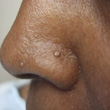
A 50-year-old woman presented with two new papules on her nose (Figure 1). A cutaneous tumor on her nose had previously been excised. She had also been successfully treated for recurrent metastatic colon cancer and endometrial cancer.
DIAGNOSIS: MUIR-TORRE SYNDROME
Muir-Torre syndrome (MTS), an autosomal dominant genodermatosis, was originally defined as the discovery of a sebaceous neoplasm and an internal malignancy; subsequently, it was suggested that the diagnostic criteria expand to also include individuals who have multiple keratoacanthomas (occasionally with sebaceous differentiation), an internal malignancy and a positive family history of MTS.1,2
There may be two different types of MTS. The most common type is a variant of hereditary non-polyposis colorectal cancer;3 this type is caused by defects in one of the mismatch repair genes: mutL homolog 1, colon cancer, nonpolyposis type 2 (E. coli) (MLH1), mutS homolog 2, colon cancer, nonpolyposis type 1 (E. coli) (MSH2) or mutS homolog 6 (E. coli) (MSH6).2 The second type of MTS shows no deficiency in mismatch repair.3
History
MTS was independently described by Muir in 1967 and Torre in 1968 as an association between sebaceous neoplasms and a visceral cancer. However, Smith was the first to describe a Maltese man with multiple facial nodules and primary malignancies in 1958; Muir based his initial report on this same patient nearly a decade later.4 In 1981, Lynch and colleagues suggested MTS was a variant of hereditary non-polyposis colorectal cancer when they found patients with sebaceous tumors to have a positive family history of colorectal cancer.3 A recent study demonstrated MTS-qualifying skin lesions in 9.2% of patients with hereditary non-polyposis colorectal cancer, further supporting the postulation that hereditary non-polyposis colorectal cancer and MTS are related.2
Epidemiology
MTS is a rare disorder with a little more than 200 cases documented to date.5,6 It shows autosomal dominant inheritance with variable expressivity and a high degree of penetrance7 and is seen in both males and females with a gender ratio of 3:2. MTS malignancies generally have a less aggressive course than the same malignancies occurring sporadically in the general population8 and, while malignancies in MTS patients tend to metastasize more often, affected individuals have shown prolonged median survival rates.2,8
Clinical Presentation Of MTS-Associated Cutaneous Tumors
The majority of skin lesions present after the diagnosis of the first internal malignancy. Cutaneous lesions present at a median age of 53 years, while the first internal malignancies are discovered at a median age of 50 years.8 The number of skin neoplasms can range from one to more than 100 and are found all over the body.9 Sebaceous neoplasms rarely occur independent of internal malignancy; therefore, if even a single tumor of sebaceous differentiation (excluding sebaceous hyperplasia) is discovered, a diagnosis of MTS should be considered.2,8
 The skin lesions associated with MTS include sebaceous adenoma [Figures 1 (above) and 2 (left)], sebaceous epithelioma (sebaceoma), sebaceous carcinoma and basal cell carcinoma with sebaceous differentiation.2 Some investigators also include keratoacanthoma (particularly those with sebaceous differentiation) as an MTS-related cutaneous neoplasm. Sebaceous hyperplasia is not specific for MTS; indeed, sebaceous hyperplasia occurs frequently in the general population.
The most common tumors found in MTS patients are sebaceous adenomas; they are seen in 68% of MTS patients10 and are usually found on the face.11 Sebaceous adenomas are lobular, benign tumors arising from sebaceous glands. Clinically, they appear as yellow or flesh-colored circumscribed papules or nodules, usually less than 0.5 cm in diameter. Histologically, mature sebocytes appear in the center and in greater numbers than germinative cells.
Sebaceous epitheliomas are also benign. They are similar in color to sebaceous adenomas but are slightly larger and may have rolled borders.10 Histologically, they differ from sebaceous adenomas in that they have greater numbers of germinative, basaloid cells than mature sebocytes.12
In contrast, sebaceous carcinomas are malignant tumors. They have a variety of clinical presentations depending on whether they are periocular or extraocular. Unfortunately, the correct diagnosis is not always initially suspected and there is a delay in proper treatment.10 The majority of sebaceous carcinomas occur in the periocular region and demonstrate aggressive behavior with regard to recurrence and metastasis.3,4,10 Microscopic examination can show a tumor with an infiltrative growth pattern and numerous mitotic figures, hyperchromatic nuclei and pleomorphism; they also demonstrate pagetoid spread into the overlying epithelium.12
Basal cell carcinomas with sebaceous differentiation are also a malignant neoplasm with clinical features and behavior similar to basal cell carcinomas without this unique pathologic feature.
Keratoacanthomas occur in middle-age and elderly individuals with a history of sun exposure. They characteristically present as 1-cm to 2-cm nodules with a central core composed of keratin.3,4,8
MTS-Associated Cancers
The most common internal malignancy in patients with MTS is colorectal carcinoma; indeed, more than half of all MTS patients have one or more primary colorectal neoplasms. The majority of these occur at or proximal to the splenic flexure, in contrast to the typical location of the distal colon that is seen in the majority of cases in the general population. Also, the malignancies are detected approximately one decade earlier in MTS patients than those occurring in the general population.4,8
Other MTS-associated visceral malignancies include, in decreasing incidence, cancers affecting genitourinary organs, breast, head and neck and small intestine. Hematologic malignancies have also been reported in MTS patients; these include Non-Hodgkin’s lymphoma, chronic lymphocytic leukemia, polycythemia vera and Hodgkin’s lymphoma.13
Pathogenesis
Germline mutations in human mismatch repair (MMR) genes are responsible for the genetically unstable malignancies seen in MTS patients. The dysfunctional MMR proteins result in the microsatellite instability present in MTS tumors.3 The vast majority of MMR defects are seen in human MSH2, though human MLH1 is responsible in about 10% of cases.14 Human MSH6 has also recently been implicated, as some sebaceous tumors have shown lack of expression of the protein.2,14 In addition, immunosuppression has been linked to revealing the MTS phenotype in certain patients, with sebaceous tumors appearing in greater numbers in individuals with profound immune suppression.3,7,9
Relationship Between MTS and Lynch Syndrome
MTS is considered by many to be a subtype of hereditary non-polyposis colorectal cancer, otherwise known as Lynch syndrome. Lynch syndrome causes a variety of visceral cancers to develop, the majority of which are colorectal and endometrial carcinomas. However, there may be skin involvement in Lynch syndrome, including sebaceous adenomas, sebaceous carcinomas and keratoacanthomas. Both MTS and Lynch syndromes are inherited in an autosomal dominant fashion and involve germline defects in the same mismatch repair genes. Lynch syndrome can even evolve into MTS in the same family. While Lynch syndrome is a much more common cause of colorectal and endometrial cancers, MTS is still quite rare. However, since MTS is considered to be a subtype of Lynch syndrome, patients with Lynch syndrome should be screened for MTS-associated skin tumors.15
Evaluation of Cutaneous Sebaceous Neoplasms
Immunohistochemistry of sebaceous neoplasms for when the tissue samples are set in paraffin is a useful screening test for MTS. Monoclonal antibodies to the MMR proteins known to be associated with MTS are utilized to detect MMR protein expression. Absence of expression on tissue specimens will be seen as a lack of nuclear staining and consistent with a possible diagnosis of MTS; for these individuals, genetic counseling, intensive cancer screening and germline testing are recommended.2,14 Such absence in MSH2 or MLH1 is predictive of microsatellite instability in 100% of patients. However, expression of these MMR proteins does not necessarily rule out DNA repair defects.14
Recommended Work-Up
Patients diagnosed with MTS or MTS-associated sebaceous lesions, as well as family members of patients, should undergo evaluation for internal malignancy. A complete colonoscopy is recommended, as the majority of MTS colorectal cancers are found in the proximal colon. Continued surveillance for malignancy throughout the life of the MTS patient is crucial, given the likelihood of these patients to develop multiple cancers. Screening recommendations for MTS patients and asymptomatic family members have been suggested and predominately include an age-appropriate evaluation as specified by the American Cancer Association, with emphasis on colorectal and genitourinary neoplasms.8
Treatment
Sebaceous adenomas can be treated with excision.7 As sebaceous carcinomas can behave aggressively, excision with microscopically controlled margins (Mohs surgery) should be considered. If more extreme disease is suspected, a head and neck surgeon, an oncologic surgeon and/or a radiation oncologist should be considered for consultation.10
Resolution
Our patient had two papules located on the left nostril (Figures 1 and 2). The anterior papule was a sebaceous adenoma. It showed loss of expression for MLH1 on immunohistochemistry with preservation of MSH2 and MSH6 expressions. The posterior papule was a “collision tumor” consisting of both a sebaceous trichofolliculoma and a sebaceous adenoma. It showed intact nuclear staining for MLH1 in the trichofolliculoma area; however, staining for MLH1 was lost in the adenoma. MSH2 and MSH6 expressions were preserved in both the trichofolliculoma and adenoma.
Prior to the recent development of her two nasal papules, our patient had been diagnosed with and treated for colon cancer with para-aortic metastases, endometrial adenocarcinoma and a benign sebaceous neoplasm on the right nostril (Figure 2). The adenocarcinoma in the colon had been diagnosed when she was 39 years old and the endometrial adenocarcinoma was found when she was 47 years old. The colon cancer showed histopathologic findings suggestive of microsatellite instability. Similar to her recent cutaneous tumors, the sebaceous neoplasm on the right nostril showed loss of expression for MLH1 with preservation of MSH2 and MSH6 expressions.
Our patient’s family history was significant for cancer, including early-onset breast and colon cancers. She has no children. Her only sibling, a brother, declined genetic testing. She is currently cancer-free.
Conclusion
MTS is a rare autosomal dominant disorder diagnosed with the discovery of: (1) a sebaceous neoplasm and an internal malignancy or (2) multiple keratoacanthomas, an internal malignancy and a positive family history of MTS. The most common type is related to hereditary non-polyposis colorectal cancer, with defects often seen in MMR genes that lead to macrosatellite instability and a predisposition to malignancy. The majority of MTS skin lesions present after the diagnosis of the first internal malignancy; however, they may precede or occur concurrently with the first internal malignancy. MTS-associated visceral cancers generally have a less aggressive course and occur at younger ages than in the general population. Loss of MMR expression, diagnosed on immunohistochemistry studies, should prompt genetic counseling, intensive cancer screening and germline testing. Patients diagnosed with MTS or with MTS-associated sebaceous skin tumors, as well as family members of MTS patients, should undergo evaluation for internal malignancy, especially colorectal and genitourinary cancers. Considering the predisposition to develop multiple cancers, continued life-long surveillance of MTS patients is essential.
Ms. Grimshaw is with the University of Texas (UT) Medical School.
Dr. Cohen is with the department of dermatology at UT Medical School, the University of Houston Health Center and the department of dermatology at the University of Texas MD Anderson Cancer Center, all in Houston, TX.
Disclosure: TThe authors have no conflicts of interest or financial disclosures to report.
 Patient Presentation
A 50-year-old woman presented with two new papules on her nose (Figure 1). A cutaneous tumor on her nose had previously been excised. She had also been successfully treated for recurrent metastatic colon cancer and endometrial cancer.
DIAGNOSIS: MUIR-TORRE SYNDROME
Muir-Torre syndrome (MTS), an autosomal dominant genodermatosis, was originally defined as the discovery of a sebaceous neoplasm and an internal malignancy; subsequently, it was suggested that the diagnostic criteria expand to also include individuals who have multiple keratoacanthomas (occasionally with sebaceous differentiation), an internal malignancy and a positive family history of MTS.1,2
There may be two different types of MTS. The most common type is a variant of hereditary non-polyposis colorectal cancer;3 this type is caused by defects in one of the mismatch repair genes: mutL homolog 1, colon cancer, nonpolyposis type 2 (E. coli) (MLH1), mutS homolog 2, colon cancer, nonpolyposis type 1 (E. coli) (MSH2) or mutS homolog 6 (E. coli) (MSH6).2 The second type of MTS shows no deficiency in mismatch repair.3
History
MTS was independently described by Muir in 1967 and Torre in 1968 as an association between sebaceous neoplasms and a visceral cancer. However, Smith was the first to describe a Maltese man with multiple facial nodules and primary malignancies in 1958; Muir based his initial report on this same patient nearly a decade later.4 In 1981, Lynch and colleagues suggested MTS was a variant of hereditary non-polyposis colorectal cancer when they found patients with sebaceous tumors to have a positive family history of colorectal cancer.3 A recent study demonstrated MTS-qualifying skin lesions in 9.2% of patients with hereditary non-polyposis colorectal cancer, further supporting the postulation that hereditary non-polyposis colorectal cancer and MTS are related.2
Epidemiology
MTS is a rare disorder with a little more than 200 cases documented to date.5,6 It shows autosomal dominant inheritance with variable expressivity and a high degree of penetrance7 and is seen in both males and females with a gender ratio of 3:2. MTS malignancies generally have a less aggressive course than the same malignancies occurring sporadically in the general population8 and, while malignancies in MTS patients tend to metastasize more often, affected individuals have shown prolonged median survival rates.2,8
Clinical Presentation Of MTS-Associated Cutaneous Tumors
The majority of skin lesions present after the diagnosis of the first internal malignancy. Cutaneous lesions present at a median age of 53 years, while the first internal malignancies are discovered at a median age of 50 years.8 The number of skin neoplasms can range from one to more than 100 and are found all over the body.9 Sebaceous neoplasms rarely occur independent of internal malignancy; therefore, if even a single tumor of sebaceous differentiation (excluding sebaceous hyperplasia) is discovered, a diagnosis of MTS should be considered.2,8
 The skin lesions associated with MTS include sebaceous adenoma [Figures 1 (above) and 2 (left)], sebaceous epithelioma (sebaceoma), sebaceous carcinoma and basal cell carcinoma with sebaceous differentiation.2 Some investigators also include keratoacanthoma (particularly those with sebaceous differentiation) as an MTS-related cutaneous neoplasm. Sebaceous hyperplasia is not specific for MTS; indeed, sebaceous hyperplasia occurs frequently in the general population.
The most common tumors found in MTS patients are sebaceous adenomas; they are seen in 68% of MTS patients10 and are usually found on the face.11 Sebaceous adenomas are lobular, benign tumors arising from sebaceous glands. Clinically, they appear as yellow or flesh-colored circumscribed papules or nodules, usually less than 0.5 cm in diameter. Histologically, mature sebocytes appear in the center and in greater numbers than germinative cells.
Sebaceous epitheliomas are also benign. They are similar in color to sebaceous adenomas but are slightly larger and may have rolled borders.10 Histologically, they differ from sebaceous adenomas in that they have greater numbers of germinative, basaloid cells than mature sebocytes.12
In contrast, sebaceous carcinomas are malignant tumors. They have a variety of clinical presentations depending on whether they are periocular or extraocular. Unfortunately, the correct diagnosis is not always initially suspected and there is a delay in proper treatment.10 The majority of sebaceous carcinomas occur in the periocular region and demonstrate aggressive behavior with regard to recurrence and metastasis.3,4,10 Microscopic examination can show a tumor with an infiltrative growth pattern and numerous mitotic figures, hyperchromatic nuclei and pleomorphism; they also demonstrate pagetoid spread into the overlying epithelium.12
Basal cell carcinomas with sebaceous differentiation are also a malignant neoplasm with clinical features and behavior similar to basal cell carcinomas without this unique pathologic feature.
Keratoacanthomas occur in middle-age and elderly individuals with a history of sun exposure. They characteristically present as 1-cm to 2-cm nodules with a central core composed of keratin.3,4,8
MTS-Associated Cancers
The most common internal malignancy in patients with MTS is colorectal carcinoma; indeed, more than half of all MTS patients have one or more primary colorectal neoplasms. The majority of these occur at or proximal to the splenic flexure, in contrast to the typical location of the distal colon that is seen in the majority of cases in the general population. Also, the malignancies are detected approximately one decade earlier in MTS patients than those occurring in the general population.4,8
Other MTS-associated visceral malignancies include, in decreasing incidence, cancers affecting genitourinary organs, breast, head and neck and small intestine. Hematologic malignancies have also been reported in MTS patients; these include Non-Hodgkin’s lymphoma, chronic lymphocytic leukemia, polycythemia vera and Hodgkin’s lymphoma.13
Pathogenesis
Germline mutations in human mismatch repair (MMR) genes are responsible for the genetically unstable malignancies seen in MTS patients. The dysfunctional MMR proteins result in the microsatellite instability present in MTS tumors.3 The vast majority of MMR defects are seen in human MSH2, though human MLH1 is responsible in about 10% of cases.14 Human MSH6 has also recently been implicated, as some sebaceous tumors have shown lack of expression of the protein.2,14 In addition, immunosuppression has been linked to revealing the MTS phenotype in certain patients, with sebaceous tumors appearing in greater numbers in individuals with profound immune suppression.3,7,9
Relationship Between MTS and Lynch Syndrome
MTS is considered by many to be a subtype of hereditary non-polyposis colorectal cancer, otherwise known as Lynch syndrome. Lynch syndrome causes a variety of visceral cancers to develop, the majority of which are colorectal and endometrial carcinomas. However, there may be skin involvement in Lynch syndrome, including sebaceous adenomas, sebaceous carcinomas and keratoacanthomas. Both MTS and Lynch syndromes are inherited in an autosomal dominant fashion and involve germline defects in the same mismatch repair genes. Lynch syndrome can even evolve into MTS in the same family. While Lynch syndrome is a much more common cause of colorectal and endometrial cancers, MTS is still quite rare. However, since MTS is considered to be a subtype of Lynch syndrome, patients with Lynch syndrome should be screened for MTS-associated skin tumors.15
Evaluation of Cutaneous Sebaceous Neoplasms
Immunohistochemistry of sebaceous neoplasms for when the tissue samples are set in paraffin is a useful screening test for MTS. Monoclonal antibodies to the MMR proteins known to be associated with MTS are utilized to detect MMR protein expression. Absence of expression on tissue specimens will be seen as a lack of nuclear staining and consistent with a possible diagnosis of MTS; for these individuals, genetic counseling, intensive cancer screening and germline testing are recommended.2,14 Such absence in MSH2 or MLH1 is predictive of microsatellite instability in 100% of patients. However, expression of these MMR proteins does not necessarily rule out DNA repair defects.14
Recommended Work-Up
Patients diagnosed with MTS or MTS-associated sebaceous lesions, as well as family members of patients, should undergo evaluation for internal malignancy. A complete colonoscopy is recommended, as the majority of MTS colorectal cancers are found in the proximal colon. Continued surveillance for malignancy throughout the life of the MTS patient is crucial, given the likelihood of these patients to develop multiple cancers. Screening recommendations for MTS patients and asymptomatic family members have been suggested and predominately include an age-appropriate evaluation as specified by the American Cancer Association, with emphasis on colorectal and genitourinary neoplasms.8
Treatment
Sebaceous adenomas can be treated with excision.7 As sebaceous carcinomas can behave aggressively, excision with microscopically controlled margins (Mohs surgery) should be considered. If more extreme disease is suspected, a head and neck surgeon, an oncologic surgeon and/or a radiation oncologist should be considered for consultation.10
Resolution
Our patient had two papules located on the left nostril (Figures 1 and 2). The anterior papule was a sebaceous adenoma. It showed loss of expression for MLH1 on immunohistochemistry with preservation of MSH2 and MSH6 expressions. The posterior papule was a “collision tumor” consisting of both a sebaceous trichofolliculoma and a sebaceous adenoma. It showed intact nuclear staining for MLH1 in the trichofolliculoma area; however, staining for MLH1 was lost in the adenoma. MSH2 and MSH6 expressions were preserved in both the trichofolliculoma and adenoma.
Prior to the recent development of her two nasal papules, our patient had been diagnosed with and treated for colon cancer with para-aortic metastases, endometrial adenocarcinoma and a benign sebaceous neoplasm on the right nostril (Figure 2). The adenocarcinoma in the colon had been diagnosed when she was 39 years old and the endometrial adenocarcinoma was found when she was 47 years old. The colon cancer showed histopathologic findings suggestive of microsatellite instability. Similar to her recent cutaneous tumors, the sebaceous neoplasm on the right nostril showed loss of expression for MLH1 with preservation of MSH2 and MSH6 expressions.
Our patient’s family history was significant for cancer, including early-onset breast and colon cancers. She has no children. Her only sibling, a brother, declined genetic testing. She is currently cancer-free.
Conclusion
MTS is a rare autosomal dominant disorder diagnosed with the discovery of: (1) a sebaceous neoplasm and an internal malignancy or (2) multiple keratoacanthomas, an internal malignancy and a positive family history of MTS. The most common type is related to hereditary non-polyposis colorectal cancer, with defects often seen in MMR genes that lead to macrosatellite instability and a predisposition to malignancy. The majority of MTS skin lesions present after the diagnosis of the first internal malignancy; however, they may precede or occur concurrently with the first internal malignancy. MTS-associated visceral cancers generally have a less aggressive course and occur at younger ages than in the general population. Loss of MMR expression, diagnosed on immunohistochemistry studies, should prompt genetic counseling, intensive cancer screening and germline testing. Patients diagnosed with MTS or with MTS-associated sebaceous skin tumors, as well as family members of MTS patients, should undergo evaluation for internal malignancy, especially colorectal and genitourinary cancers. Considering the predisposition to develop multiple cancers, continued life-long surveillance of MTS patients is essential.
Ms. Grimshaw is with the University of Texas (UT) Medical School.
Dr. Cohen is with the department of dermatology at UT Medical School, the University of Houston Health Center and the department of dermatology at the University of Texas MD Anderson Cancer Center, all in Houston, TX.
Disclosure: TThe authors have no conflicts of interest or financial disclosures to report.
Patient Presentation
A 50-year-old woman presented with two new papules on her nose (Figure 1). A cutaneous tumor on her nose had previously been excised. She had also been successfully treated for recurrent metastatic colon cancer and endometrial cancer.
DIAGNOSIS: MUIR-TORRE SYNDROME
Muir-Torre syndrome (MTS), an autosomal dominant genodermatosis, was originally defined as the discovery of a sebaceous neoplasm and an internal malignancy; subsequently, it was suggested that the diagnostic criteria expand to also include individuals who have multiple keratoacanthomas (occasionally with sebaceous differentiation), an internal malignancy and a positive family history of MTS.1,2
There may be two different types of MTS. The most common type is a variant of hereditary non-polyposis colorectal cancer;3 this type is caused by defects in one of the mismatch repair genes: mutL homolog 1, colon cancer, nonpolyposis type 2 (E. coli) (MLH1), mutS homolog 2, colon cancer, nonpolyposis type 1 (E. coli) (MSH2) or mutS homolog 6 (E. coli) (MSH6).2 The second type of MTS shows no deficiency in mismatch repair.3
History
MTS was independently described by Muir in 1967 and Torre in 1968 as an association between sebaceous neoplasms and a visceral cancer. However, Smith was the first to describe a Maltese man with multiple facial nodules and primary malignancies in 1958; Muir based his initial report on this same patient nearly a decade later.4 In 1981, Lynch and colleagues suggested MTS was a variant of hereditary non-polyposis colorectal cancer when they found patients with sebaceous tumors to have a positive family history of colorectal cancer.3 A recent study demonstrated MTS-qualifying skin lesions in 9.2% of patients with hereditary non-polyposis colorectal cancer, further supporting the postulation that hereditary non-polyposis colorectal cancer and MTS are related.2
Epidemiology
MTS is a rare disorder with a little more than 200 cases documented to date.5,6 It shows autosomal dominant inheritance with variable expressivity and a high degree of penetrance7 and is seen in both males and females with a gender ratio of 3:2. MTS malignancies generally have a less aggressive course than the same malignancies occurring sporadically in the general population8 and, while malignancies in MTS patients tend to metastasize more often, affected individuals have shown prolonged median survival rates.2,8
Clinical Presentation Of MTS-Associated Cutaneous Tumors
The majority of skin lesions present after the diagnosis of the first internal malignancy. Cutaneous lesions present at a median age of 53 years, while the first internal malignancies are discovered at a median age of 50 years.8 The number of skin neoplasms can range from one to more than 100 and are found all over the body.9 Sebaceous neoplasms rarely occur independent of internal malignancy; therefore, if even a single tumor of sebaceous differentiation (excluding sebaceous hyperplasia) is discovered, a diagnosis of MTS should be considered.2,8
The skin lesions associated with MTS include sebaceous adenoma [Figures 1 (above) and 2 (left)], sebaceous epithelioma (sebaceoma), sebaceous carcinoma and basal cell carcinoma with sebaceous differentiation.2 Some investigators also include keratoacanthoma (particularly those with sebaceous differentiation) as an MTS-related cutaneous neoplasm. Sebaceous hyperplasia is not specific for MTS; indeed, sebaceous hyperplasia occurs frequently in the general population.
The most common tumors found in MTS patients are sebaceous adenomas; they are seen in 68% of MTS patients10 and are usually found on the face.11 Sebaceous adenomas are lobular, benign tumors arising from sebaceous glands. Clinically, they appear as yellow or flesh-colored circumscribed papules or nodules, usually less than 0.5 cm in diameter. Histologically, mature sebocytes appear in the center and in greater numbers than germinative cells.
Sebaceous epitheliomas are also benign. They are similar in color to sebaceous adenomas but are slightly larger and may have rolled borders.10 Histologically, they differ from sebaceous adenomas in that they have greater numbers of germinative, basaloid cells than mature sebocytes.12
In contrast, sebaceous carcinomas are malignant tumors. They have a variety of clinical presentations depending on whether they are periocular or extraocular. Unfortunately, the correct diagnosis is not always initially suspected and there is a delay in proper treatment.10 The majority of sebaceous carcinomas occur in the periocular region and demonstrate aggressive behavior with regard to recurrence and metastasis.3,4,10 Microscopic examination can show a tumor with an infiltrative growth pattern and numerous mitotic figures, hyperchromatic nuclei and pleomorphism; they also demonstrate pagetoid spread into the overlying epithelium.12
Basal cell carcinomas with sebaceous differentiation are also a malignant neoplasm with clinical features and behavior similar to basal cell carcinomas without this unique pathologic feature.
Keratoacanthomas occur in middle-age and elderly individuals with a history of sun exposure. They characteristically present as 1-cm to 2-cm nodules with a central core composed of keratin.3,4,8
MTS-Associated Cancers
The most common internal malignancy in patients with MTS is colorectal carcinoma; indeed, more than half of all MTS patients have one or more primary colorectal neoplasms. The majority of these occur at or proximal to the splenic flexure, in contrast to the typical location of the distal colon that is seen in the majority of cases in the general population. Also, the malignancies are detected approximately one decade earlier in MTS patients than those occurring in the general population.4,8
Other MTS-associated visceral malignancies include, in decreasing incidence, cancers affecting genitourinary organs, breast, head and neck and small intestine. Hematologic malignancies have also been reported in MTS patients; these include Non-Hodgkin’s lymphoma, chronic lymphocytic leukemia, polycythemia vera and Hodgkin’s lymphoma.13
Pathogenesis
Germline mutations in human mismatch repair (MMR) genes are responsible for the genetically unstable malignancies seen in MTS patients. The dysfunctional MMR proteins result in the microsatellite instability present in MTS tumors.3 The vast majority of MMR defects are seen in human MSH2, though human MLH1 is responsible in about 10% of cases.14 Human MSH6 has also recently been implicated, as some sebaceous tumors have shown lack of expression of the protein.2,14 In addition, immunosuppression has been linked to revealing the MTS phenotype in certain patients, with sebaceous tumors appearing in greater numbers in individuals with profound immune suppression.3,7,9
Relationship Between MTS and Lynch Syndrome
MTS is considered by many to be a subtype of hereditary non-polyposis colorectal cancer, otherwise known as Lynch syndrome. Lynch syndrome causes a variety of visceral cancers to develop, the majority of which are colorectal and endometrial carcinomas. However, there may be skin involvement in Lynch syndrome, including sebaceous adenomas, sebaceous carcinomas and keratoacanthomas. Both MTS and Lynch syndromes are inherited in an autosomal dominant fashion and involve germline defects in the same mismatch repair genes. Lynch syndrome can even evolve into MTS in the same family. While Lynch syndrome is a much more common cause of colorectal and endometrial cancers, MTS is still quite rare. However, since MTS is considered to be a subtype of Lynch syndrome, patients with Lynch syndrome should be screened for MTS-associated skin tumors.15
Evaluation of Cutaneous Sebaceous Neoplasms
Immunohistochemistry of sebaceous neoplasms for when the tissue samples are set in paraffin is a useful screening test for MTS. Monoclonal antibodies to the MMR proteins known to be associated with MTS are utilized to detect MMR protein expression. Absence of expression on tissue specimens will be seen as a lack of nuclear staining and consistent with a possible diagnosis of MTS; for these individuals, genetic counseling, intensive cancer screening and germline testing are recommended.2,14 Such absence in MSH2 or MLH1 is predictive of microsatellite instability in 100% of patients. However, expression of these MMR proteins does not necessarily rule out DNA repair defects.14
Recommended Work-Up
Patients diagnosed with MTS or MTS-associated sebaceous lesions, as well as family members of patients, should undergo evaluation for internal malignancy. A complete colonoscopy is recommended, as the majority of MTS colorectal cancers are found in the proximal colon. Continued surveillance for malignancy throughout the life of the MTS patient is crucial, given the likelihood of these patients to develop multiple cancers. Screening recommendations for MTS patients and asymptomatic family members have been suggested and predominately include an age-appropriate evaluation as specified by the American Cancer Association, with emphasis on colorectal and genitourinary neoplasms.8
Treatment
Sebaceous adenomas can be treated with excision.7 As sebaceous carcinomas can behave aggressively, excision with microscopically controlled margins (Mohs surgery) should be considered. If more extreme disease is suspected, a head and neck surgeon, an oncologic surgeon and/or a radiation oncologist should be considered for consultation.10
Resolution
Our patient had two papules located on the left nostril (Figures 1 and 2). The anterior papule was a sebaceous adenoma. It showed loss of expression for MLH1 on immunohistochemistry with preservation of MSH2 and MSH6 expressions. The posterior papule was a “collision tumor” consisting of both a sebaceous trichofolliculoma and a sebaceous adenoma. It showed intact nuclear staining for MLH1 in the trichofolliculoma area; however, staining for MLH1 was lost in the adenoma. MSH2 and MSH6 expressions were preserved in both the trichofolliculoma and adenoma.
Prior to the recent development of her two nasal papules, our patient had been diagnosed with and treated for colon cancer with para-aortic metastases, endometrial adenocarcinoma and a benign sebaceous neoplasm on the right nostril (Figure 2). The adenocarcinoma in the colon had been diagnosed when she was 39 years old and the endometrial adenocarcinoma was found when she was 47 years old. The colon cancer showed histopathologic findings suggestive of microsatellite instability. Similar to her recent cutaneous tumors, the sebaceous neoplasm on the right nostril showed loss of expression for MLH1 with preservation of MSH2 and MSH6 expressions.
Our patient’s family history was significant for cancer, including early-onset breast and colon cancers. She has no children. Her only sibling, a brother, declined genetic testing. She is currently cancer-free.
Conclusion
MTS is a rare autosomal dominant disorder diagnosed with the discovery of: (1) a sebaceous neoplasm and an internal malignancy or (2) multiple keratoacanthomas, an internal malignancy and a positive family history of MTS. The most common type is related to hereditary non-polyposis colorectal cancer, with defects often seen in MMR genes that lead to macrosatellite instability and a predisposition to malignancy. The majority of MTS skin lesions present after the diagnosis of the first internal malignancy; however, they may precede or occur concurrently with the first internal malignancy. MTS-associated visceral cancers generally have a less aggressive course and occur at younger ages than in the general population. Loss of MMR expression, diagnosed on immunohistochemistry studies, should prompt genetic counseling, intensive cancer screening and germline testing. Patients diagnosed with MTS or with MTS-associated sebaceous skin tumors, as well as family members of MTS patients, should undergo evaluation for internal malignancy, especially colorectal and genitourinary cancers. Considering the predisposition to develop multiple cancers, continued life-long surveillance of MTS patients is essential.
Ms. Grimshaw is with the University of Texas (UT) Medical School.
Dr. Cohen is with the department of dermatology at UT Medical School, the University of Houston Health Center and the department of dermatology at the University of Texas MD Anderson Cancer Center, all in Houston, TX.
Disclosure: TThe authors have no conflicts of interest or financial disclosures to report.


 Patient Presentation
 The skin lesions associated with MTS include sebaceous adenoma [Figures 1 (above) and 2 (left)], sebaceous epithelioma (sebaceoma), sebaceous carcinoma and basal cell carcinoma with sebaceous differentiation.2 Some investigators also include keratoacanthoma (particularly those with sebaceous differentiation) as an MTS-related cutaneous neoplasm. Sebaceous hyperplasia is not specific for MTS; indeed, sebaceous hyperplasia occurs frequently in the general population.