Restoring Mobility in Complex Diabetic Peripheral Arterial Disease
Successful Endovascular Atheroplasty With the Santreva-ATK System and Sirolimus-Balloon Angioplasty
Successful Endovascular Atheroplasty With the Santreva-ATK System and Sirolimus-Balloon Angioplasty
Key Summary
- This case report involved a 72-year-old woman with diabetes, Rutherford 3 claudication, ankle-brachial index (ABI) 0.72, and a 5-cm distal superficial femoral artery occlusion.
- Santreva-ATK endovascular atheroplasty (AngioSafe) plus sirolimus-coated balloon angioplasty achieved <10% residual stenosis without flow-limiting dissection, embolization, or stenting. ABI improved to 0.98, and the patient was discharged home within 24 hours.
- At 10 months, the artery remained patent without restenosis and walking was unrestricted and pain-free. The authors emphasize multidisciplinary selection, expert operators, and ultrasound surveillance.
© 2026 HMP Global. All Rights Reserved.
Any views and opinions expressed are those of the author(s) and/or participants and do not necessarily reflect the views, policy, or position of Vascular Disease Management or HMP Global, their employees, and affiliates.
VASCULAR DISEASE MANAGEMENT. 2026;23(9):E135-E137
Introduction
Peripheral arterial disease (PAD) is one of the most debilitating vascular complications associated with longstanding diabetes mellitus (DM). Diabetic PAD typically affects lower extremity vessels with a propensity for long, diffuse, and calcified lesions in the femoropopliteal and infrapopliteal arterial segments. As arterial stiffening and luminal narrowing progress, patients experience severe reduction in walking capacity, transitioning from intermittent claudication (IC) to severe short-distance IC, and eventually chronic limb-threatening ischemia (CLTI).1
In individuals with diabetes, short-distance claudication—where a patient can only ambulate inside their home for a few meters before halting due to severe ischemic calf pain—radically diminishes independence and quality of life. Open surgical revascularization (bypass grafting) in older patients with diabetes carries significant perioperative risks, wound-healing complications, and prolonged hospital stays. Conversely, conventional balloon angioplasty alone often fails in calcified lesions due to acute elastic recoil, high pressures causing severe arterial wall dissection, and suboptimal vessel preparation.2
Case Presentation
A 72-year-old woman with a 10-year history of type 2 DM, arterial hypertension, and hyperlipidemia presented with longstanding lower-extremity IC affecting her left leg. Over the preceding 6 months, her functional capacity had declined severely. At her baseline evaluation, pain-free walking distance was restricted to less than 10 meters, confining her exclusively to minimal movement inside her home. Moving from room to room triggered severe, cramp-like calf pain (Rutherford category 3/severe lifestyle-limiting claudication approaching rest pain).
The baseline left ankle-brachial index (ABI) was severely reduced at 0.72, while duplex ultrasound (DUS) and preprocedural contrast-enhanced computed tomography angiography revealed a chronic total occlusion (CTO) of the left distal superficial femoral artery measuring approximately 5 cm in length. Single-vessel peroneal artery runoff to the foot was preserved.
Given the clinical characteristics of the disease (severe IC, CTO, and single-vessel runoff), the patient’s relatively young age, and the presence of calcified diabetic arteriopathy, an endovascular strategy focusing on intraluminal crossing, plaque modification (vessel preparation), and sirolimus-coated balloon angioplasty was chosen to decrease periprocedural morbidity, avoid mandatory metallic stent implantation, enhance vessel patency, and preserve stent implantation and surgical bypass as future treatment options, if necessary. Conventional balloon angioplasty alone often fails in calcified CTOs due to acute elastic recoil, high pressures causing severe arterial wall dissection, and suboptimal luminal gain. Using modern endovascular devices able to debulk the atheroma and alter vessel compliance prior to balloon inflation, operators can achieve maximum luminal gain at lower inflation pressures, minimizing barotrauma and avoiding metallic stent implantation.
Based on the hospital’s vascular multidisciplinary team (MDT) decision, the incorporation of wireless intraluminal crossing and simultaneous plaque modification using the Santreva-ATK endovascular atheroplasty system (AngioSafe) was an excellent option for this patient. Of note, such procedures should only be performed by vascular experts in high-volume centers of excellence, following a case-based MDT decision, to minimize severe, limb-threatening complications such as acute thromboembolic events.
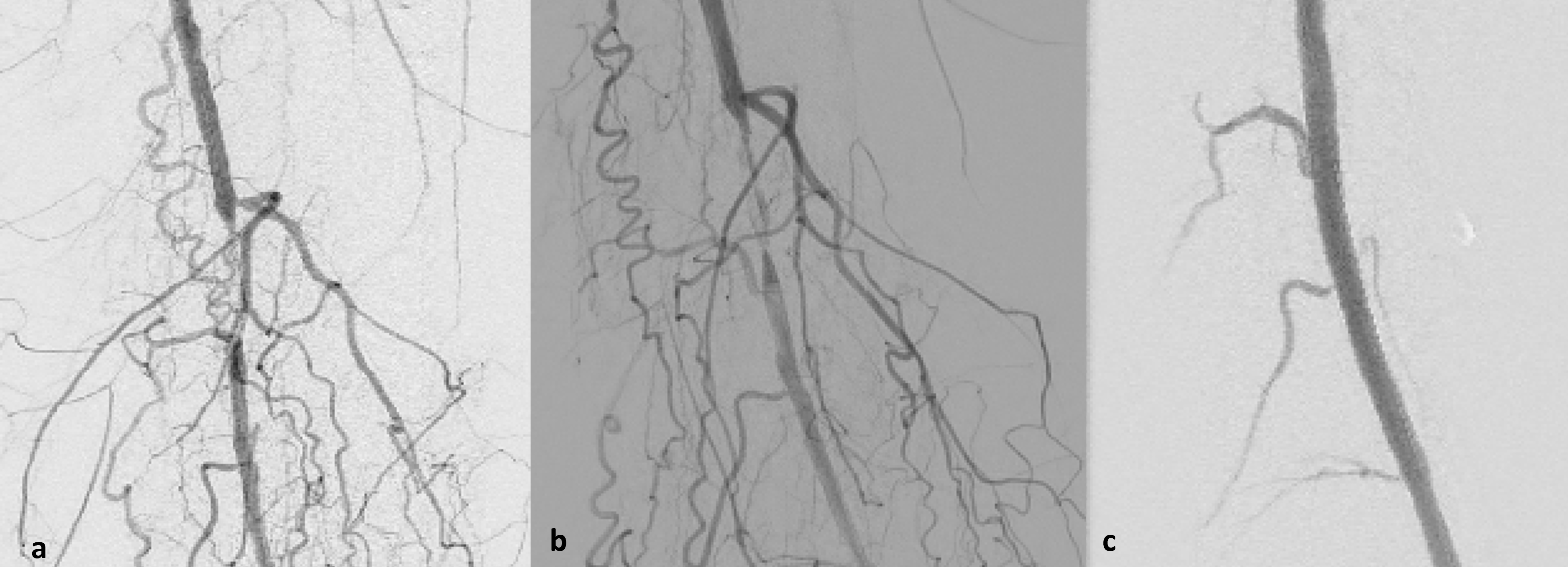
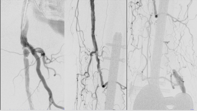
Written informed consent was obtained by the patient prior to the procedure. Under local anesthesia and ultrasound guidance, an antegrade common femoral artery access was obtained using a 7F sheath, and the lesion was depicted with digital subtraction angiography (DSA) (Figure, a). Subsequently, wireless intraluminal lesion crossing and simultaneous atheroplasty was performed using the Santreva-ATK endovascular revascularization catheter (Figure, b), followed by a 0.014-inch guidewire crossing that was positioned at the distal popliteal artery. Intravascular ultrasound (IVUS) was used to verify the intraluminal CTO crossing and provide accurate vessel sizing. A 6 x 80-mm sirolimus-coated balloon (Selution SLR, Cordis), sized 1:1 vessel: balloon diameter, was inflated at low pressure (max 10 atm) for 180 seconds to enable maximum luminal gain, avoid significant vessel-wall dissection, and enhance patency.3,4 Postprocedural angiography and IVUS confirmed complete luminal restoration with < 10% residual stenosis, no flow-limiting dissection, and no embolization to the single outflow vessel runoff to the foot. No stent deployment was required (Figure, c). The access site was sealed using a vascular closure device. The next morning, physical examination confirmed warm peripheral extremities, excellent capillary refill, and an ABI of 0.98. The patient was able to walk along the hospital corridor completely free of ischemic pain and was discharged home on dual antiplatelet and statin therapy (total hospitalization time < 24 hours). The treated lesion remains patent without restenosis according to scheduled regular DUS imaging follow-up. Conclusively, treatment with the Santreva-ATK endovascular atheroplasty endovascular catheter followed by sirolimus balloon angioplasty achieved complete revascularization, enabling next-day hospital discharge and sustained, unrestricted, pain-free walking at the 10-month follow-up. n
Affiliations and Disclosures
Stavros Spiliopoulos, MD, PhD, Stavros Grigoriadis, MD, MSc, PhD, and Konstantinos Palialexis, MD, MSc, PhD are from the 2nd Department of Radiology, Interventional Radiology Unit, Medical School, National and Kapodistrian University of Athens, “Attikon” University General Hospital, Athens, Greece; Constantine N. Antonopoulos, MD, PhD, and George Sfyroeras, MD, PhD, are from the 1st Vascular Surgery Department, School of Medicine, National and Kapodistrian University of Athens, “Attikon” University Hospital, Athens, Greece.
The authors report no financial relationships or conflicts of interest regarding the content herein.
Manuscript accepted August 25, 2026.
Address for correspondence: Stavros Spiliopoulos, MD, PhD, 2nd Department of Radiology, Interventional Radiology Unit, Attikon University General Hospital, 1st Rimini St, GR 12461, Chaidari, Athens, Greece. Email: stavspiliop@med.uoa.gr
References
1. Mazzolai L, Teixido-Tura G, Lanzi S, et al; ESC Scientific Document Group. 2024 ESC guidelines for the management of peripheral arterial and aortic diseases. Eur Heart J. 2024;45(36):3538-3700. doi:10.1093/eurheartj/ehae179
2. Saratzis A, Patrone L, Secemsky EA, et al; VPAD collaborators. Use of vessel preparation in endovascular peripheral arterial disease (PAD) interventions: a global qualitative analysis. J Endovasc Ther. 2026:15266028261424732. doi:10.1177/15266028261424732
3. Agarwal G, Kamal R, Saeed D, et al. Should interventional radiologists incorporate intravascular ultrasound (IVUS) into their daily practice? A systematic review and meta-analysis of IVUS in peripheral arterial endovascular interventions. Cardiovasc Intervent Radiol. 2026;49(8):1441-1451. doi:10.1007/s00270-026-04509-0
4. Teichgräber U, Ingwersen M, Lehmann T, et al; SIRONA Study Group. Comparison of sirolimus- vs paclitaxel-coated balloon angioplasty for femoropopliteal artery disease: the SIRONA randomized noninferiority trial. J Am Coll Cardiol. 2026;S0735-1097(25)10433-6. doi:10.1016/j.jacc.2025.12.017