Case Studies in Addressing Jones Fractures in Athletes
-
Figure 1a
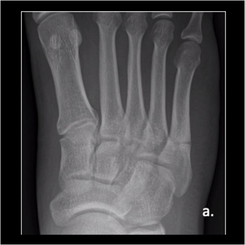
Figure 1a. A 23-year-old male trainee twisted his foot while running, presenting a week or 2 later. Here is an injury film.
-
Figure 1b
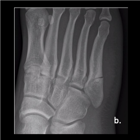
Figure 1b. A 23-year-old male trainee twisted his foot while running, presenting a week or 2 later. Here is an injury film.
-
Figure 1c
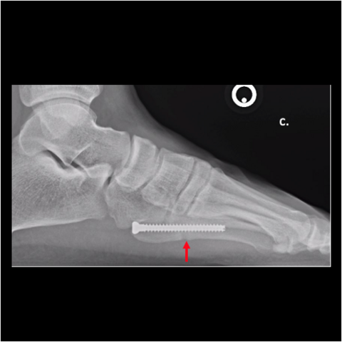
Figure 1c. We fixed his fracture with an intramedullary, fully-threaded, cannulated 4.5mm screw. He returned to training at 3 months. As shown here, the fracture looked healed at 6 weeks postop. Despite the patient having no pain, I must caution surgeons to not allow running or sports until the plantar cortex is solid because of the risk for refracture.
-
Figure 1d
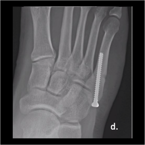
Figure 1d. We fixed his fracture with an intramedullary, fully-threaded, cannulated 4.5mm screw. He returned to training at 3 months. As shown here, the fracture looked healed at 6 weeks postop.
-
Figure 1e
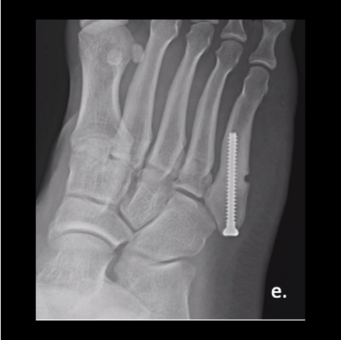
Figure 1e. We fixed his fracture with an intramedullary, fully-threaded, cannulated 4.5mm screw. He returned to training at 3 months. As shown here, the fracture looked healed at 6 weeks postop.
-
Figure 2a
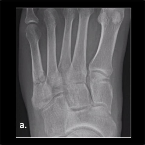
Figure 2a. A 30-year-old male soldier was injured playing basketball, as shown in this film taken upon injury. We fixated his fracture with a solid screw system. He returned to full duty in 4 months and was pain-free in days after his surgery.
-
Figure 2b
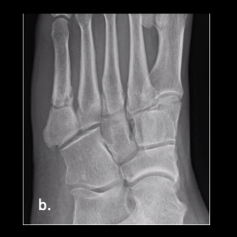
Figure 2b. A 30-year-old male soldier was injured playing basketball, as shown in this film taken upon injury. We fixated his fracture with a solid screw system. He returned to full duty in 4 months and was pain-free in days after his surgery.
-
Figure 2c
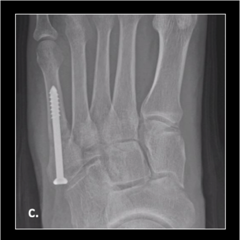
Figure 2c. Here is the 30-year-old soldier’s foot 6 weeks post-injury.
-
Figure 2d
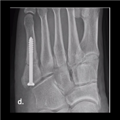
Figure 2d. Here is the 30-year-old soldier’s foot 6 weeks post-injury.
-
Figure 2e
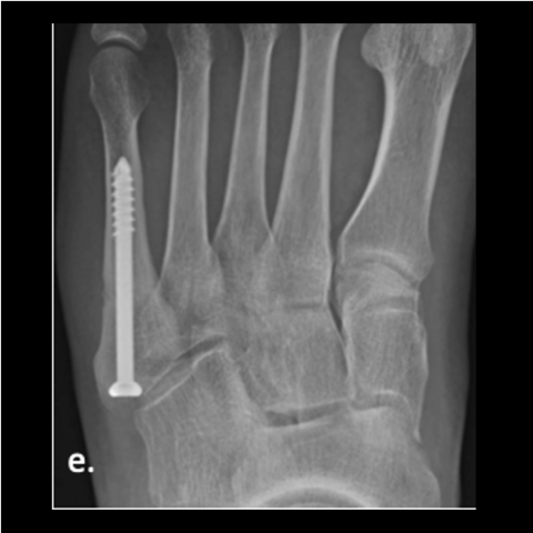
Figure 2e. Here is the 30-year-old soldier’s foot 3 months post-injury.
-
Figure 2f
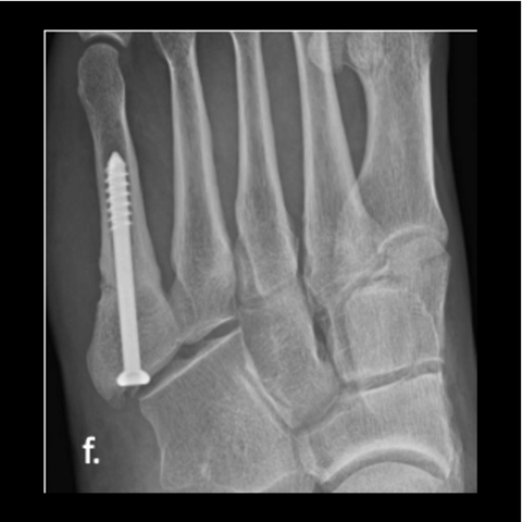
Figure 2f. Here is the 30-year-old soldier’s foot 3 months post-injury.
-
Figure 2g
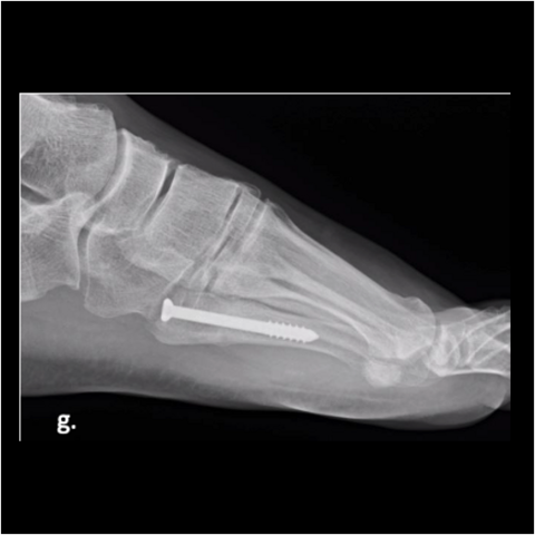
Figure 2g. Here is the 30-year-old soldier’s foot 3 months post-injury.
-
Figure 3a
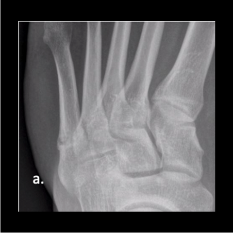
Figure 3a. A 28-year-old Army officer injured during combatives was able to walk 2 days post-surgery. He started wearing his Army boots 2 weeks after surgery and completed Sapper School 10 weeks after surgery. He then went to Special Forces training within 6 months postop. The patient never had any pain after surgery. Here is the foot when the patient got injured.
-
Figure 3b
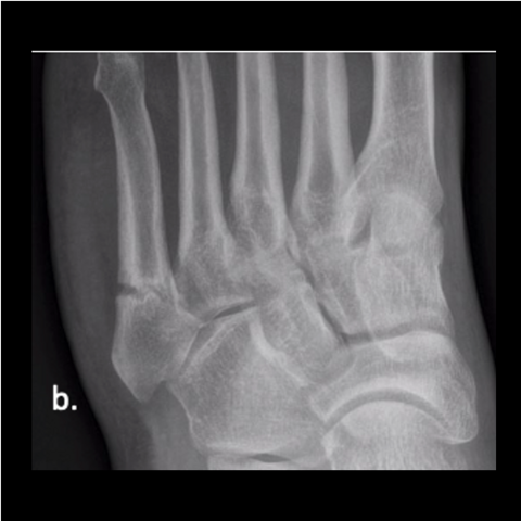
Figure 3b. A 28-year-old Army officer injured during combatives was able to walk 2 days post-surgery. He started wearing his Army boots 2 weeks after surgery and completed Sapper School 10 weeks after surgery. He then went to Special Forces training within 6 months postop. The patient never had any pain after surgery. Here is the foot when the patient got injured.
-
Figure 3c
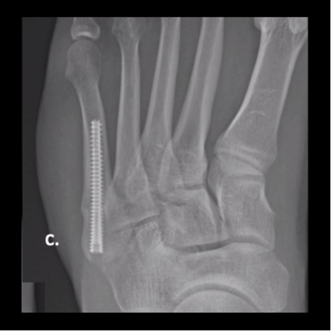
Figure 3c. Here is the 28-year-old’s foot at 6 weeks.
-
Figure 3d
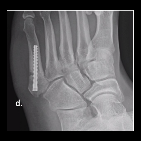
Figure 3d. Here is the 28-year-old’s foot at 6 weeks.
-
Figure 3e
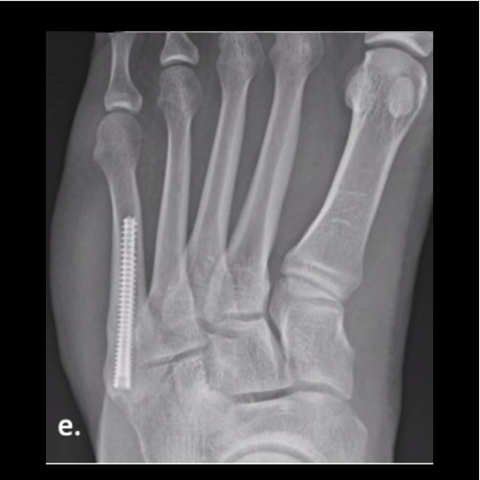
Figure 3e. Here is the 28-year-old’s foot at 3 months.
-
Figure 3f
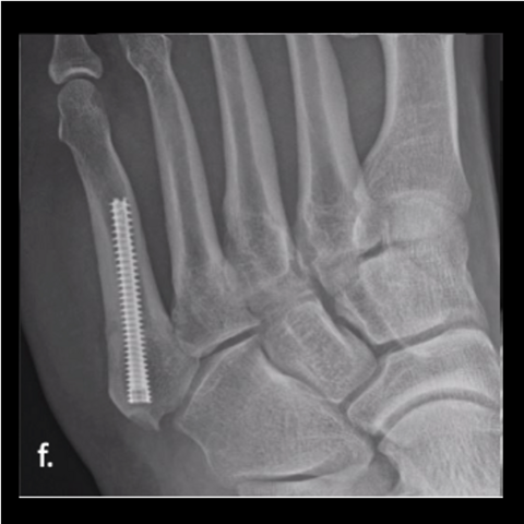
Figure 3f. Here is the 28-year-old’s foot at 3 months.
-
Figure 3g
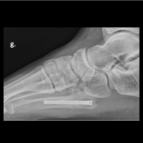
Figure 3g. Here is the 28-year-old’s foot at 3 months.
-
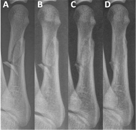
Figure 4a
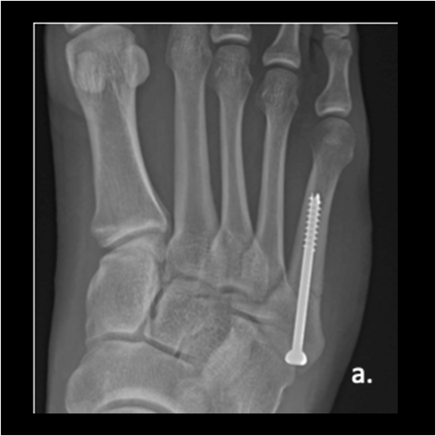
Figure 4a. A 24-year-old male soldier refractured 1 year after his original surgery. He had returned to Airborne status with at least 15 jumps since his original surgery, with no issues running. Here is the 24-year-old soldier’s foot 6 months post-surgery.
-
Figure 4b
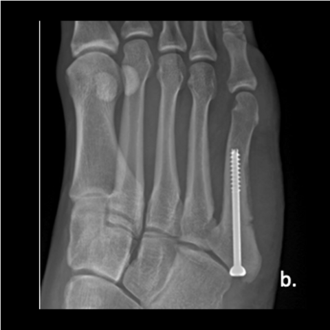
Figure 4b. Here is the 24-year-old soldier’s foot 6 months post-surgery.
-
Figure 4c
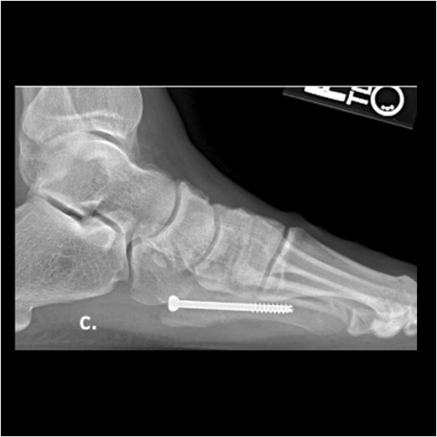
Figure 4c. Here is the 24-year-old soldier’s foot 6 months post-surgery.
-
Figure 4d
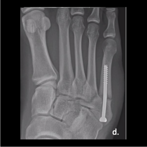
Figure 4d. The 24-year-old soldier jumped off the back of a truck, refracturing his metatarsal. Here is the foot a year post-surgery after jumping off the truck.
-
Figure 4e
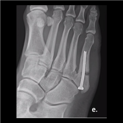
Figure 4e. The 24-year-old soldier jumped off the back of a truck, refracturing his metatarsal. Here is the foot a year post-surgery after jumping off the truck.
-
Figure 4f
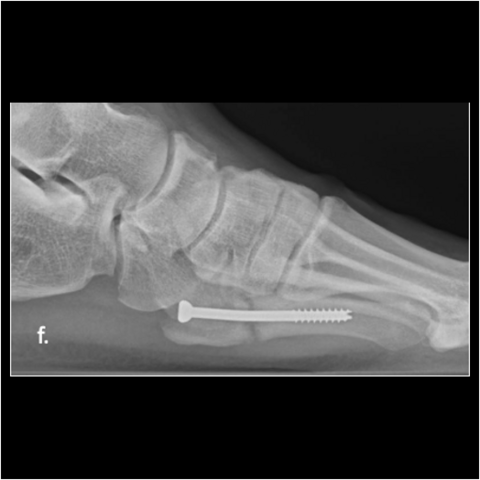
Figure 4f. The 24-year-old soldier jumped off the back of a truck, refracturing his metatarsal. Here is the foot a year post-surgery after jumping off the truck.
-
Figure 5a
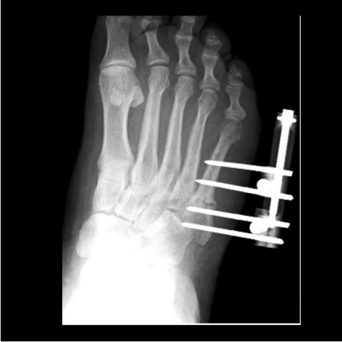
Figure 5a. The 24-year-old required hardware removal and application of external fixation for gradual compression. Most cases heal within 6 weeks with gradual compression weekly.
-
Figure 5b
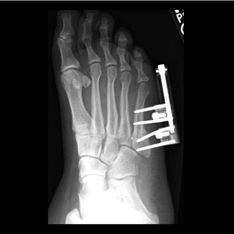
Figure 5b. The 24-year-old required hardware removal and application of external fixation for gradual compression. Most cases heal within 6 weeks with gradual compression weekly.
-
Figure 5c
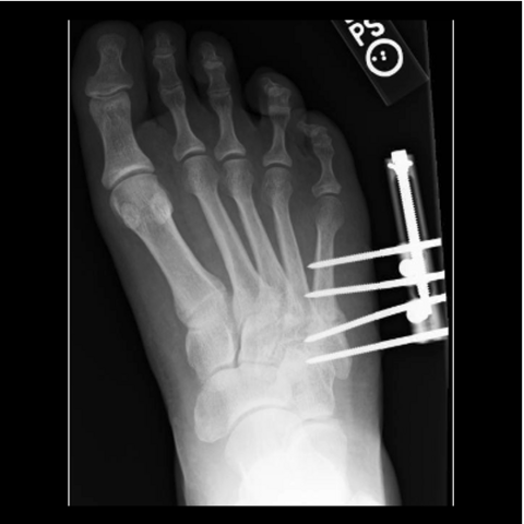
Figure 5c. The 24-year-old required hardware removal and application of external fixation for gradual compression. Most cases heal within 6 weeks with gradual compression weekly.
-
Figure 5d
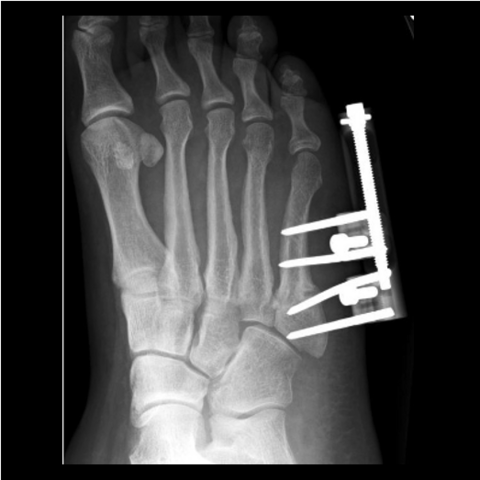
Figure 5d. The 24-year-old required hardware removal and application of external fixation for gradual compression. Most cases heal within 6 weeks with gradual compression weekly.
-
Figure 6a
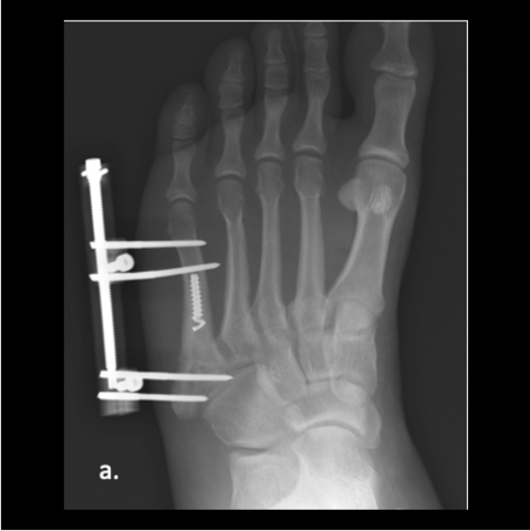
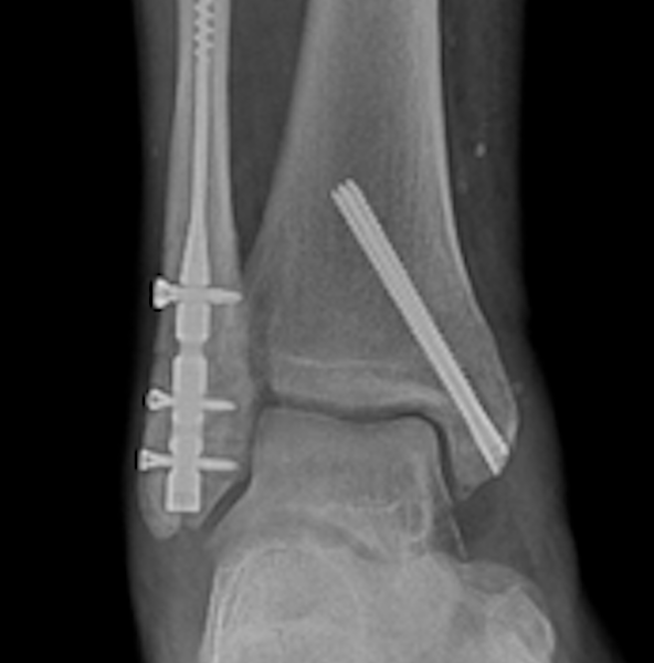
Figure 6a. A 26-year-old male soldier underwent intra-medullary screw fixation and during the surgery, his bone was so dense that the screw broke once engaged in the canal. An external fixator became the backup with gradual compression over 6 weeks. After fixator removal, the patient decided to go jogging before he was cleared and refractured. Since screw fixation was not an option, the patient refused to repeat external fixation and plate fixation was utilized. The soldier took 6 months to resume full duty and within a year required the plate to be removed. Here one can see a broken screw during surgery, ex-fix applied in surgery having to work around the broken screw.
-
Figure 6b
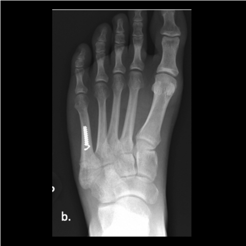
Figure 6b. Here is the foot 8 weeks post-surgery after removing ex fix and went running before being cleared leading to re-fracture. Re-injury films not shown.
-
Figure 6c
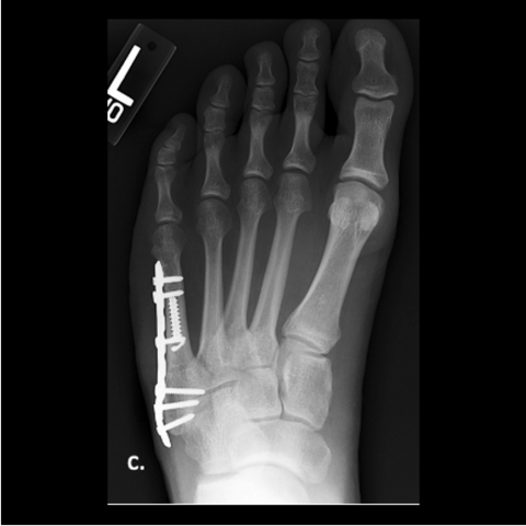
Figure 6c. Plating of the fracture since screw fixation was not possible, and the patient refused external fixation for a second time.
-
Figure 6d
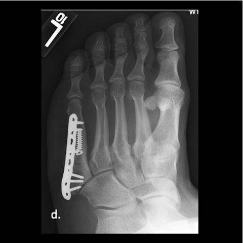
Figure 6d. Plating of the fracture since screw fixation was not possible, and the patient refused external fixation for a second time.
-
Figure 6e
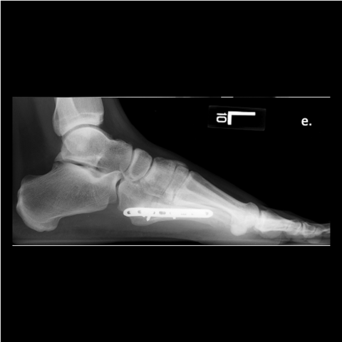
Figure 6e. Plating of the fracture since screw fixation was not possible, and the patient refused external fixation for a second time.
Click here to read a related Sports Medicine column.