Ultrasound as a Transformative Tool in Dermatology
For decades, dermatologists have relied on clinical examination, dermoscopy, and histopathology to evaluate disease above and below the skin surface. Although useful, visualization with ultrasound required a reversal to radiology. However, advances in handheld, high-frequency ultrasound technology have opened a new frontier in dermatologic diagnosis and management by allowing clinicians to visualize structures in real time, directly at the point of care.
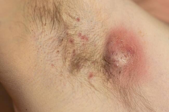
Once considered a niche imaging modality, ultrasound is increasingly finding applications across medical, surgical, and cosmetic dermatology. From assessing inflammatory skin diseases and monitoring treatment response to guiding procedures and improving surgical planning, ultrasound offers clinicians a deeper understanding of disease activity that may not be apparent on physical examination alone. Its value may be especially significant in conditions such as hidradenitis suppurativa (HS), where subclinical inflammation, fistulous tracts, and fluid collections can remain hidden beneath seemingly normal skin, contributing to delayed diagnosis and undertreatment.
Despite its growing promise, widespread adoption of dermatologic ultrasound faces challenges, including training requirements, workflow integration, reimbursement considerations, and the need for standardized protocols. Yet many early adopters believe that similar to the history of dermoscopy, ultrasound technology will evolve from a niche technology used by select experts into an indispensable component of modern dermatologic practice.
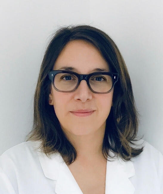
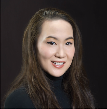
In this interview, board-certified dermatologist and Mohs surgeon Jane Yoo, MD, MPP, FAAD, FACMS, discusses the expanding role of ultrasound in dermatology, highlighting its impact on complex inflammatory diseases, particularly HS, where imaging can reveal disease severity that clinical examination alone may underestimate. She shares insights from real-world patient care, including a striking case in which ultrasound identified previously undetected HS activity during a live educational workshop. Dr Yoo also explores emerging innovations, such as elastography, Doppler imaging, and artificial intelligence (AI)-assisted interpretation, while offering practical advice for clinicians interested in incorporating ultrasound into their practices. Together, these perspectives paint a compelling picture of how ultrasound may help define the future of dermatologic care.
EXPANDING THE DERMATOLOGIC TOOLKIT
The Dermatologist: How would you characterize the role of ultrasound in dermatology today—adjunctive tool or emerging standard of care?
Dr Yoo: Handheld ultrasound has the potential to become the standard of care in dermatology. It is useful for a broad range of medical, cosmetic, and surgical indications that most dermatologists face daily. In my own practice, I will excise a melanoma on 1 patient, then manage the biologic treatment for a patient with eczema, then see another for a cosmetic consult. So, I have affectionately coined the handheld ultrasound my fourth eye (if my dermatoscope is my third eye). It is great to have additional data points to make clinical decisions, especially when you can visualize what is going on beneath the surface
The Dermatologist: Where do you see the largest clinical impact of ultrasound?
Dr Yoo: There are times when what you see on the surface does not tell the entire story; for example, in HS. Having the ability to immediately visualize deeper layers aids in diagnosis and treatment. So, ultrasound fills a much-needed gap.
The Dermatologist: What are the most compelling use cases in inflammatory or complex skin diseases?
Dr Yoo: In addition to HS, ultrasound has been helpful to track disease progression and treatment for morphea. It can detect subcutaneous involvement and fibrosis that might otherwise be missed on physical examination. In patients with psoriasis, especially those with joint symptoms, you can identify enthesitis before getting the rheumatologist involved. These are patients who would benefit tremendously from earlier, more directed interventions.
HS: A CASE STUDY IN CLINICAL VALUE
The Dermatologist: HS is often cited as a condition where ultrasound can be transformative. What does ultrasound reveal that the physical examination may miss?
Dr Yoo: The physical examination shows you the tip of the iceberg. You can visualize and palpate nodules, abscesses, and tracts. However, ultrasound can reveal tunnels and fistulous tracts beneath the surface in tissue that looks relatively normal from the skin surface. It shows you fluid collections, dermal thickening, and altered echogenicity, which are early structural changes that precede the visible fl are. Patients come in thinking they have 1 or 2 problem areas, but the ultrasound shows a much more complex picture. The patient is at a more severe Hurley stage than you think, and this alters your treatment plan.
The Dermatologist: At the 2026 Masterclasses in Dermatology Annual Meeting, you discovered undiagnosed HS disease activity in a participant during an ultrasound workshop. Can you tell us more about that experience and what key sonographic features clinicians should recognize?
Dr Yoo: We were doing a live demonstration of ultrasound with patients with HS, and I was imaging the left back of the neck of a patient who stated that she had some pain in this area. Although there were no signs of disease activity on physical examination, there were some classic sonographic findings on ultrasound, including fluid-filled cysts with posterior acoustic enhancement. This was subclinical HS that had never been identified.
The Dermatologist: How can ultrasound help reduce diagnostic delay or disease underestimation in HS and do you see it playing a role in monitoring treatment response or guiding therapeutic decisions?
Dr Yoo: HS is a condition in which there is a diagnostic delay of not months but years. The beauty of having a device with you in the office is that you can pick up disease even before it shows up on the skin. This means that ultrasound compresses the timeline dramatically and can place patients on the appropriate kinds of treatment, including biologics. It is equally powerful in monitoring treatment response because you can see fistulous tracts resolve and fluid collections decrease in size, as well as track inflammatory activity over time. The therapeutic decision-making process and patient care are significantly enhanced.
INTEGRATION AND IMPLEMENTATION
The Dermatologist: Barriers to adoption include discomfort with image interpretation and lack of training. How steep is the learning curve for dermatologists?
Dr Yoo: The learning curve can be quite steep because it is a different way of looking at skin than we are used to. When I began, I forced myself to image 1 thing daily, even on myself, until I got comfortable with it. Like any new technology, expertise takes time and patience. It took me back to the days of learning dermatopathology. When you begin, everything looks the same, but then you hone your eye to focus on the things that matter and, little by little, it starts to make sense.
The Dermatologist: Handheld, high-frequency ultrasound devices are gaining traction. How have they changed the accessibility of ultrasound in dermatology?
Dr Yoo: The technology has become far more accessible: the cost of a hand-held ultrasound device is not much more than the dermatoscope that is in every dermatologist’s white coat pocket. High-frequency probes can go up to 20 MHz and provide the necessary resolution to visualize superfi cial structures with remarkable detail. Now we need to advocate for CPT coding for handheld ultrasound to get reimbursed for the extra services we are providing for patients.
The Dermatologist: How can dermatology practices realistically incorporate ultrasound into a busy clinic setting?
Dr Yoo:Like anything else, use it strategically: presurgical planning/mapping with depth assessment, HS or psoriasis follow ups, vascular complication prevention, and filler dissolution. It still takes me some time to incorporate ultrasound into my busy workflow. But, like dermoscopy, it gets quicker over time and the improvement in patient evaluation and treatment makes it well worth it. Ultimately, it becomes something you cannot imagine practicing without.
The Dermatologist: Which patient populations or visit types are best suited for ultrasound integration?
Dr Yoo:It depends on your patient population. I see medical, surgical, and cosmetic patients in my practice. Some dermatologists are strictly cosmetic or surgical, or a combination thereof. Ultrasound can be adapted to suit the needs of your patient population, such as patients with HS, complex inflammatory disease, or soft tissue lesions where depth or character matters; patients undergoing procedures or aesthetic treatments where anatomy influences safety; and selected oncology patients where lesion depth affects planning. These are the settings where ultrasound is most likely to change treatment and management.
The Dermatologist: How do patients respond to the use of ultrasound during visits? Does it enhance understanding or engagement?
Dr Yoo: Patients are enthusiastic when they can see what is happening beneath the surface of their skin. Individuals with HS have been dismissed and undertreated for years, so it is fulfi lling to see them feel validated. It builds trust, improves engagement, and makes shared decision-making a truly wonderful experience.
THE ROAD AHEAD: WHAT IS NEXT FOR ULTRASOUND IN DERMATOLOGY?
The Dermatologist: Could ultrasound eventually rede ne how we stage or classify certain dermatologic conditions?
Dr Yoo: The Hurley Staging System is purely clinical; it does not account for subclinical fistulous tracts or early structural disease that ultrasound detects. The Sonographic Operating Score for HS and other ultrasound-based scoring systems are more sensitive and dynamic. I expect we will see ultrasound-integrated staging criteria formalized in guidelines within the next decade, not just for HS but potentially for morphea, cutaneous lymphoma, and advanced skin tumors.
The Dermatologist: What emerging applications or innovations in dermatologic ultrasound are you most excited about?
Dr Yoo: Elastography because it allows you to measure tissue stiff ness to differentiate fibrosis from active inflammation, which has huge implications for morphea and scarring diseases. New Doppler applications for monitoring vascular involvement in autoimmune conditions. And AI integration with ultrasound is already here, so there is AI-assisted image interpretation now available.
The Dermatologist: What research gaps still need to be addressed to support broader adoption?
Dr Yoo: We still need more standardization. This includes uniform scanning protocols, validated scoring systems, reproducibility data, and outcomes studies showing how ultrasound changes management and improves patient results.
The Dermatologist: What advice do you give clinicians who are considering incorporating ultrasound into their practice?
Dr Yoo: Start small and pick 1 or 2 clinical scenarios where ultrasound can add value to potentially change your management of the patient. Understand anatomy, get hands-on training through workshops and conferences, and build on the skill you already have. Most importantly, do not be afraid to get started.
Conclusion
As dermatologic ultrasound continues to evolve, clinicians do not need to become imaging experts overnight to begin realizing its benefits. Dr Yoo encourages dermatologists to start small, identifying where ultrasound can meaningfully influence patient care. Whether evaluating HS, monitoring complex inflammatory disease, planning surgical procedures, or enhancing the safety of aesthetic treatments, focused applications can help clinicians build confidence while demonstrating immediate clinical value.
Like dermoscopy before it, ultrasound requires dedicated training, repetition, and a willingness to embrace a new way of seeing disease. Hands-on workshops, anatomy-focused education, and consistent daily practice can help shorten the learning curve and accelerate proficiency. As handheld devices become more affordable and accessible, integration into routine clinical workflows is becoming increasingly feasible for a wide range of practice settings.
Looking ahead, the future of ultrasound in dermatology appears to be exceptionally promising. Emerging technologies, such as elastography, advanced Doppler imaging, and AI-assisted interpretation, have the potential to further enhance diagnostic accuracy and treatment monitoring. As research continues to establish standardized protocols, validated scoring systems, and outcomes-based evidence, ultrasound may become an increasingly important component of dermatologic care, helping clinicians detect disease earlier, personalize treatment decisions, and improve patient outcomes across medical, surgical, and cosmetic dermatology.