Using Imaging to Choose Between Endovascular Therapy and Surgical Bypass

The Iowa Clinic, West Des Moines, Iowa
During the “Diagnosis and Imaging in CLTI” session at the Amputation Prevention Symposium on Wednesday morning, Eric C. Scott, MD, of The Iowa Clinic in West Des Moines, presented a case-based discussion on how advanced vascular imaging should guide the decision between an endovascular-first or surgical bypass-first approach for patients with peripheral arterial disease and chronic limb-threatening ischemia. Rather than advocating for a single treatment strategy, Dr. Scott emphasized that selecting the optimal revascularization approach depends on a comprehensive understanding of vascular anatomy, lesion morphology, conduit availability, and disease distribution—information that can only be obtained through appropriate imaging.
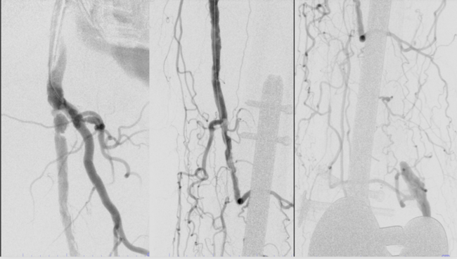
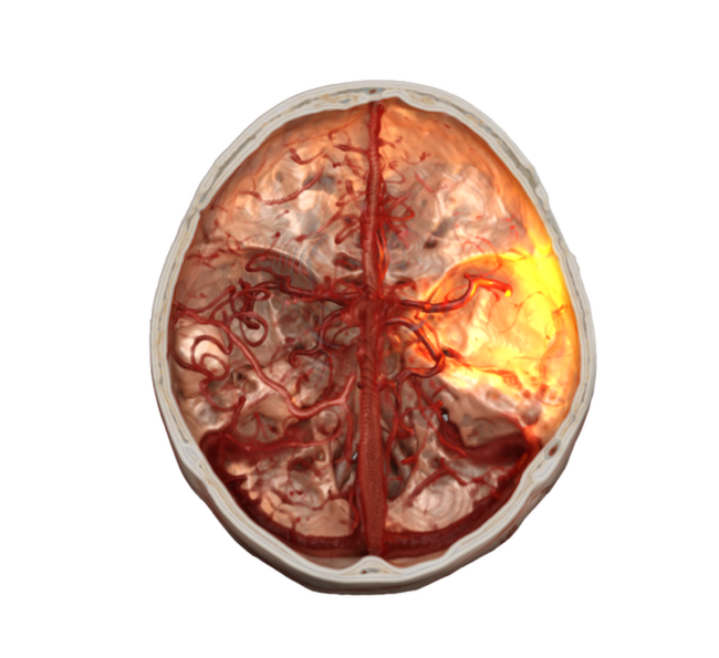
Throughout the presentation, Dr Scott demonstrated how different imaging modalities complement one another. Duplex ultrasound serves as an effective screening tool, while computed tomography angiography (CTA) provides detailed 3-dimensional (3D) visualization, allows for precise measurement of arterial diameters, and displays calcification better than any other modality, while its 3D reconstructions best display important collateral pathways. Intravascular ultrasound (IVUS), digital subtraction angiography, and operator-performed bedside ultrasound each provide additional insights that frequently alter procedural planning and improve treatment selection.
Using multiple clinical cases, Dr Scott illustrated how imaging findings can shift management toward either open surgery or endovascular intervention. For complex common femoral artery disease, particularly heavily calcified or exophytic lesions, imaging often supports femoral endarterectomy or bypass rather than aggressive endovascular recanalization. He stressed that prolonged chronic total occlusion crossing attempts may compromise long-term patency and highlighted the importance of assessing the great saphenous vein before intervention when surgical bypass remains a possibility.
The presentation also highlighted the diagnostic challenges of popliteal artery disease, where imaging plays a critical role in distinguishing atherosclerotic disease from atherosclerotic disease, thromboembolization, popliteal entrapment syndrome, cystic adventitial disease, and popliteal aneurysm. These diagnoses often require fundamentally different treatment strategies, ranging from surgical decompression or bypass to endovascular repair or conservative management depending on the underlying pathology and patient presentation.
For tibioperoneal disease, Dr Scott underscored that duplex ultrasound and CTA may be insufficient for identifying acceptable bypass targets. He described digital subtraction angiography as the gold standard for tibioperoneal and pedal imaging, and demonstrated how pedal access angiography can reveal distal target vessels not seen with conventional imaging.
The central message of the session was that imaging should not simply confirm disease but actively direct revascularization strategy. CTA, IVUS, bedside US, and pedal access angiography can enhance the understanding of patient anatomy and lesion morphology. Dr Scott concluded that the most informative imaging is often the imaging performed directly by the treating physician during procedural planning and intervention, allowing therapy to be tailored to each patient's unique anatomy and disease pattern.