






Fifth Ray Amputation: Resection Planning, Tendon Balance, and Offloading
In this interview related to her ASPS conference session, Vilayvanh Saysoukha, DPM, discusses her perspectives on considerations for fifth ray amputation, from determining the extent of resection to planning soft tissue closure and tendon management. She also shares her approach to postoperative surveillance and footwear to address future pressure points.
Key Takeaways
-
Resection planning considers the extent and location of infection, soft tissue and bone viability, imaging findings, pre-existing deformities, and prior amputations on the same foot.
-
Planning for a functional foot includes assessing soft tissue coverage and tendon balance, particularly the loss of the peroneus brevis attachment following total fifth ray amputation.
-
Follow-up includes monitoring the flap for ischemia or infection, followed by individualized footwear and offloading, with particular attention to potential pressure points at the remaining fifth metatarsal base after partial resection.
When a fifth ray amputation is being considered, what factors help determine the appropriate level of resection?
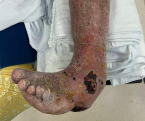
The appropriate level of resection is determined by the clinical appearance of the soft tissue and bone and correlating with magnetic resonance imaging (MRI) when possible. Another consideration is the level of osteomyelitis. Is the infection in the head, shaft, or base of the metatarsal? Does the patient have other amputations on the same foot? Are there any pre-existing deformities? For example, if the base of the fifth metatarsal is infected, a total ray resection is warranted.
How do you plan for a durable, functional foot after the procedure, particularly when it comes to soft tissue coverage, future pressure points, and tendon insertions?
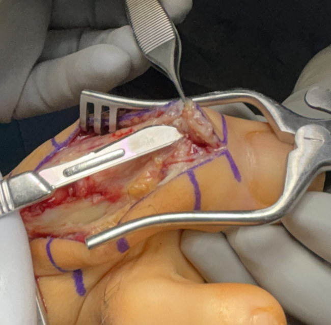
The location of the soft tissue defect from the wound determines the type of closure that can occur, if any. If enough healthy and viable soft tissue exists for primary closure, resecting more bone can make closure easier. If a total fifth ray amputation is appropriate for controlling the source of infection, one has to consider the peroneus brevis tendon losing its attachment. A varus deformity of the foot can occur after a fifth ray amputation when the patient begins to weight-bear due to the overpowering of the tibialis anterior tendon. Articles have been published on transferring the peroneus brevis tendon to the cuboid, lateral cuneiform, or even the base of the fourth metatarsal if infection is absent.1
If the cuboid is free of infection and there is no underlying rigid rearfoot deformity, I will usually perform the fifth ray amputation, tag the peroneus brevis tendon with a suture or temporarily suture the peroneus brevis tendon to the peroneus longus and place retention sutures or staples on the skin flaps. I will bring the patient back to the OR, transfer the peroneus brevis tendon to the cuboid bone with a PEEK screw, apply a skin substitute graft for soft tissue reinforcement and achieve primary closure. Utilizing a JP drain or similar can mitigate hematoma or seroma formation. Other published cases discuss a peroneus brevis and longus tenodesis.2 Anecdotally, I have heard feedback from other surgeons performing a split tibialis tendon transfer, but I cannot find any research or case study for that procedure. If a foot deformity or other pathology exists, a tibiotalocalcaneal arthrodesis may be appropriate. If the patient has had other digital or metatarsal amputations on the same foot, a transmetatarsal amputation may be best for achieving a more functional foot.
What do you watch most closely during follow-up, and how do you adjust offloading or footwear to reduce the risk of another wound?
During close follow-up, I monitor the skin flap for signs of ischemia or infection. Once the amputation flap is healed, we plan on appropriate shoe gear and bracing if necessary. The footwear consists of a double depth diabetic shoe (extra width, extra depth) and a Plastazote insert. For partial fifth ray amputations, the styloid process and the base of the fifth metatarsal can become pre-ulcerative. An accommodative Plastazote insert to offload the region is critical for reducing the risk of another wound.
Dr. Saysoukha is a quadruple-certified podiatric foot and ankle surgeon, entrepreneur, and healthcare leader focused on advancing patient care through innovation, education, and evidence-based practice. She serves as CEO and Founder of Premier Foot & Ankle Centers of Tennessee, where she leads a patient-centered organization specializing in minimally invasive surgery, limb preservation, reconstructive surgery, and wound care.
References
-
Schoenhaus J, Jay RM, Schoenhaus H. Transfer of the peroneus brevis tendon after resection of the fifth metatarsal base. J Am Podiatr Med Assoc. 2004;94(6):594-603. doi:10.7547/0940594.
-
Schweinberger MH, Roukis TS. Balancing of the transmetatarsal amputation with peroneus brevis to peroneus longus tendon transfer. J Foot Ankle Surg. 2007;46(6):510-514. doi:10.1053/j.jfas.2007.05.012.
© 2026 HMP Global. All Rights Reserved.
Any views and opinions expressed are those of the author(s) and/or participants and do not necessarily reflect the views, policy, or position of Podiatry Today or HMP Global, their employees, and affiliates.