


Treating Calcaneal Bone Cysts in Athletes
If you see teenagers and young adults in your clinic, you will likey also see calcaneal bone cysts. Although the frequency is highly debated within the literature, not a week goes by that I don’t see a bone cyst somewhere in the foot, ankle, or leg. During my 30 years of practicing with the Army, the number of patients who actually presented complaining of calcaneal body pain is zero. I have seen more pathologic fractures in soldiers than I have seen patients with pain in that specific area.
Whether calcaneal bone cysts are interosseous lipomas, unicameral, or aneurysmal probably doesn’t make that much difference to the patient, or frankly to us as the surgeon. As long as the lesion is within the cortical confines of the calcaneus then we know that the cysts are likely benign. Like most of you, I have found chondroblastomas, osteosarcomas, hemangiomas, or giant cell tumors of the calcaneus to be very rare and I have seen maybe one of each in 30 years.
I performed a yet unpublished review of all the calcaneal bone cysts (33 cases) seen over the past 6 years at Ft. Leonard Wood (Table 1). I found only one study that had more cases, with 47 in their series.1 My series had ages ranging from 8–66 years. The majority were athletes in some form, either teenagers in high school, military recruits, or active duty soldiers. Twenty-seven of the 33 were in the military. Only 2 patients actually presented with persistent heel pain and one had a pathologic fracture. All the rest were diagnosed simply because they were being X-rayed for some other condition: bunions, ankle sprain, flat feet, etc. Most tumors of the foot are found incidentally on imaging and are benign per Temple and Pruza in 2021.2 Amazingly, none of the 33 presented with plantar fasciitis with the lesions ranging from a small cyst to something quite large as shown in Figure 1. Of the 33, only 3 patients required calcaneal bone curettage and grafting. Many of the larger lesions that involved trainees who were medically discharged from service.
Pogoda in 2004 stressed that cysts reaching a critical size, defined as 100% intracalcaneal cross section in the coronal plane and at least 30% in the sagittal plane, are at risk for becoming symptomatic and at risk for fracture.1 Using guidance from authors like Pogoda, many large lesions within this series were deemed too high risk to continue training, so medical discharge was chosen, because we cannot operate on trainees electively.
As with every bone lesion seen within our clinic regardless of symptoms, patients received education on their lesion. Depending on the situation, not all lesions underwent advanced imaging. Again, magnetic resonance imaging (MRI) was preferred to computed tomography (CT) simply because the MRI assists with differentiating unicameral from aneurysmal cysts by illuminating the contents of the lesion. On occasion we would address the cyst if patients were undergoing elective surgery of that foot or ankle. Two of the 3 patients who underwent primary elective procedures received curettage and grafting. In both cases, we elected to do so simply because the size of the lesion was significant enough that with the patients’ activities as an athlete and as a soldier, they were at a much higher risk of pathologic fracture. The third patient was treated due to chronic heel pain.
Deciding on Observation, Injection, or Surgical Intervention
There are currently 3 lines of thought regarding treatment: observation, injection, and surgical intervention. When considering which course of treatment is best for an individual patient, the surgeon must consider the fracture risk without treatment, the risk of complications with treatment, and the recurrence rate with each treatment approach.3
Unfortunately, throughout the past 30 years of my practice, one only has to review the literature for all the ways to address a calcaneal bone cyst. Whether it be injections of steroids, sclerosing agents or other options, the gold standard is good old-fashioned curettage and grafting. I probably haven’t done more than 75 bone cysts/tumors in my career, I have never wavered from grafting with bone. I have never been one to utilize any of the synthetic options. Whether it be cement, ceramic or calcium products, despite hundreds of articles in support, all it takes is one bad outcome, and there are many citations to ruin your day.4-8 So I have avoided all synthetic fillers and really don’t want to dwell on any of them. I believe that if you are an experienced user and you are comfortable using a particular product, then go for it; however, if you are a novice, one should not experiment.
One of the newest trends I am very excited about is endoscopic debridement—not that patients have a lot of complaints with a 2cm incision. However, the idea of a minimal incision approach with direct visualization within the cyst sounds far more accurate than blindly curetting a hole that is often irregular in shape solely under C-arm guidance. I find it is very easy to miss sections within a multilobulated cyst.
Innami and colleagues in 2011 were among the first to promote the use of endoscopic debridement of bone cysts in athletes.9 In their series, they injected a synthetic bone filler post-debridement, finding adequate results. Yildirim in 2011 performed a comparison study between endoscopic and open, finding similar results again utilizing a synthetic filler.10 Jalan in 2021 stressed the use of autograft as having the most predictable results of grafting specifically utilizing iliac crest with their three cases.8 Amongst the literature for any and all bone cysts regardless of the calcaneus, I have observed that autograft, then allograft seems to be the preferred technique despite a plethora of citations touting synthetic fillers.
When a Teen Athlete Presents With an Asymptomatic Bone Cyst
A 15-year-old female athlete—softball, basketball, and volleyball—presented with hallux valgus and an asymptomatic bone cyst (Figure 2 and Figure 3). The patient underwent MRI evaluation (the MRIs were not available for this article). MRI revealed a fluid filled aneurysmal bone cyst involved almost the entire calcaneal body from medial to lateral. As the X-rays show, the lesion is a classic neutral triangle area with expansive size.
The family was counseled that such a large lesion would be very susceptible to pathologic fracture and with all the patient’s basketball and volleyball jumping, an injury was likely. With her already receiving Division II scholarship offers, we elected to address the cyst when we addressed her bunion surgery. The lesion was grafted after adequate curettage, which found very little contents. The lesion was grafted with finely morselized cancellous bone chips and acelluar matrix powder of mesenchymal stem cells derived from pig intestine. She was followed 4 years and is now playing DII softball pain-free.
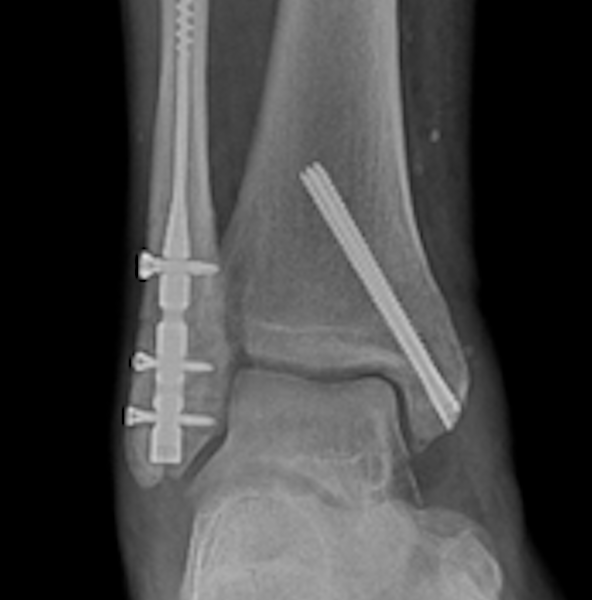
A Case of a Soldier With a Unicameral Bone Cyst
A 24-year-old male active duty soldier presented with chronic heel pain, not from his bone cyst but his Haglund’s deformity (Figure 4, Figure 5 and Figure 6). The soldier underwent MRI, which showed that the lesion was filled with fat indicative of a unicameral bone cyst. Like the last case, the lesion involved the entire neutral triangle from medial to lateral and plantar to dorsal.
I recommended that we address this at the same time as his Haglund’s surgery. The lesion was debrided, finding almost no material or fluid within the lesion. The lesion was grafted using the same technique. The patient was followed 18 months until he moved to another assignment. He resumed full duty and resumed running pain-free.
What Happens After Inadequate Debridement and a Lack of Graft Incorporation?
A 19-year-old Marine with persistent heel pain (Figure 7 and Figure 8). He underwent curettage and bone grafting by another provider. He left Ft. Leonard Wood after 4 months to Japan. The Marine was fairly pain-free until he was over a year post-surgery having increasing heel pain especially since he was back to full running and sports. Repeated CT and MRIs were performed, which clearly showed inadequate debridement and lack of graft incorporation.
It is of course very easy to look backwards and see that the lesion was not sufficiently debrided. Within the figures 7–8, I have highlighted the cortical margins, which are still easily visible postoperatively. MRI 16 months later clearly shows the cortices were not sufficiently decorticated and an internal layer of tissue is still present. The Marine required repeated curettage and grafting. The failure here seemed to be in the preparation, not with the grafting.
Key Points on Calcaneal Bone Cysts
It is very critical to follow calcaneal cyst patients as long as possible—at least 12–18 months—to ensure full bone incorporation in cases where allograft or synthetic fillers have been utilized. Synthetic products will often take far less time to consolidate, I find, and if complications occur they will do so within the first 6 months.
Regardless of the type of cyst found, it is very critical to curette the bone down to bleeding bone. In some cases, a lining of tissue may be present. Failure to remove any tissue inside can and will lead to recurrence or worse resorption of the grafting material. Often, I utilize a small K-wire or burr to denude the inner cortical rim of the lesion. This step is very important, I find, to promote revascularization and incorporation of the bone; otherwise you will not see complete incorporation of the graft and development of normal trabecular patterns as shown in Case #3.
Whether you utilize a window or endoscopic technique to gain entrance, in my experience, there is far too much emphasis on restoring the window. I simply take a drill bit, make a window, and am not concerned about closing the window. In some cases, the window may help allow any bleeding or drainage that can occur post-curettage to leak outward into a drain/hemovac.
Pathology reports are probably not as critical, I feel, especially in cases in which the contents are scarce. If and when there are significant tissue contents, pathology specimens are imperative.
In Conclusion
Bone cysts are rarely symptomatic but always problematic as they grow in size. Regardless of type, the potential for pathologic fracture is always present amongst our athletes and military. Evaluation and treatment are routinely an elective process, but when the lesion is large enough, it is very hard to neglect especially when you are already addressing that limb surgically.
A. Douglas Spitalny, DPM is a Staff Podiatrist at General Leonard Wood Army Hospital in Ft. Leonard Wood, MO.
References
1. Pogoda P, Priemel M, Linhart W, et al. Clinical relevance of calcaneal bone cysts: a study of 50 cysts in 47 patients. Clin Orthop Relat Res. 2004 Jul;(424):202-10.
2. Temple EW, Prusa RD. Calcaneal bone tumors. Clin Podiatr Med Surg. 2021 Apr;38(2):227-233.
3. Gottlich C, Fisher JC, Campano D, Diab M. Management of calcaneal cysts in the pediatric population: a review. J Am Acad Orthop Surg Glob Res Rev. 2023 Mar 10;7(3):e22.00248.
4. Hoshi M, Oebisu N, Iwai T, et al. Possible pathogenesis of calcaneal bone cysts. Arch Orthop Trauma Surg. 2020 Oct;140(10):1303-1310.
5. Saraph V, Zwick EB, Maizen C, Schneider F, Linhart WE. Treatment of unicameral calcaneal bone cysts in children: review of literature and results using a cannulated screw for continuous decompression of the cyst. J Pediatr Orthop. 2004 Sep-Oct;24(5):568-73.
6. Hoshi M, Iwai T, Oebisu N, Shimatani A, Takada N, Nakamura H. Pathological fracture of a solitary bone cyst in the calcaneus: a case series and literature review. Arch Orthop Trauma Surg. 2023;143(3):1155-1162.
7. Glaser DL, Dormans JP, Stanton RP, Davidson RS. Surgical management of calcaneal unicameral bone cysts. Clin Orthop Relat Res. 1999;(360):231-7.
8. Jalan D, Gupta A, Elhence A, Nalwa A, Bharti JN, Elhence P. Primary aneurysmal bone cyst of the calcaneum: A report of three cases and review of literature. Foot (Edinb). 2021;47:101795.
9. Innami K, Takao M, Miyamoto W, Abe S, Nishi H, Matsushita T. Endoscopic surgery for young athletes with symptomatic unicameral bone cyst of the calcaneus. Am J Sports Med. 2011 ;39(3):575-81.
10. Yildirim C, Akmaz I, Sahin O, Keklikci K. Simple calcaneal bone cysts: a pilot study comparing open versus endoscopic curettage and grafting. J Bone Joint Surg Br. 2011 Dec;93(12):1626-31.
11. Nishimura A, Matsumine A, Kato K, et al. Endoscopic versus open surgery for calcaneal bone cysts: a preliminary report. J Foot Ankle Surg. 2016;55(4):782-7.


