What Are These Pigmented, Pedunculated Lesions?
Case Report
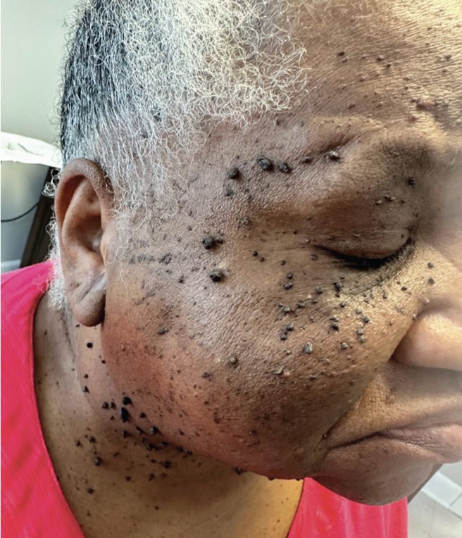
A 60-year-old African American woman presented to the clinic with multiple hyperpigmented, raised papules distributed across the face and neck. The lesions had been present for several years, progressively increasing in number with age. The patient was otherwise healthy, with no associated symptoms or cosmetic concerns. On physical examination, numerous well-circumscribed, pedunculated, hyperpigmented papules measuring 1 mm to 5 mm in diameter were noted on the face and neck. Additionally, a solitary flesh-colored, cystic lesion on the neck was clinically consistent with a benign epidermoid cyst. Given the asymptomatic nature of the lesions, the patient declined any intervention at this time.
What is your diagnosis?
Scroll below to find out!
Diagnosis: Dermatosis Papulosa Nigra
Dermatosis papulosa nigra (DPN) is a benign cutaneous condition that predominantly affects individuals of African descent, although it has also been reported among Filipino,Vietnamese, European, and Mexican individuals.1 A strong genetic predisposition has been demonstrated, with familial inheritance observed in more than 50% of cases.1 The condition disproportionately affects women, with a reported 2:1 female-to-male ratio, and both lesion count and size increase with age.2 In the United States, approximately one-third of African American individuals present with DPN, with lower prevalence among those with lighter skin pigmentation.1 Onset typically occurs during adolescence, with lesions progressively increasing in number and size over time. A quality-of-life study found that DPN moderately affects patients, often causing embarrassment and self-consciousness.3
Clinical Presentation and Histology
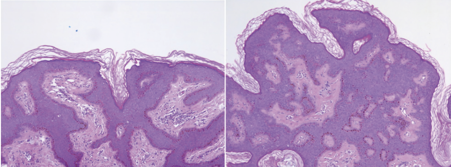
Clinically, DPN presents as hyperpigmented or flesh-colored papules on the face and neck that are frequently symmetrically distributed, most often in individuals with darker skin types (Fitzpatrick III–VI).1,4 Histopathologic features include irregular acanthosis, papillomatosis, hyperkeratosis, and keratin-filled invaginations of the epidermis.1 Elongated, interconnected reteridges with basal layer hyperpigmentation are characteristic, while papillomatous acanthotic structures often demonstrate a prominent fibrous stroma¹ (Figure 2). The papules are histologically indistinguishable from small seborrheic keratoses (SK) and are frequently associated with fibroblast growth factor receptor 3 (FGFR3) mutations in sun-exposed skin. However, the presence of enlarging patches rather than multiple small papules makes SK a less likely diagnosis in this context. Some investigators argue that DPN should be considered a clinical variant of SK, given these histologic similarities.2
Pathogenesis
While the precise molecular etiology of DPN remains unclear, it is thought to be caused by a defect in the nevoid development of the pilosebaceous follicle.4 Ultraviolet radiation has also been suggested as a potential contributing factor, possibly promoting cellular proliferation in genetically predisposed individuals.5
Activating mutations in FGFR3 and alterations in the phosphatidylinositol-4,5-bisphosphate 3-kinase (PI3K) pathway have been implicated in lesion development, although no single causative gene has been defi nitively identifi ed.6 Oncogenic FGFR3 mutations have similar pathogenesis to SK in that protein kinase B activation leads to inhibition of p53-mediated apoptosis, causing keratinocyte accumulation in growing lesions.2
Although no studies have confirmed it, hormonal involvement is possible and may play a role in regulating cell division.2 Estrogen interaction with FGFR3 could amplify contributing factors that lead to DPN, which may help explain its predilection for women.2 Further investigation is needed to explore this potential link.
Differential Diagnosis
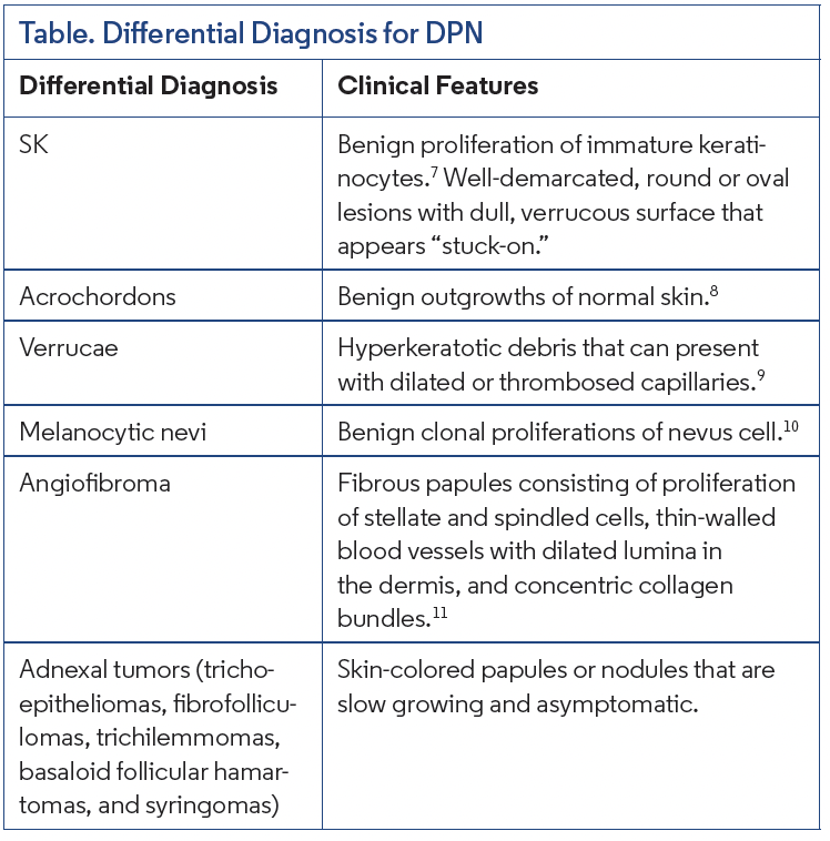
DPN must be distinguished from several other benign cutaneous conditions, including SK, acrochordons (skin tags), verrucae, melanocytic nevi, angiofibromas, and adnexal tumors7-11 (Table).
Clinical Associations With DPN
A pigmented transverse nasal band has been associated with DPN,as well as other skin conditions.12 Systemic associations have also been reported. In a retrospective study of 55 patients with DPN, 72.7% were found to have cardiovascular disease (CVD).13 Lesion localization was influenced by the presence of CVD, with diffuse or proliferative forms showing stronger associations.13 Although causality has not been established, these findings suggest DPN may serve as a cutaneous marker for systemic disease in certain populations.
While rare, paraneoplastic associations have been described. One case reported a female patient presenting with eruptive DPN lesions in a “Christmas-tree” distribution who was subsequently diagnosed with colon adenocarcinoma.14 This presentation resembled the Leser-Trélat sign. If DPN represents a variant of Leser-Trélat, it could indicate a novel paraneoplastic syndrome, with possible associations not only with gastrointestinal adenocarcinomas but also hematologic malignancies, such as leukemia and lymphoma.15
Treatment and Complications
Treatment of DPN is generally elective and pursued for cosmetic concerns. Common therapeutic modalities include snip excision, light curettage with or without anesthesia, and light electrodesiccation.1 Cryotherapy is less favored due to the risk of hypopigmentation. Potential complications of lesion removal include scarring, dyspigmentation, and secondary infection. While ablative fractional carbon dioxide laser therapy has been investigated as a treatment modality, its efficacy and long-term outcomes require further study.1 Although there are higher risks of complications, such as infection, erythema, scarring, and dyspigmentation, a case presented by Ali et al. advocated for the treatment of DPN with CO2 laser due to higher patient satisfaction and a low recurrence rate.16
Our Patient
After clinical diagnosis, the patient was offered shave removal with electrodesiccation, but she declined any intervention. She was counseled regarding the benign nature of her condition, possible cosmetic treatment options, and the low but potential risks of recurrence and pigmentary changes if lesions were removed.
Conclusion
DPN is a benign epidermal neoplasm characterized by hyperpigmented or skin-colored papules primarily affecting the face and neck. Although histologically similar to SK, DPN preferentially affects individuals with darker skin phototypes and demonstrates a strong genetic predisposition, predominant in women. Diagnosis is primarily clinical and may be supported by dermoscopic evaluation, which typically reveals features such as irregular acanthosis, papillomatosis, and epidermal hyperkeratosis. Given its benign nature, intervention is typically reserved for aesthetic concerns.
Continued study of the molecular mechanisms underlying DPN, particularly the roles of FGFR3 mutations, PI3K pathway activation, and hormonal modulation, may provide new insights into its pathogenesis and management. Further research is warranted to elucidate potential systemic associations and optimize therapeutic strategies.
© 2026 HMP Global. All Rights Reserved.
Any views and opinions expressed are those of the author(s) and/or participants and do not necessarily reflect the views, policy, or position of the Dermatology Learning Network or HMP Global, their employees, and affiliates.
Dr Henry is a postdoctoral research scholar at the University of Iowa in Iowa City, IA. Dr Khachemoune is a dermatologist at Premier Dermatology in Ashburn, VA, and the Derm DX section editor.
Disclosure: The authors report no relevant financial relationships.
References
1. Xiao A, Muse ME, Ettefagh L. Dermatosis papulosa nigra. In: StatPearls [Internet]. StatPearls Publishing; 2023.
2. Metin SA, Lee BW, Lambert WC, Parish LC. Dermatosis papulosa nigra: a clinically and histopathologically distinct entity. Clin Dermatol. 2017;35(5):491-496. doi:10.1016/j.clindermatol.2017.06.001
3. Uwakwe LN, Souza BDE, Subash J, McMichael AJ. Dermatosis papulosa nigra: a quality-of-life survey study. J Clin Aesthet Dermatol. 2020;13(2):17-19.
4. Leung AKC, Barankin B, Lam JM, Leong KF. An atlas of lumps and bumps, part 4. Consultant. 2021;61(5):e20-e22. doi:10.25270/con.2021.05.00001
5.Taylor SC, Averyhart AN, Heath CR. Postprocedural wound-healing efficacy following removal of dermatosis papulosa nigra lesions in an African American population: a comparison of a skin protectant ointment and a topical antibiotic. J Am Acad Dermatol. 2011;64(3 Suppl):S30-S35. doi:10.1016/j.jaad.2010.11.009
6. Qiu CC, Brown AE, Lobitz GR, Shanker A, Hsu S. The color of skin: black diseases of the skin, nails, and mucosa. Clin Dermatol. 2019;37(5):447-467. doi:10.1016/j. clindermatol.2019.08.003
7. Hafner C, Vogt T. Seborrheic keratosis. J Dtsch Dermatol Ges. 2008;6(8):664-677. doi:10.1111/j.1610-0387.2008. 06788.x
8.Banik R, Lubach D. Skin tags: localization and frequencies according to sex and age. Dermatologica. 1987;174(4):180-183. doi:10.1159/000249169
9.Bae JM, Kang H, Kim HO, Park YM. Diff erential diagnosis of plantar wart from corn, callus and healed wart with the aid of dermoscopy. Br J Dermatol. 2009;160(1):220-222. doi:10.1111/j.1365-2133.2008. 08937.x
10. Frischhut N, Zelger B, Andre F, Zelger BG. The spectrum of melanocytic nevi and their clinical implications [published correction appears in J Dtsch Dermatol Ges. 2022;20(7):1054]. J Dtsch Dermatol Ges. 2022;20(4):483-504. doi:10.1111/ddg.14776
11.Macri A, Kwan E, Tanner LS. Cutaneous angiofi broma. In: StatPearls [Internet]. StatPearls Publishing; 2022
12. Zawar V, Daga S, Pawar M, Kumavat S. Pigmented transverse nasal band: a distinct presentation. J Cosmet Dermatol. 2019;18(1):301-302. doi:10.1111/jocd. 678
13. Mongo S, Macaire B, Kouikani F, Letomo K, Kombo B, Mbolla B. Papulosa nigra dermatosis another factor associated with cardio-vascular diseases? World J Cardiovascular Diseases. 2023;13(12):819-826. doi:10.4236/wjcd.2023.1312070
14. Duncan N, Usatine RP, Heath CR. Key features of dermatosis papulosa nigra vs seborrheic keratosis. Cutis. 2025;115(2):70-71. doi:10.12788/cutis.1170
15. Schwartzberg JB, Ricotti CA Jr, Ballard CJ, Nouri K. Eruptive dermatosis papulosa nigra as a possible sign of internal malignancy. Int J Dermatol. 2007;46(2):186187. doi:10.1111/j.1365-4632.2007.02767.x
16. Ali FR, Bakkour W, Ferguson JE, Madan V. Carbon dioxide laser ablation of dermatosis papulosa nigra: high satisfaction and few complications in patients with pigmented skin. Lasers Med Sci. 2016;31(3):593-595. doi:10.1007/s10103-016