What Is the Cause of This Color Change on the Neck?
Case Report
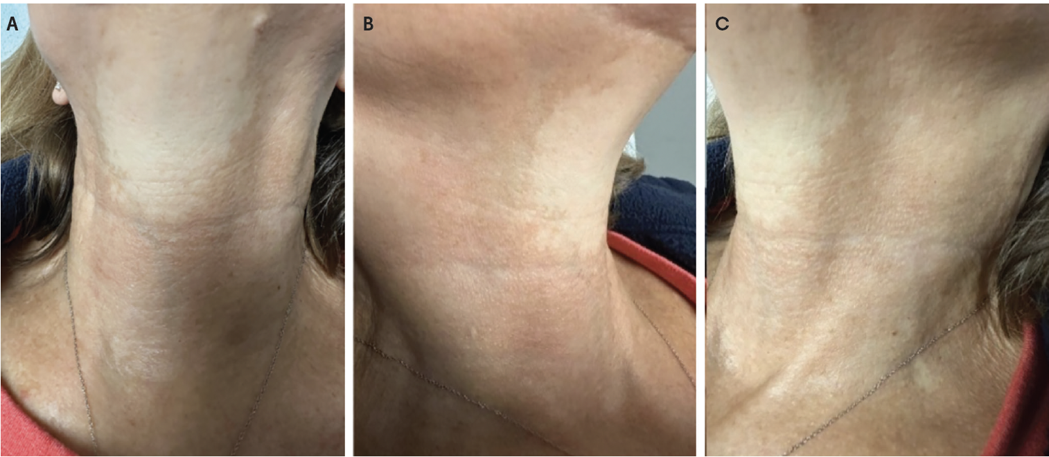
A 60-year-old woman presented with a several-year history of progressive color change on the neck. Past medical history was notable for nonmelanoma skin cancer; she was otherwise healthy. She did not report any drug use or family history. Dermatologic examination revealed confluent, reticulated, reddish-brown patches with central hypopigmented atrophic areas on the lateral aspects of the neck, with relative sparing of the submental triangle (Figure 1 A–C).
What is your diagnosis?
Scroll below to find out!
Diagnosis: Poikiloderma of Civatte
Poikiloderma of Civatte (PC) is a chronic dermatologic condition that pertains to a category of melanodermas.1 Most commonly seen in middle-aged or older individuals, PC is a benign disorder characterized by a combination of vascular and pigmentary changes, most notably mottled erythema, hyperpigmentation, telangiectasia, and epidermal atrophy. Typically occurring symmetrically in sun-exposed areas, particularly the V of the chest and the lateral aspects of the cheeks and neck, sparing of the submental triangle is regarded as a highly distinctive characteristic of PC.2 Although its precise etiopathogenesis remains incompletely understood, chronic ultraviolet exposure is regarded as the principal causative factor. However, several additional contributors have been implicated, including lighter skin phototypes (Fitzpatrick I– III); exposure to photosensitizing agents, such as cosmetics or fragrances; hormonal influences, particularly decreased estrogen levels; and potential genetic susceptibility.
Clinical Presentation
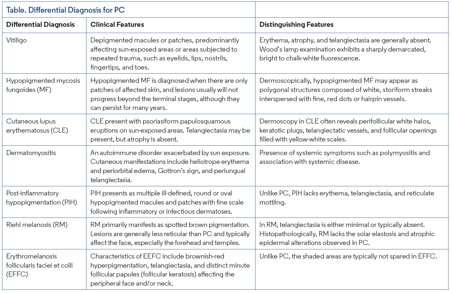
Clinically, PC presents as confluent, reticulated, reddish-brown patches with central hypopigmented atrophic areas. It is clinically categorized into erythemato-telangiectatic, pigmented, and mixed types, determined by the predominant characteristic, with the mixed type being the most frequently documented presentation.2 The coexistence of epidermal atrophy, telangiectasia, and hyperpigmentation is particularly helpful in distinguishing PC from other disorders in the differential diagnosis (Table). PC is generally asymptomatic, although some patients may report mild burning, pruritus, episodic flushing, or increased skin sensitivity. While diagnosis is usually clinical, a biopsy may occasionally be required to confirm the diagnosis or exclude other conditions. However, dermoscopy yields a distinctive image that aligns closely with histologic findings, frequently exhibiting a predominant reticular (fishnet-like) pattern, observed in 53.6% of cases. Key dermoscopic features include linear, irregular vessels; rhomboidal/polygonal vessels; dotted/globular vessels; and distinctive, converging, curved vessels, sometimes described as “flying-seagull-like,” which may be characteristic of the erythemato-telangiectatic type.2
Histopathology
The histopathologic findings are distinctive yet not pathognomonic. The most significant and consistent characteristic, deemed the sine qua non finding, is marked solar elastosis of the papillary dermis, which is separated from the epidermis by a grenz zone.3,4 Other characteristic findings include dilated superficial dermal vessels (telangiectasia), mild epidermal atrophy (the epidermis is often flattened and thin, with effacement of the rete ridges), and dermal melanophages.3 Epidermal changes also frequently include hyperkeratosis. The presence of dermal melanophages saturated with melanin, due to melanin incontinence, and irregularly distributed melanin granules in the lower epidermis are also distinctive features.3,4
Management
As it is considered a dermatologic condition rather than a disease, active medical intervention is not routinely required. The focus of management is primarily on preventive measures; essentially, strict photoprotection and the avoidance of potentially sensitizing agents, including perfumes and cosmetics.3 When cosmetic improvement is desired, various laser and light-based therapies may be employed. The therapeutic management of PC remains challenging, yet the optimal intervention should address both the vascular and pigmented components simultaneously. Options include intense pulsed light (IPL), pulsed dye laser (PDL), and potassium titanyl phosphate laser.3 Furthermore, to address textural components and photodamage, fractionated ablative and nonablative lasers can be used. IPL improves dyspigmentation and telangiectasia. While PDL is highly effective for the vascular component, caution is warranted when treating the cervical skin, a delicate area prone to atrophy and scarring. Therefore, the application of low fluences (≤5 J/cm2 on a 10-mm spot size) is recommended to reduce the risk of severe, persistent depigmentation.3,5 The 577-nm pro-yellow laser has emerged as a promising and safe option for PC, effectively targeting both hemoglobin and melanin chromophores.5,6 Topical agents, such as hydroquinone and retinoids, may serve as adjunctive therapy, especially for the pigmented subtype.
Our Patient
In this case, the patient presented with reticulated hyperpigmented macules and patches on the lateral and posterior aspects of the neck, with relative sparing of the submental region. A clinical diagnosis of PC was established based on these characteristic findings. Histopathologic examination is generally not required when the presentation is typical. The patient was counseled that PC is a benign dermatologic condition and not associated with systemic morbidity. We explained that its pathogenesis is multifactorial but predominantly related to chronic ultraviolet exposure, with potential contributory factors, including photosensitizing agents such as fragrances. Management strategies, including photoprotection and optional therapeutic interventions, were reviewed. Potential treatments discussed included vascular and pigment-targeting laser modalities, topical depigmenting agents such as hydroquinone, topical corticosteroids, and topical retinoids. The patient declined active treatment. She was advised to use broad-spectrum sunscreen daily and avoid applying perfume directly to the affected areas.
Conclusion
PC is a chronic dermatologic condition characterized by redbrown, reticular hyperpigmented patches distributed over sunexposed areas, most commonly the lateral neck, cheeks, and upper chest, with relative sparing of the submental triangle. Its true prevalence is difficult to determine, as individuals with mild disease may not seek medical attention. The condition is attributed to long-term ultraviolet-induced photodamage and occurs most frequently in postmenopausal women. Patient counseling should emphasize rigorous photoprotection, including broad-spectrum sunscreen use and physical barriers. Therapeutic approaches primarily involve light- and laser-based modalities.
References
1. Lautenschlager S, Itin PH. Reticulate, patchy and mottled pigmentation of the neck. Acquired forms. Dermatology. 1998;197(3):291-296. doi:10.1159/000018016
2. Katoulis AC, Sgouros D, Bozi E, et al. Diagnosis and differential diagnosis of poikiloderma of Civatte: a dermoscopy cohort study. Dermatol Pract Concept. 2023;13(1):e202307. doi:10.5826/dpc.1301a7
3. Katoulis AC, Rigopoulos D, Tzima K, Stavrianeas NG. Poikiloderma of Civatte: a review. Expert Rev Dermatol. 2014;7(4):377-382. doi:10.1586/edm.12.34
4. Katoulis AC, Stavrianeas NG, Panayiotides JG, et al. Poikiloderma of Civatte: a histopathological and ultrastructural study. Dermatology. 2007;214(2):177-182. doi:10.1159/000098580
5. Sarac G, Kapicioglu Y, Cenk H. A new treatment option for poikiloderma of Civatte: 577 nm pro-yellow laser. J Cosmet Dermatol. 2022;21(1):316-319. doi:10.1111/jocd.14609
6. Temiz SA, Ataseven A, Dursun R, Özer İ. Successful treatment of poikiloderma of Civatte with a 577-nm pro-yellow laser. J Cosmet Dermatol. 2020;19(10):27692770. doi:10.1111/jocd.13650