Surgeon-Performed Localization and the Push for Ultrasound Training in Modern Breast Cancer Treatment
Please share your name, title, and a brief overview of your professional history.
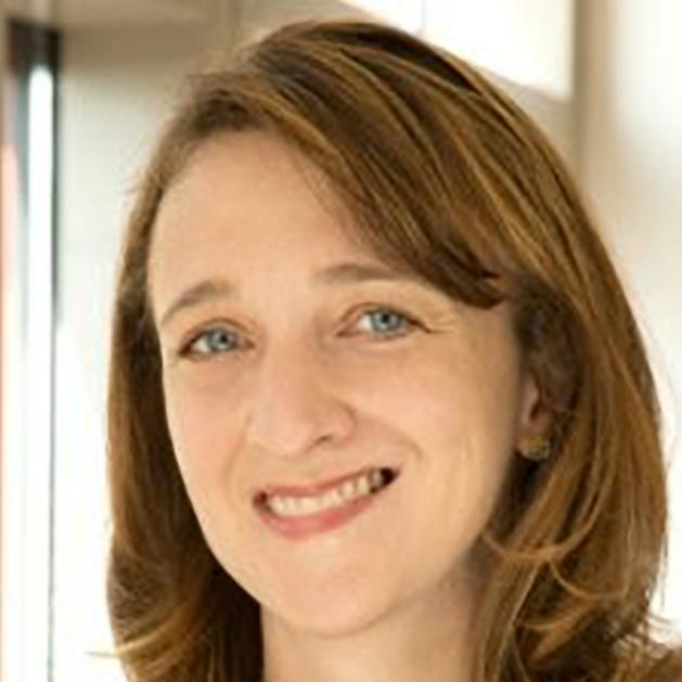
 Nimmi S. Kapoor, MD: My name is Nimmi S. Kapoor, and I’m an associate professor of surgery at David Geffen School of Medicine, University of California Los Angeles (UCLA). After earning an undergraduate degree at UCLA, I went to medical school at Weill Cornell Medical College and stayed in New York for a general surgery residency at NewYork-Presbyterian Hospital—Cornell, where I also had invaluable training at Memorial Sloan Kettering Cancer Center. While at Cornell, I spent 2 years conducting translational research in endocrine surgery and breast surgical oncology. After 11 years in NYC, I returned to California to complete a breast surgical oncology fellowship at John Wayne Cancer Center and Cedars-Sinai Medical Center.
Departing from my heavy academic training, I took my first job with RadNet at Breastlink, a community-based multidisciplinary center consisting of breast surgeons, medical oncologists, radiologists, and plastic surgeons. It was here, not in my academic training, that I truly gained skills in breast ultrasound from my mentor, Dr John West. Dr West taught me how to scan the breast and how to do vacuum-assisted core needle breast biopsies. Soon, I was able to start performing ultrasound-guided intraoperative breast tumor localizations as well. Tumor localization is a necessary step when performing lumpectomy, or breast-conserving surgery, for small breast cancers and other lesions. Typically, and throughout all of my academic training, radiologists perform the localization procedure before surgery in an awake patient under local anesthesia. Fortunately, these skills did not take long to acquire and were very patient-centric as patients could have the procedure during their breast cancer surgery while asleep, rather than awake with radiology.
When RadNet asked me to help them open a Breastlink site in San Fernando Valley, I had no idea just how valuable my breast ultrasound skills would be. The hospitals I would serve, only 8 to 12 miles from UCLA, lacked some basic infrastructure, including specimen mammography, on-site radiologists, and breast-dedicated radiologists. Surprisingly, I found it not only patient-centric but also safest to start performing most tumor localizations myself so I could prevent errors that I was noticing when performed through the current local systems. After spending 7 years improving the local infrastructures and advancing patient care systems, it was not lost on me that the skills I learned with Dr West were not learned in any of my otherwise rigorous training environments and that I needed to find a way to bring these essential breast ultrasound skills to academic training so that all breast surgeons could be equipped with them.
I joined UCLA in 2023 with the mission to improve the way breast surgery is taught and practiced, and since then I have been doing just that.
Please share a brief overview of your research exploring whether surgeon-performed intraoperative wire localization allows for lower margin positivity rates compared to radiologist-performed preoperative localization in early breast cancer.
Dr Kapoor: Fortunately, many of my colleagues at UCLA were interested in expanding their breast ultrasound skills, but they first wanted more data. We decided to do a few retrospective studies to make sure surgeon-performed intraoperative wire localization was safe and at least as effective as standard radiologist-performed preoperative localization.
What were some of the key findings of your research? Did anything surprise you and your colleagues?
Dr Kapoor: The first study evaluated margin positivity and complications after surgeon-performed intraoperative wire localization. In 137 patients with breast cancer, only 7.3% of patients had positive margins, and most complications were minor seromas. This was much better than the national averages of margin positivity after breast conservation, which ranged from 15% to 20% in contemporary studies. As a result, our research team did a comparative study and identified consecutive patients with unifocal breast cancer who underwent wire-localization either with standard radiologist-performed preoperative wire localization (POWL) or with intraoperative surgeon-performed intraoperative ultrasound-guided wire localization (IOWL). We expected to find similar results in margin positivity between the groups, thinking that we were all breast-dedicated surgeons with similar training, removing similar “shave-cavity” margins, and with excellent breast-dedicated radiologists performing the localizations.
Instead, we found striking differences between the 2 groups, with the IOWL group having significantly lower positivity than the POWL group at 7.1 % vs 23.9 %, respectively, P = 0.002. This significance remained even after adjusting for key variables associated with margin positivity, such as presence of DCIS, tumor size, and volume of tissue removed (aOR 0.34; 95 % CI, 0.13-0.93). Importantly, there was also a decrease in the rate of margin re-excision between the groups (5.9 % vs 18.5 %, IOWL vs POWL, respectively; P = 0.011).
Were there any notable differences in operative time, workflow efficiency, or patient satisfaction between IOWL and POWL approaches?
Dr Kapoor: Good question. Since the comparative study was retrospective and we could not capture metrics of workflow efficiency or patient satisfaction, we opened a prospective clinical survey study of “dual localization,” where we have patients undergo standard preoperative localization and then surgeons attempt intraoperative localization. This allows for a “back-up” method of localization in case a surgeon is unable to locate the lesion. Preliminary, unpublished patient-survey response data reveal that all patients undergo some level of burden with preoperative localization, including metrics in time, pain, and suffering. Surgeon-response preliminary survey data show that 100% of lesions were able to be localized with ultrasound, and most intraoperative localizations took less than 5 minutes to perform.
Do you foresee challenges or limitations in implementing IOWL more widely, especially in institutions without access to intraoperative ultrasound or trained surgical personnel?
Dr Kapoor: Yes, there are many institution-specific challenges with implementing IOWL. These challenges range from training and ultrasound access to logistical workflow and “turf wars” between surgery and radiology. We have outlined some of these challenges and ways to approach the many hurdles that surgeons may face in adopting IOWL in “The Evolution of Breast Ultrasound in Surgical Practice: Current Applications, Missed Opportunities, and Future Directions.” In the meantime, at UCLA, we developed a successful cadaver-based course to teach breast ultrasound-guided procedures to our residents, fellows, and breast surgeons, and now have an annual course called “Breast Ultrasound, Intra-operative Wire Localization, and Oncoplastic Skills Course for Surgeons” available at UCLA for any surgeon to come and learn breast ultrasound skills.
Although numerous courses—such as those from the American College of Surgeons, the American Society of Breast Surgeons, and now UCLA—offer opportunities to develop breast ultrasound skills, I believe it is crucial that every breast surgery training program provides access to ultrasound in both the clinic and the operating room. Additionally, we need more surgeon educators proficient in these fundamental techniques to effectively train our future surgeons. These changes will benefit both patients and surgeons, which is why I remain committed to advocacy.
© 2025 HMP Global. All Rights Reserved.
Any views and opinions expressed are those of the author(s) and/or participants and do not necessarily reflect the views, policy, or position of the Journal of Clinical Pathways or HMP Global, their employees, and affiliates.