






Beyond Negative Diagnostics: The Role of Clinical Judgment in Limb Salvage Surgery
This case examines how surgeons can integrate clinical progression, patient-specific risk, and inconclusive diagnostic findings when determining whether urgent intervention is warranted.
Key Takeaways
-
No single laboratory, imaging, microbiological, or operative finding can independently confirm or exclude every clinically significant diabetic foot infection.
-
Discordance between a concerning clinical presentation and negative or inconclusive diagnostic findings requires continued reassessment rather than reliance on any one result.
-
When the potential consequences of delay are substantial, operative exploration may have both diagnostic and therapeutic value—but the decision should reflect the patient’s complete clinical picture and individualized risk.
Diagnostic testing can narrow clinical uncertainty, but it does not always resolve it. In patients with diabetes and a rapidly evolving inflammatory presentation, foot and ankle surgeons may face a consequential question: Should they continue to pursue diagnostic confirmation, or intervene before a potentially limb-threatening process progresses? This case explores how clinical findings, patient-specific risk, and inconclusive diagnostic results informed the decision to proceed with operative exploration.
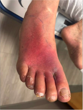
A 71-year-old male with a history of type 2 diabetes mellitus, hypertension, and hyperlipidemia presented to the hospital with progressive right foot pain, erythema, swelling, fatigue, and subjective constitutional symptoms for approximately 3 days.
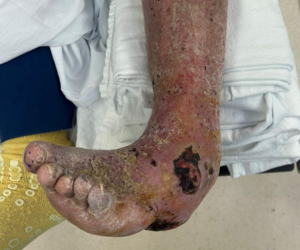
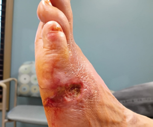
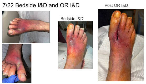
Vitals included a blood pressure of 147/76, pulse was 100 bpm, respirations were 23/minute, and oxygen saturation was 98%. The patient was afebrile. Initial evaluation demonstrated a marked systemic inflammatory response, including significant leukocytosis, elevated C-reactive protein, elevated erythrocyte sedimentation rate, and hyperglycemia. Physical examination localized the inflammatory process to the right first web space and dorsal forefoot, where diffuse erythema, tenderness, and swelling raised concern for an evolving deep space diabetic foot infection (Image 1). Pedal pulses were nonpalpable and protective sensation was diminished. Given the patient’s diabetes, significant inflammatory response, and rapidly progressive clinical findings, we could not safely exclude the possibility of a limb-threatening infection despite the absence of definitive diagnostic confirmation.
Initial diagnostic evaluation was comprehensive. Plain radiographs demonstrated extensive vascular calcifications and an incidental retained radiopaque foreign body within the plantar-lateral heel, while computed tomography confirmed its location without demonstrating communication with the clinically involved first web space. Because the foreign body was anatomically separated from the region of maximal inflammation, its contribution to the patient’s presentation remained uncertain. Nevertheless, the combination of progressive local findings over only 3 days and significant systemic inflammation prompted urgent surgical consultation due to concern of risk for tissue loss, osteomyelitis, systemic deterioration, and ultimately major amputation. During his hospital course, vascular and infectious disease teams also contributed.
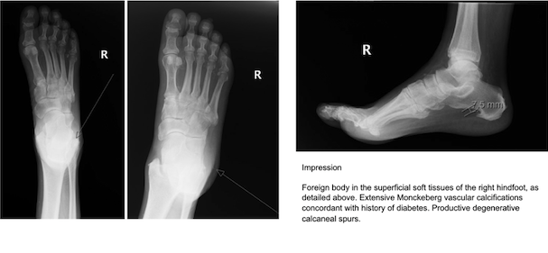
Initial incision and drainage took place at bedside. Urgent operative exploration of the first web space was undertaken one day after admission based on the patient’s overall clinical presentation rather than on imaging findings alone. Contrary to preoperative concern, exploration failed to identify purulent material, a drainable abscess, gross tissue necrosis, or obvious deep extension of infection. However, an insulin needle was found in the bottom of his foot, not in the area of the first webspace. The operative site was thoroughly irrigated and left open to facilitate continued wound management with negative-pressure wound therapy. Subsequent magnetic resonance imaging demonstrated no evidence of osteomyelitis or residual drainable abscess, and microbiological cultures remained negative throughout the patient's hospitalization. Despite an extensive diagnostic workup that included serial clinical examinations, laboratory investigations, multimodal imaging, operative exploration, local wound care and microbiological analysis, no definitive etiology explaining the patient's inflammatory presentation was identified.
The patient was hospitalized for 2 weeks, and there was an attempt at hyperbaric oxygen therapy, which the patient did not desire to continue with. Thus, his eventual discharge was with negative pressure wound therapy and follow up with the podiatric surgeon in one week. His inflammatory markers decreased significantly, and his overall clinical condition improved. The wounds demonstrated gradual healing, and healing is still in progress at time of publication.
Thoughts on the Key Clinical Challenges
Rather than diminishing the significance of the case, these findings represent a vital educational contribution. The central challenge in this case was not diagnosing a rare organism or identifying an unusual imaging abnormality, but determining the appropriate management of a patient whose clinical presentation strongly suggested a limb-threatening diabetic foot infection while every major diagnostic modality failed to provide definitive confirmation. This discordance between clinical assessment and objective diagnostic findings is rarely emphasized in the literature despite representing a scenario likely encountered by many foot and ankle surgeons.
The evaluation of diabetic foot infections has evolved considerably over the past several decades. Contemporary management incorporates laboratory investigations, advanced imaging, microbiological cultures, histopathological evaluation when appropriate, and standardized clinical classification systems to improve diagnostic accuracy. Collectively, these tools have significantly enhanced the ability to identify osteomyelitis, deep soft tissue infection, septic arthritis, and other limb-threatening complications. However, no individual diagnostic modality possesses sufficient diagnostic accuracy to independently establish or exclude clinically significant diseases in every patient. Diagnostic uncertainty therefore remains an inherent aspect of diabetic foot surgery rather than an uncommon exception.
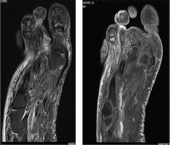
This case demonstrates that negative diagnostic studies should not automatically be interpreted as evidence that clinically significant pathology is absent. Although postoperative MRI demonstrated no osteomyelitis or drainable abscess, operative exploration revealed no purulence, and microbiological cultures remained negative, these findings did not invalidate the initial clinical concern that prompted emergency intervention. Instead, we feel that they illustrate the limitations of individual diagnostic modalities when interpreted independently. Each investigation contributed valuable information, yet none fully explained the patient's presentation.
From a surgical perspective, the decision to proceed urgently to the operating room was justified by the patient’s overall risk profile rather than by any single diagnostic finding. Patients with diabetes mellitus are particularly vulnerable to rapid progression of deep infection because impaired immune function, peripheral neuropathy, and microvascular disease may obscure early manifestations while accelerating tissue destruction. In this context, we felt that waiting for unequivocal diagnostic confirmation may have exposed the patient to unacceptable risk. Surgical exploration therefore functioned as both a therapeutic intervention and a diagnostic procedure, excluding several limb-threatening conditions that could not be confidently ruled out by noninvasive testing alone.
Perhaps the most important lesson of this case is that clinical judgment remains the cornerstone of surgical decision-making when diagnostic certainty cannot be achieved. Laboratory studies, advanced imaging, microbiological cultures, and operative findings are invaluable tools that refine diagnostic reasoning, but they cannot replace careful bedside assessment, serial clinical evaluation, and an appreciation of disease progression. Evidence-based surgery requires thoughtful integration of all available information rather than reliance on any single diagnostic result.
Ultimately, this case reinforces a principle that extends well beyond diabetic foot surgery: the purpose of diagnostic testing is to reduce uncertainty, not to eliminate it. When objective investigations and the patient's clinical condition appear discordant, surgeons must integrate all available evidence while recognizing the potentially devastating consequences of delayed intervention. In high-risk patients, timely operative exploration may remain the most appropriate course of action even when comprehensive diagnostic evaluation fails to establish a definitive diagnosis. The broader implication is that successful limb salvage surgery depends not only on advanced diagnostic technology, but also on sound clinical judgment, careful risk assessment, and the willingness to act decisively when the patient's condition warrants intervention, despite persistent diagnostic uncertainty.
Drs. Russell, Saleem, and Frias are all first-year residents at Morristown Medical Center in Morristown, NJ.
The authors have no non-financial or commercial, proprietary, or financial interest in the products or companies described in the manuscript. The author(s) did not receive grants or a consultant in honorarium to conduct the study, write the manuscript, or otherwise assist in the development of the above-mentioned manuscript.
© 2026 HMP Global. All Rights Reserved.
Any views and opinions expressed are those of the author(s) and/or participants and do not necessarily reflect the views, policy, or position of Podiatry Today or HMP Global, their employees, and affiliates.