A Practical Alternative to NPWT in Rural Limb Preservation: A Case Report
This case report highlights the use of a regulated oxygenated negative pressure-assisted dressing (RO-NPT) as a potential viable, cost-conscious solution for complex wound management.
Key Takeaways
1. RCAD may offer a practical NPWT alternative in low-resource settings. The case demonstrates that rapid capillary action dressings can deliver comparable wound healing benefits when traditional NPWT devices are unavailable or cost-prohibitive.
2. This modality aims to support wound healing through multilayer capillary transport. RCAD supports granulation tissue formation, reduces exudate, and maintains an optimal wound environment—key factors in promoting healing.
3. Features of RCAD include simplified application. The technique uses accessible materials and straightforward application steps, making it an option for clinicians working in rural or resource-constrained environments.
Lower extremity ulceration remains a major driver of morbidity, healthcare cost, and limb loss, particularly among patients with diabetes, neuropathy, peripheral arterial disease, and complex multimorbidity.1-4 Negative pressure wound therapy (NPWT) is among the most frequently utilized advanced modalities in wound care. Mechanistically, NPWT removes exudate, reduces edema, decreases bacterial burden, and produces microdeformation at the wound surface, collectively supporting granulation tissue formation and wound contraction.5-7 Clinical literature, including systematic reviews and meta-analyses in diabetic foot ulcer populations, supports improved healing rates and reduced amputation risk for NPWT compared with conventional dressings.8-10
Although NPWT is an effective modality choice for complex and exudative wounds, practical barriers unrelated to clinical education often limit its use in rural settings. Home health availability in rural communities may be inconsistent, insufficient, or even unavailable to support the frequent dressing changes, device troubleshooting, and patient monitoring required for NPWT. In addition, logistical challenges related to equipment delivery, supply replacement, and timely canister or dressing changes can delay initiation or interrupt therapy. These barriers may result in prolonged wound stagnation or necessitate alternative treatment strategies despite the theoretical suitability of NPWT. Consequently, clinicians practicing in rural environments must often rely on advanced dressing technologies that provide effective exudate control without dependence on device-based infrastructure. Rural hospitals and clinics with limited administrative infrastructure or without durable medical equipment (DME) licensing capacity present billing and equipment distribution challenges.11-13 These real-world barriers create a gap between “ideal” wound therapy and what is achievable for patients living far from specialty centers.
Rapid capillary action dressings (RCADs) represent a class of advanced wound dressings designed to manage moderate-to-heavy exudate through multilayer capillary transport rather than powered suction.14,15 These dressings utilize hydrophilic fiber architectures to continuously draw wound fluid away from the wound bed and redistribute it into absorbent layers, thereby reducing periwound maceration, stabilizing the wound environment, and supporting autolytic debridement.15-17 By maintaining a controlled moisture balance without reliance on mechanical components, alarms, or powered devices, RCADs address several practical limitations associated with traditional NPWT, particularly in outpatient, rural, and resource-limited settings.
Vacutex (Protex Healthcare) is one such RCAD. Its mechanism, as described generally above, enables sustained exudate management and maintenance of a stable wound environment without reliance on pumps, alarms, tubing, or canisters, thereby potentially reducing staff burden and dependence on home health infrastructure.15,16 Clinical reports and case series have described its use in managing difficult and nonhealing wounds, demonstrating effective exudate control, support of granulation tissue formation, and favorable patient tolerance in both inpatient and outpatient settings.18 Manufacturer technical descriptions further characterize the dressing as generating a consistent low-level pressure gradient through continuous capillary fluid transfer, approximating NPWT-like effects without mechanical suction.15
Although RCADs do not generate true suction-mediated microdeformation, their ability to provide continuous fluid transfer, protect the wound bed, and maintain a stable healing environment has led to increasing interest in their use as a pragmatic alternative when NPWT is unavailable, delayed, or impractical.14,17 This approach may be particularly relevant in rural healthcare systems, where durable medical equipment access, administrative constraints, and workforce limitations frequently create a gap between ideal wound care strategies and real-world feasibility.11-13
This case report describes the use of this particular RCAD in a rural practice setting for an ulceration that would have been ideal for NPWT but NPWT could not be obtained due to local DME constraints. The wound responded rapidly with improved exudate control, robust granulation tissue formation, and closure over a short time course, with high patient satisfaction.
Case Summary
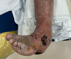
A 61-year-old male with insulin-using type 2 diabetes mellitus, peripheral polyneuropathy, venous insufficiency, hyperlipidemia, and gastroesophageal reflux disease presented to the emergency department with an acute onset of a left foot infection. The patient reported a 3-day history of a rapidly worsening ulceration over the lateral aspect of the left fifth metatarsal head, accompanied by increasing pain, swelling, erythema, and lymphangitic streaking extending proximally. He endorsed feeling generally unwell but denied fever, chills, nausea, vomiting, chest pain, or shortness of breath. Despite attempted self-care at home, the ulceration and surrounding cellulitis progressed, prompting urgent evaluation after clinicians identified the ulcer as grossly infected during an outpatient clinic visit earlier the same day.
On examination, the patient had bilateral palpable dorsalis pedis and posterior tibial pulses with brisk capillary refill. The team noted moderate nonpitting edema and increased temperature in the left foot, ankle, and lower leg. Neurologic assessment demonstrated diminished protective sensation consistent with diabetic peripheral neuropathy. Dermatologic examination revealed diffuse erythema of the left lower extremity with streaking. An ulceration at the lateral fifth metatarsal head measured 4.5 × 2.3 × 0.5 cm, with exposed bone, tendon, and fascia with purulent drainage and local signs of infection. The wound had irregular borders, moderate exudate, and no tunneling or undermining. The ulceration was exquisitely painful on examination.
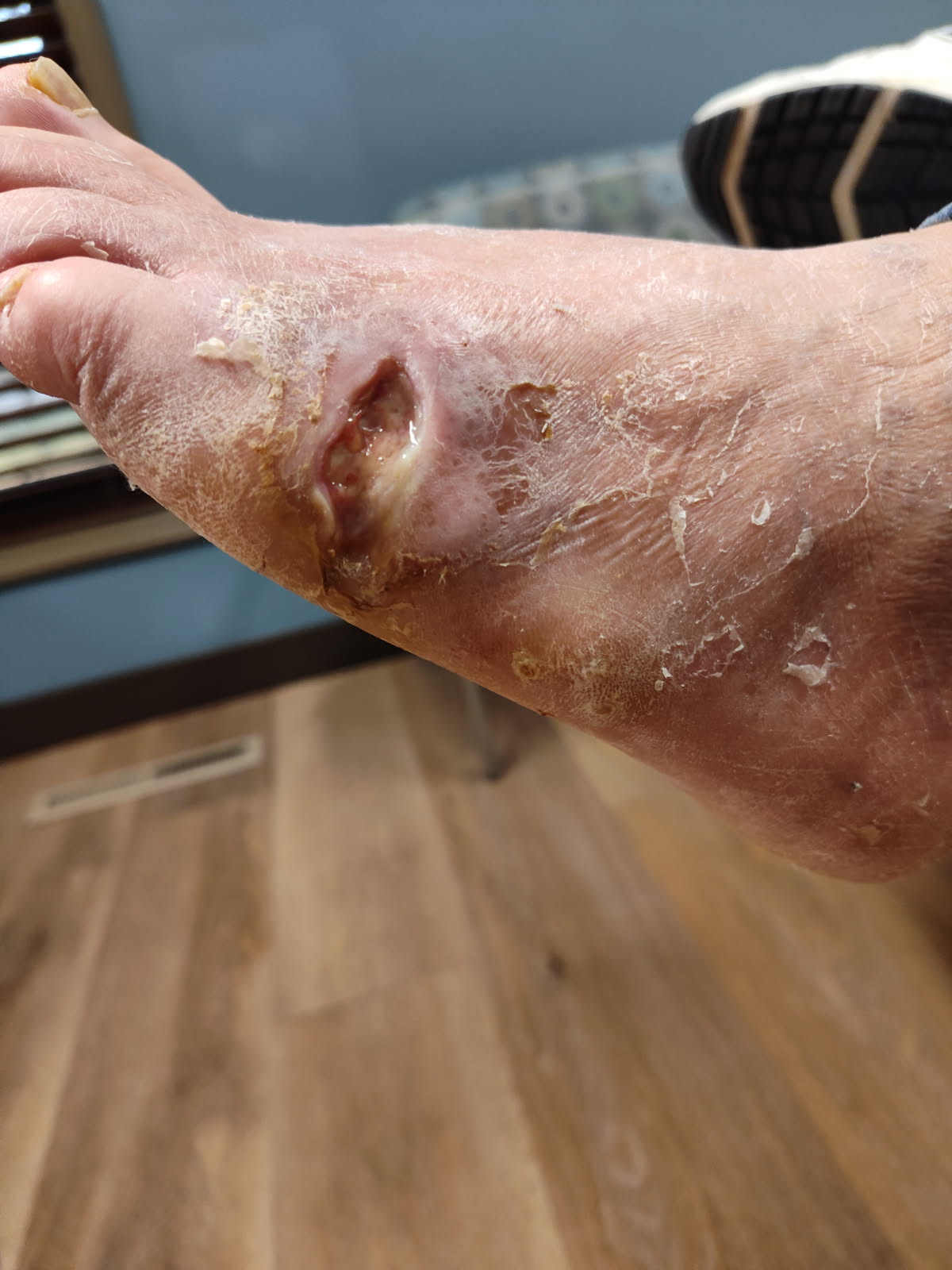
Laboratory evaluation demonstrated leukocytosis (white blood cells 14.0 ×10³/µL) and significantly elevated C-reactive protein (17.0 mg/dL), consistent with acute infection. Plain radiographs of the left foot showed no evidence of soft tissue gas, acute osseous abnormality, or radiographic osteomyelitis. The patient underwent bedside irrigation and sharp excisional debridement with evacuation of infected and nonviable tissue, and collection of a deep wound culture swab was obtained. Vital signs were within normal limits with no tachycardia, hypotension, fever, or respiratory compromise to suggest sepsis. The patient was discharged after ED treatment with intravenous antibiotics (vancomycin) due to overall clinical stability and absence of criteria for inpatient admission. At time of disposition, the patient was alert, hemodynamically stable, and without persistent hypotension, hypoxia, or altered mental status. Although the patient did have leukocytosis and an elevated C-reactive protein (CRP), lactic acid was normal, procalcitonin was below the threshold concerning for bacterial sepsis, and there was no evidence of end‑organ dysfunction. Imaging demonstrated no osteomyelitis, abscess, gas, or other acute surgical pathology.
The patient demonstrated clinical stabilization following IV fluids, vancomycin, antipyretics, and electrolyte repletion. Given the lack of severe sepsis or shock, ability to tolerate oral antibiotics, and availability of close outpatient podiatry follow‑up with clear return precautions, the team deemed outpatient management appropriate.
A bone biopsy did not take place during the ED or early post‑ED course due to the absence of radiographic evidence of osteomyelitis, the patient’s clinical stability, and lack of indication for emergent operative intervention. Although bone exposure was present, exposure alone does not mandate immediate biopsy in the absence of supportive imaging or failure of initial therapy. ED imaging demonstrated only nonspecific soft‑tissue edema without abscess, gas, or osseous destruction.
Debridement performed in the ED and subsequent outpatient visits included excision of nonviable soft tissue, with no intentional osseous resection and no bone fragments removed. Initial management focused on source control and antimicrobial therapy, including use of oral antibiotics with known bone penetration (doxycycline). We considered bone biopsy a next‑step diagnostic option to pursue if the patient failed to improve, if imaging later demonstrated osteomyelitis, or if surgical intervention became indicated.
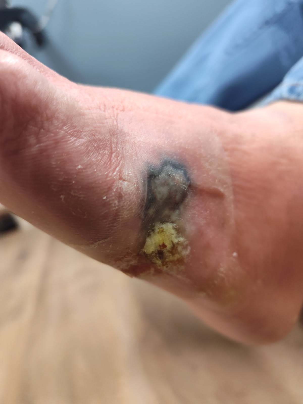
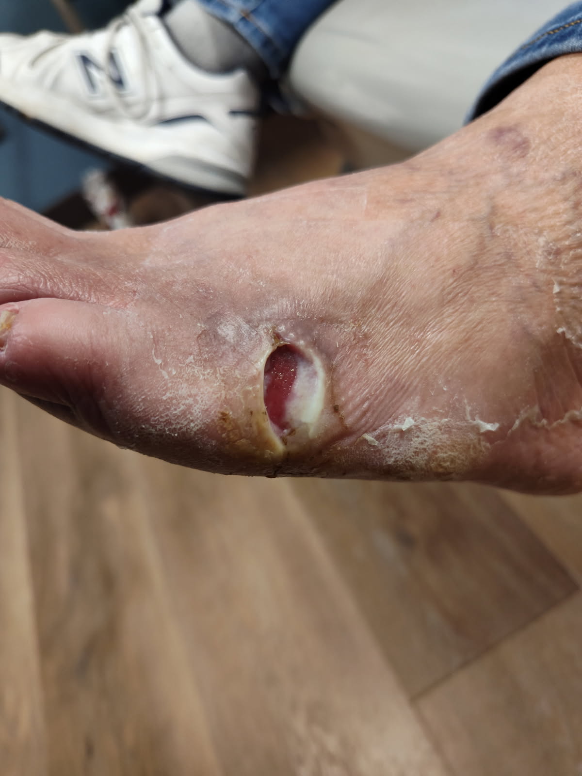
At the initial follow-up 1 week after being discharged from the emergency department, the ulcer at the plantar-lateral aspect of the left fifth metatarsal neck measured 3.2 × 2.4 × 0.4 cm and demonstrated a complex wound bed with exposed periosteum and fascia, along with approximately 40% fibrotic tissue and scant serous drainage; the patient reported pain rated 5/10. All obvious signs of local infection had resolved.
During the follow-up we cleansed the wound with chlorhexidine soap, thoroughly rinsed with sterile saline, and sharply debrided to remove all nonviable tissue. Following debridement, we gently crosshatched the wound bed and wound edges with a scalpel until achieving pinpoint bleeding, with the goal of stimulating local perfusion and promoting granulation tissue formation.
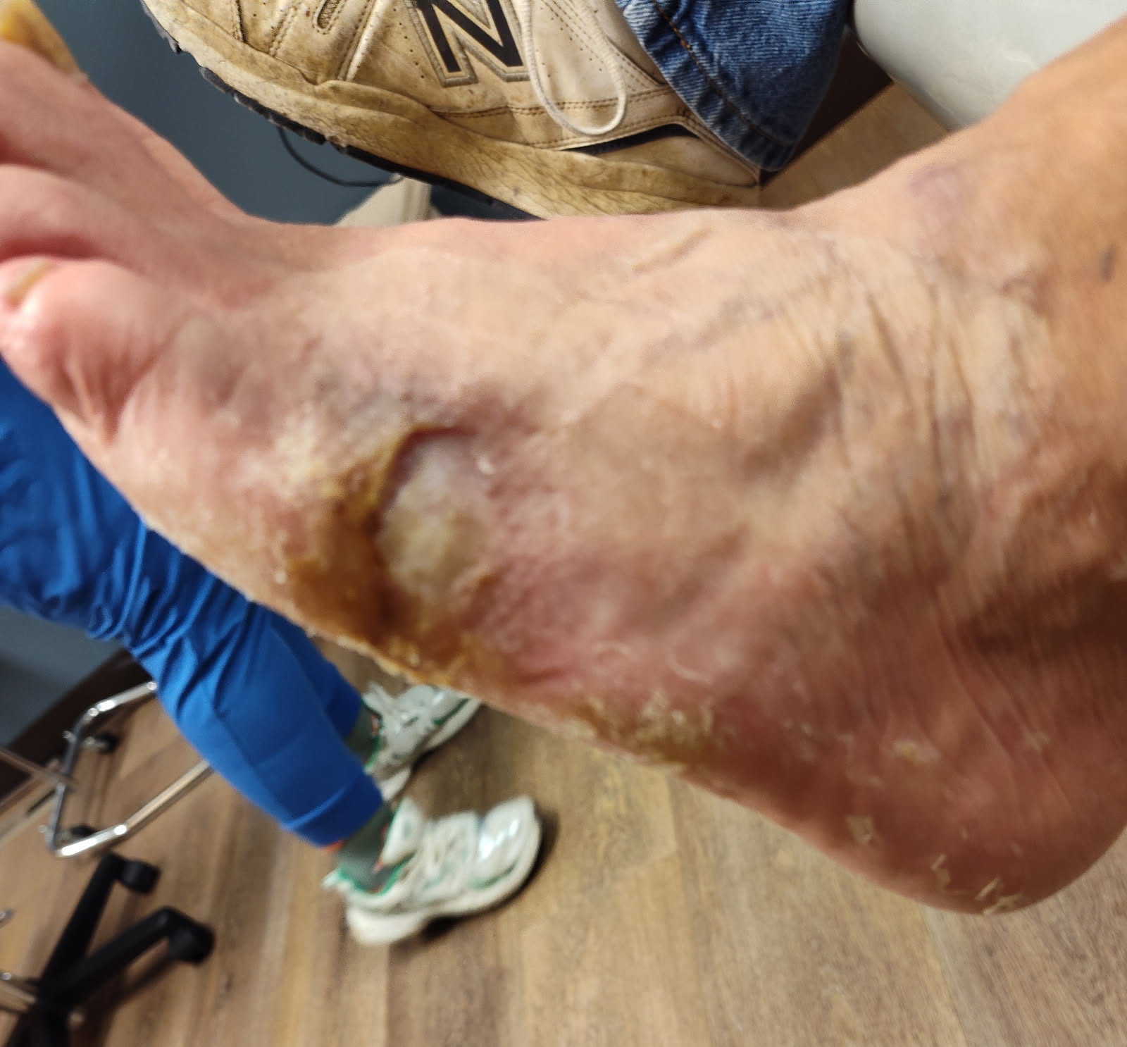
Given the presence of exposed periosteum and deep soft tissue structures following debridement, we considered negative pressure wound therapy (NPWT) as a preferred modality to promote granulation tissue formation and manage wound exudate. However, NPWT was not available in this rural hospital setting due to institutional and logistical limitations, including the inability to provide outpatient NPWT through local durable medical equipment pathways. As a result, we instead initiated a rapid capillary action dressing as an alternative advanced wound care strategy with goals including support of exudate control, protecting the wound bed, and facilitating granulation tissue development in the absence of device-based therapy. We applied this dressing directly to the wound bed and secured it in place with dry sterile dressings. Offloading recommendations included a controlled ankle motion (CAM) boot and crutches.
The patient was to perform dressing changes at home every 72 hours. We saw him weekly, during which time we cleansed wound, sharply debrided of all nonviable tissue, and mechanically stimulated the wound bed and margins via crosshatching prior to reapplication of advanced dressings.
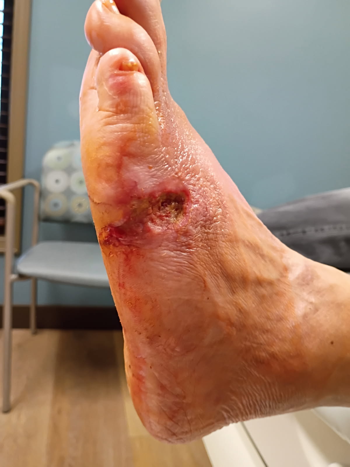
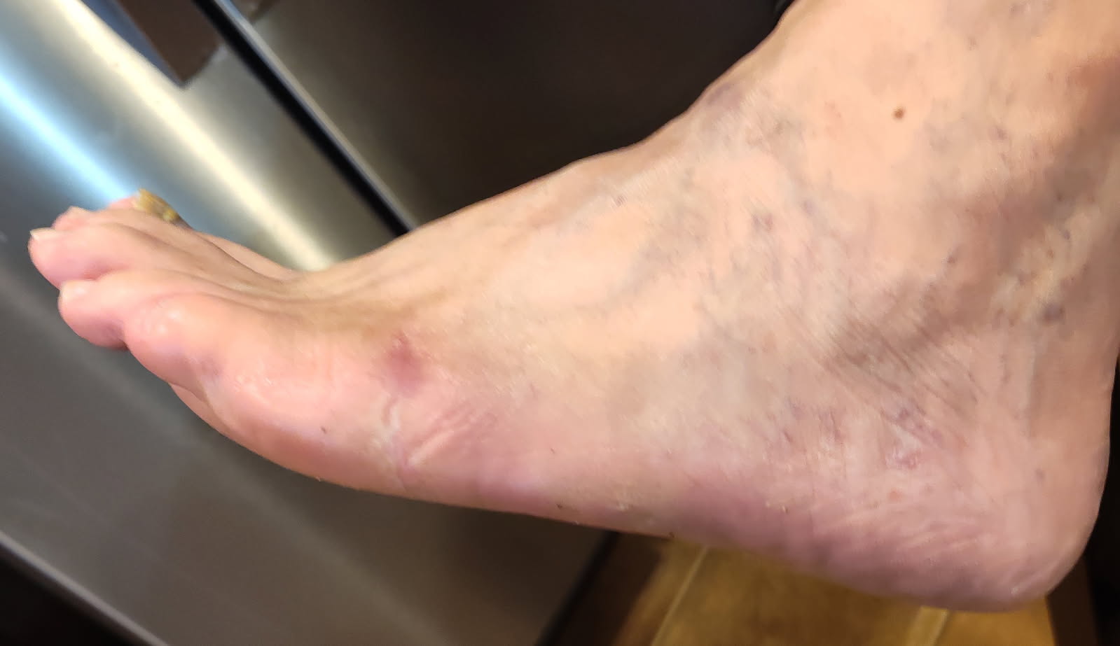
Two weeks following the initial emergency department visit, the wound demonstrated a substantial reduction in size to 1.9 × 1.1 × 0.2 cm, with marked improvement in tissue quality, including progressive granulation tissue formation over previously exposed structures and reduced pain (2/10). Improvement continued 4 days later with further contraction to 1.6 × 0.9 × 0.2 cm and a fully granular wound bed without residual exposure of deep structures. The next week, the ulcer measured 1.2 × 0.7 × 0.1 cm, remained 100% granular, and was pain-free with no drainage or signs of infection. By the following week, the wound had nearly completely epithelialized, measuring 0.6 × 0.2 × 0.1 cm, with a healthy granular base, intact periwound skin, and complete resolution of pain, drainage, and local inflammatory signs. Six weeks after the initial emergency department visit, the wound had fully closed.
Discussion
Negative pressure wound therapy (NPWT) has become a cornerstone in the management of complex lower-extremity wounds, particularly those characterized by significant exudate, irregular wound geometry, and exposure of deep structures such as tendon, fascia, or bone. In the present case, the ulceration demonstrated multiple features traditionally considered ideal indications for NPWT, including exposed periosteum and deep soft tissue following debridement, moderate exudate burden, and a need for rapid granulation tissue formation to facilitate wound closure. Under ideal circumstances, NPWT would have been a reasonable escalation strategy to support wound bed preparation and healing progression.
However, this case highlights a common and underreported reality in rural wound care settings: access to NPWT is not always determined by clinical need alone. In many rural hospitals and outpatient practices, NPWT implementation is constrained by limited home health availability, supply chain delays, and institutional barriers related to durable medical equipment (DME) licensing, billing infrastructure, and device distribution. Even when NPWT is clinically indicated, these logistical challenges may delay initiation or preclude its use entirely, potentially prolonging wound stagnation and increasing the risk of complications. As such, rural clinicians are often required to identify alternative strategies that can approximate key therapeutic goals of NPWT without reliance on device-based infrastructure.
Rapid capillary action dressings (RCADs) represent one such alternative. In this case, we specifically selected this option to address the dual challenges of exudate management and granulation over exposed deep structures in the absence of NPWT availability. Following initiation of RCAD therapy, the wound demonstrated rapid and sustained improvement, with progressive granulation tissue coverage of previously exposed periosteum and fascia, consistent reduction in wound dimensions, and eventual complete epithelialization within approximately 6 weeks. Importantly, this favorable clinical trajectory occurred without recurrent infection, despite the severity of the initial presentation.
The shared-care model employed in this case may also be particularly relevant to rural practice. The patient performed dressing changes at home every 72 hours, reducing dependence on home health services, while weekly in-clinic visits allowed for wound cleansing, serial sharp debridement, and mechanical stimulation of the wound bed and margins. This approach balanced patient autonomy with clinician oversight and may represent a pragmatic framework for advanced wound management in settings where frequent professional dressing changes are not feasible.
While the findings of this case cannot establish equivalence between RCADs and NPWT, they suggest that RCADs may serve as a viable alternative in selected patients when NPWT is unavailable, delayed, or impractical. This is especially pertinent in rural healthcare environments, where structural barriers frequently limit access to advanced device-based therapies. Further prospective studies comparing RCADs and NPWT in well-defined wound populations, including diabetic foot ulcers with exposed deep structures, are needed to better define patient selection criteria, outcomes, and cost-effectiveness.
Conclusion
This case demonstrates the successful use of a rapid capillary action dressing in the management of a complex diabetic foot ulcer with exposed periosteum and deep soft tissue structures in a rural healthcare setting where NPWT was not feasible. Despite clinical features that would traditionally favor NPWT, the wound progressed rapidly to complete closure using RCAD therapy in combination with serial sharp debridement, offloading, and close follow-up. These findings suggest that RCADs may provide a practical and effective alternative for exudate control and granulation support when access to NPWT is limited by logistical or institutional constraints. For clinicians practicing in rural or resource-limited environments, RCADs may represent a valuable addition to the advanced wound care armamentarium, helping bridge the gap between ideal therapy and real-world feasibility.
Arthur Evensen, DPM, CWSP, is a podiatric surgeon at Golden Valley Memorial Healthcare in Clinton, Missouri. He is board certified by the American Board of Wound Management and the American Board of Podiatric Medicine, with a clinical emphasis on diabetic limb salvage and advanced wound care. Jamie Moore, AGNP, is an adult-gerontology nurse practitioner at Golden Valley Memorial Healthcare in Clinton, Missouri. Gabrielle Miller is a medical assistant at Golden Valley Memorial Healthcare. Brooke Mulloy is a medical assistant at Golden Valley Memorial Healthcare.
Disclosures
The authors disclose that artificial intelligence (AI)–based tools were used in the preparation of this manuscript solely to assist with literature organization, language refinement, and reference formatting. All clinical interpretation, conceptual synthesis, and editorial oversight were performed by the authors. Human review and responsibility for accuracy, integrity, and originality were maintained throughout.
The authors declare no financial conflicts of interest related to this work. Vacutex dressings used in the study were provided by Protex Healthcare; however, the company had no involvement in study design, data collection, data interpretation, or manuscript preparation. The author received no financial compensation, incentives, or direction from Protex Healthcare or any other manufacturer, and all clinical decisions and conclusions were made independently based on patient care considerations.
References
1. Armstrong DG, Boulton AJM, Bus SA. Diabetic foot ulcers and their recurrence. N Engl J Med. 2017 Jun 15;376(24):2367-2375. doi: 10.1056/NEJMra1615439. PMID: 28614678
2. Hicks CW, Selvarajah S, Mathioudakis N, et al. Burden of infected diabetic foot ulcers on hospital admissions and costs. Ann Vasc Surg. 2016 May;33:149-58. doi: 10.1016/j.avsg.2015.11.025. Epub 2016 Feb 22. PMID: 26907372; PMCID: PMC6048950.
3. Lavery LA, Davis KE, Berriman SJ, et al. WHS guidelines update: Diabetic foot ulcer treatment guidelines. Wound Repair Regen. 2016 Jan-Feb;24(1):112-26. doi: 10.1111/wrr.12391. PMID: 26663430.
4. Snyder RJ, Cardinal M, Dauphinée DM, Stavosky J. A post-hoc analysis of reduction in diabetic foot ulcer size at 4 weeks as a predictor of healing by 12 weeks. Ostomy Wound Manage. 2010 Mar 1;56(3):44-50. PMID: 20368673.
5. Morykwas MJ, Argenta LC, Shelton-Brown EI, McGuirt W. Vacuum-assisted closure: a new method for wound control and treatment: animal studies and basic foundation. Ann Plast Surg. 1997 Jun;38(6):553-62. doi: 10.1097/00000637-199706000-00001. PMID: 9188970.
6. Saxena V, Hwang CW, Huang S, Eichbaum Q, Ingber D, Orgill DP. Vacuum-assisted closure: microdeformations of wounds and cell proliferation. Plast Reconstr Surg. 2004;114(5):1086-1098. doi:10.1097/01.prs.0000135330.51408.97
7. Orgill DP, Bayer LR. Negative pressure wound therapy: past, present and future. Int Wound J. 2013 Dec;10 Suppl 1(Suppl 1):15-9. doi: 10.1111/iwj.12170. PMID: 24251839; PMCID: PMC7950903.
8. Dumville JC, Hinchliffe RJ, Cullum N, et al. Negative pressure wound therapy for treating foot wounds in people with diabetes mellitus. Cochrane Database Syst Rev. 2013 Oct 17;(10):CD010318. doi: 10.1002/14651858.CD010318.pub2. Update in: Cochrane Database Syst Rev. 2018 Oct 17;10:CD010318. doi: 10.1002/14651858.CD010318.pub3. PMID: 24132761.
9. Liu Z, Dumville JC, Hinchliffe RJ, et al. Negative pressure wound therapy for treating foot wounds in people with diabetes mellitus. Cochrane Database Syst Rev. 2018 Oct 17;10(10):CD010318
10. Du H, Jiang T, Mao R, Yang X, Chen Z. The role of negative pressure therapy in diabetic foot ulcer: a meta-analysis. J Diabetes. 2026 Apr;18(4):e70226. doi: 10.1111/1753-0407.70226. PMID: 41952430; PMCID: PMC13062642.
11. Kirsner RS, Romanelli M. Use of advanced technologies across the wound care spectrum: prologue. Int Wound J. 2016 Sep;13 Suppl 3(Suppl 3):5-7. doi: 10.1111/iwj.12633. PMID: 27547957; PMCID: PMC7950111.
12. Ji S, Liu X, Huang J, et al. Consensus on the application of negative pressure wound therapy of diabetic foot wounds. Burns Trauma. 2021 Jun 21;9:tkab018. doi: 10.1093/burnst/tkab018. PMID: 34212064; PMCID: PMC8240517.
13. National Rural Health Association. Strategies for smarter wound healing in rural communities. Rural Health Voices (blog). October 28, 2025. Accessed May 8, 2026. https://www.ruralhealth.us/blogs/2025/10/strategies-for-smarter-wound-healing-in-rural-communities
14. Thomas S. A guide to the selection of dressings. World Wide Wounds. July 1997. Accessed May 8, 2026. http://www.worldwidewounds.com/1997/july/Thomas-Guide/Dress-Select.html
15. Protex Healthcare NV. VACUTEX™ rapid capillary action wound dressing: instructions for use. Protex Healthcare NV; [date not stated]. Accessed May 8, 2026. https://protexhealthcare.com/wp-content/uploads/vacutex-IFU-individueel_ENG.pdf
16. Deeth M, Pain L. VACUTEX: a dressing designed for patients, tailored by nurses. Br J Nurs. 2001 Feb 22-Mar 7;10(4):268-71. doi: 10.12968/bjon.2001.10.4.12354. PMID: 12170652.
17. Vowden K, Vowden P. Understanding exudate management and the role of exudate in the healing process. Br J Community Nurs. 2003;8(11 Suppl):4-13. doi: 10.12968/bjcn.2003.8.sup5.12607. PMID: 15115218.
18. Jannsen AHJ. Promising results in wound care with a new rapid capillary action dressing (VACUTEX™): case series study. Wounds International. 2021;12(3). Accessed May 8, 2026. https://woundsinternational.com/wp-content/uploads/2023/02/7ee9b8b3d2b8a0e376c5c01cf4c4b225.pdf
© 2026 HMP Global. All Rights Reserved.
Any views and opinions expressed are those of the author(s) and/or participants and do not necessarily reflect the views, policy, or position of Podiatry Today or HMP Global, their employees, and affiliates.