Custom 3D-Printed Navicular Implant for Complex Fracture Reconstruction: A Case Report
A custom 3D-printed total navicular implant offered an alternative reconstructive approach for an 86-year-old patient with a severely comminuted navicular fracture and osteoporosis. At 12 months, the patient maintained alignment, returned to full activity, and experienced no implant-related complications.
Key Takeaways
- Custom 3D printing may expand options for complex navicular fractures. Patient-specific implants can restore native anatomy and medial column alignment when severe comminution or poor bone quality makes traditional fixation challenging.
- Implant design can address both structural and functional needs. In this case, the titanium implant incorporated porous surfaces for osseointegration, a smooth talar interface to preserve motion, fixation points for stability, and an anchor for posterior tibial tendon reattachment.
- Early results are promising, but long-term evidence is limited. The patient returned to full activity without complications at 12 months, but questions remain regarding implant longevity, adjacent joint wear, stress shielding, cost, and appropriate patient selection.
The midfoot plays a critical role in load transmission and stability, comprising the medial and lateral longitudinal columns. The medial column includes the talus, navicular, cuneiforms, and first 3 metatarsals.1,2 The navicular occupies a central position within the medial column and plays an important role in maintaining medial longitudinal arch integrity and transmitting forces through the midfoot.
Navicular pathology can arise from a variety of etiologies, including trauma, stress fractures, degenerative deformity, osteonecrosis, Müller-Weiss disease, and Köhler disease.2 The vascular supply to the navicular arises from the dorsalis pedis and medial plantar arteries, with a relatively hypo vascular central “watershed” zone.2,3 This limited perfusion contributes to delayed healing and increased risk of osteonecrosis.
Displaced and comminuted navicular fractures are particularly difficult to manage. Surgical options include arthrodesis, open reduction and internal fixation (ORIF), bone grafting, and vascularized grafts.4,5 Classification systems such as the Saxena, Sangeorzan, and Watson-Jones classifications help guide treatment decisions based on fracture pattern and severity.
The concept of additive manufacturing or 3D printing first arose in the 1980s with Charles Hull developing the first 3D printer in 1986.6,7 Since its introduction, additive manufacturing has gained traction in orthopedic applications. Custom 3D-printed implants allow for precise anatomical reconstruction and have been increasingly utilized in complex foot and ankle cases.4,5,8,9 This report presents the use of a patient-specific total navicular replacement in the setting of a severe traumatic fracture.
Clinical Presentation and Treatment Planning
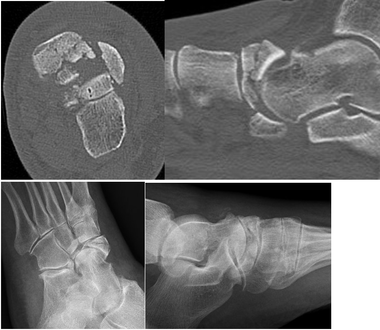
An 86-year-old female with a history of osteoporosis and prior right total hip arthroplasty presented with acute left foot pain following a fall while at home. She was unable to bear weight due to pain. Initial evaluation at an urgent care facility included radiographs and computed tomography (CT) of the left lower extremity, which demonstrated a displaced, comminuted intra-articular navicular fracture (Figure 1). The patient denied tobacco, alcohol, or illicit drug use, and her family history was noncontributory. At pre-injury baseline, the patient was fully ambulatory. Physical examination revealed significant left dorsal midfoot edema and ecchymosis. Neurovascular status was intact, with palpable dorsalis pedis and posterior tibial pulses. Midtarsal joint motion was limited due to pain, while ankle and subtalar joint motion remained within normal limits.
Radiographs demonstrated a comminuted navicular body fracture with talonavicular and naviculocuneiform joint disruption. Additional findings included decreased calcaneal inclination and increased talar declination angles. CT imaging confirmed fracture comminution without additional osseous involvement. Given the severity of the fracture, underlying osteoporosis, and the patient’s desire for early mobilization and return to function, a decision was made to proceed with total navicular replacement using a custom 3D-printed implant. A contralateral CT scan assisted with implant design. The surgical team and engineers collaboratively developed the titanium implant, incorporating screw fixation points, a porous surface adjacent to the cuneiforms to promote osseous ingrowth, and a smooth talar interface to preserve joint motion. This included an 8-mm medial anchor point for posterior tibial tendon reattachment.
Notes on the Operative Technique
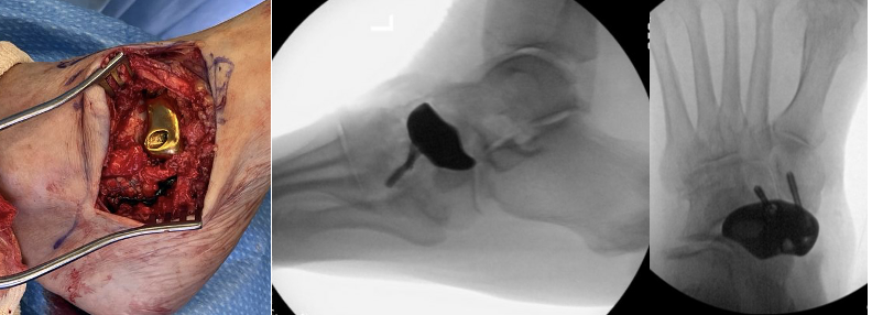
With the patient in a supine position, the surgeon made a curvilinear incision over the talonavicular joint extending dorsally across the navicular and cuneiforms. Subperiosteal dissection continued down to the naviculocuneiform joint. After detaching the posterior tibial tendon, the navicular was assessed, revealing significant comminution. The surgeon then completely excised the navicular. Next, we prepared the cuneiform joint surfaces with cartilage removal and subchondral fenestration. The implant was prepared on the back table by soaking the implant in an antibiotic solution. It was then inserted, along with a combination of autograft from the excised navicular and allograft material. Fixation consisted of screws placed through the implant into the medial and intermediate cuneiforms. A suture through the pre-designed anchor point allowed for posterior tibial tendon reattachment. Intraoperative fluoroscopy confirmed appropriate alignment, fixation stability, and restoration of medial column length (Figure 2). Range of motion testing demonstrated satisfactory implant stability.
Considerations During the Postoperative Course
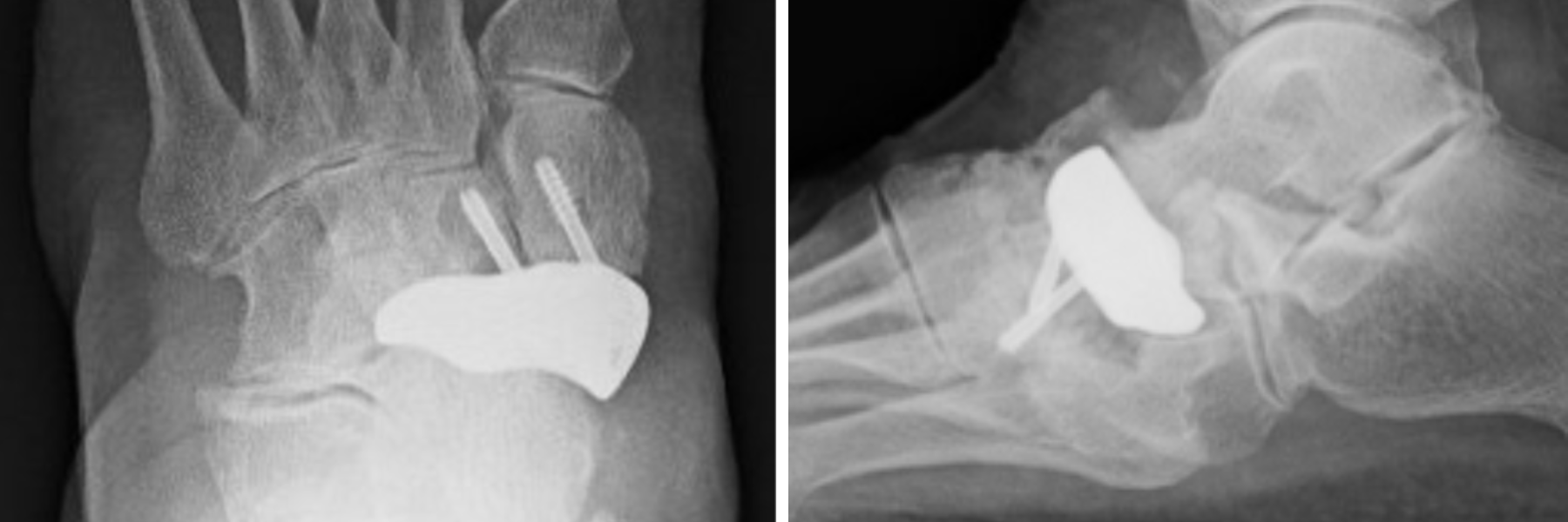
Postoperatively, the patient maintained strict non-weight-bearing status in a posterior splint. At 2 weeks postop, she transitioned to a controlled ankle motion (CAM) boot while continuing non-weight-bearing and initiating passive range-of-motion exercises. Partial weight-bearing began at 5 weeks, followed by progression to full weight-bearing in supportive footwear with an ankle brace at 2 months. At follow-up, the patient demonstrated a pain-free range of motion of the ankle, subtalar, and midtarsal joints, and was able to perform a single-limb heel rise without discomfort and with appropriate inversion. Radiographs showed mild heterotopic ossification without clinical symptoms. At 12 months postop, imaging confirmed maintained alignment with no evidence of hardware loosening or lucency (Figure 3). The patient returned to full activity without complications.
Discussion
There are several interventions used in the surgical management of navicular pathology; however, management of complex navicular fractures remains challenging due to limited vascularity and risk of osteonecrosis. ORIF aims to restore anatomic alignment but is often limited in cases of severe comminution or poor bone quality, where fixation failure and loss of reduction are concerns. Primary arthrodesis offers reliable pain relief and stability, but sacrifices joint motion and may alter normal gait mechanics. Vascularized bone grafting has demonstrated success in addressing osteonecrosis by restoring blood supply; however, these procedures are technically demanding, associated with donor-site morbidity, and limited by graft size and contour mismatch.4 Traditional techniques such as vascularized bone grafting provide biologic reconstruction but are technically demanding and limited by graft size and donor site morbidity. Vascularized bone flaps have served as a viable method to provide vascular supply, but these techniques have size limitations and require additional surgical training.8 The use of custom 3D printed total implants is an option surgeons may choose with the goals of restoring anatomic alignment and preserving mobility.
The path to creating a custom 3D implant incorporates a systematic process that allows for collaboration between the surgeon, the patient, and engineers. This process can be divided into 4 major steps, including data acquisition, pre-processing, printing, and post-processing.10 During this process, it is important to additionally take into consideration the architecture of the material used in order to achieve biocompatibility, an optimal surface for cell attachment, and have comparable properties to natural bone to minimize stress shielding effects. Titanium was used in this case as it demonstrates good osteoconductivity and biocompatibility.8
Recent advancements in additive manufacturing have introduced patient-specific 3D-printed implants as an alternative for complex osseous reconstruction. These implants contribute to precise restoration of native anatomy while preserving joint articulation when feasible. Several reports in the orthopedic literature have demonstrated promising outcomes using custom 3D-printed implants in the foot and ankle. For example, Adams and colleagues described a navicular replacement following failed vascularized grafting, with maintained alignment and return to pre-injury activity at 4-year follow-up. Similarly, a case series involving patient-specific titanium implants for limb salvage and deformity correction have reported favorable functional outcomes and implant stability.7
Compared with these reports, the present case further supports the feasibility of total navicular replacement using a 3D-printed implant, particularly in an elderly patient with osteoporosis and severe fracture comminution. A key advantage we noted in this case was the ability to tailor implant design to the patient’s anatomy, including the incorporation of porous surfaces to promote osseointegration, smooth articular interfaces to preserve motion, and fixation points to enhance stability. Additionally, the inclusion of a dedicated anchor point for posterior tibial tendon reattachment represents an important functional consideration that may not be achievable with traditional reconstructive techniques.
Biomechanically, preservation of medial column length and alignment is critical to maintaining normal foot function. The use of a custom implant allowed for restoration of this alignment while avoiding the stiffness associated with arthrodesis. Early mobilization and progression to weight-bearing in this patient further highlight the potential functional advantages of this approach.
Despite these promising findings, several limitations must be acknowledged. The current literature on 3D-printed total navicular replacement remains limited to case reports and small series, with a lack of long-term outcome data. Questions remain regarding implant longevity, wear characteristics at adjacent articulations, and the risk of stress shielding due to differences in material properties compared with native bone. Additionally, the cost and resource requirements associated with custom implant fabrication may limit widespread adoption.
Another important consideration is patient selection. Absolute contraindications include active infection, while relative contraindications, such as uncontrolled diabetes, poor vascular status, and tobacco use, may impair osseointegration and healing. Careful preoperative planning and multidisciplinary collaboration remain essential to optimize outcomes.
Future directions should focus on prospective studies evaluating long-term clinical and radiographic outcomes, as well as comparative analyses against established surgical techniques. Cost-effectiveness studies and continued refinement of implant materials and design—particularly with respect to elastic modulus and surface architecture—will be critical in advancing the clinical utility of this technology.
In Conclusion
This study highlighted the use of a total navicular 3D implant in a patient with a significant injury. After 1 year, the patient continues to have improved function with return to the pre-injury level of weight-bearing. Further studies can examine the incidence of delayed complications such as stress shielding, opposing surface cartilage wear, and implant failure. It is prudent to consider the difference in stiffness and elastic modulus between different materials during the implant design process. Future scientific research should include proper data augmentation algorithms to enhance the quality of raw image datasets, as well as optimizing printing parameters.
Dr. Varshney is a third-year podiatric resident at Scripps Mercy Hospital in San Diego, CA.
Dr. Pham is a foot and ankle surgeon at Sharp Memorial Hospital in San Diego, CA.
The authors have no non-financial or commercial, proprietary, or financial interest in the products or companies described in the manuscript. The author(s) did not receive grants or a consultant honorarium to conduct the study, write the manuscript or otherwise assist in the development of the above-mentioned manuscript.
References
1. Sangeorzan BJ, Benirschke SK. Fractures and dislocations of the midfoot. In: Banks AS, Downey MS, Martin DE, Miller SJ, eds. McGlamry's Comprehensive Textbook of Foot and Ankle Surgery. 4th ed. Wolters Kluwer; 2013. Chapter 105.
2. DiGiovanni CW. Fractures of the navicular. Foot Ankle Clin. 2004;9(1):25-63.
3. Coulibaly MO, Jones CB, Sietsema DL, Schildhauer TA. Results and complications of operative and nonoperative navicular fracture treatment. Injury. 2015;46(8):1669-1677.
4. Gilbert BJ, Horst F, Nunley JA. Potential donor rotational bone grafts using vascular territories in the foot and ankle. J Bone Joint Surg Am. 2004;86(9):1857-1873.
5. Adams SB, Danilkowicz RM. Talonavicular joint-sparing 3D-printed navicular replacement for osteonecrosis of the navicular. Foot Ankle Int. 2021;42(9):1197-1204.
6. ASTM International Committee F42 on Additive Manufacturing Technologies. Standard Terminology for Additive Manufacturing Technologies. ASTM F2792-10. ASTM International; 2009.
7. Dekker TJM, Steele JR, Federer AE, Hamid KS, Adams SB. Use of patient-specific 3D-printed titanium implants for complex foot and ankle limb salvage, deformity correction, and arthrodesis procedures. Foot Ankle Int. 2018;39(8):916-921.
8. Fishman FG, Adams SB, Easley ME, Nunley JA II. Vascularized pedicle bone grafting for nonunions of the tarsal navicular. Foot Ankle Int. 2012;33(9):734-739.
9. Constantino JR, Perler AD. Total navicular and cuboid replacement utilizing patient-specific 3D-printed implants for treatment of osteonecrosis of the midfoot: a case report and technique guide. Ann 3D Print Med. 2024;13:100140.
10. Wu Y, Liu J, Kang L, et al. An overview of 3D-printed metal implants in orthopedic applications: present and future perspectives. Heliyon. 2023;9:e15856.
© 2026 HMP Global. All Rights Reserved.
Any views and opinions expressed are those of the author(s) and/or participants and do not necessarily reflect the views, policy, or position of Podiatry Today or HMP Global, their employees, and affiliates.