L-methylfolate, Methylcobalamin, and Pyridoxal-5′-Phosphate in Wagner Grade 2 Diabetic Foot Ulcers: A Retrospective Case Series
This small retrospective case series suggests that adjunctive use of a prescription medical food with standard wound care was associated with favorable healing outcomes in patients with Wagner grade 2 diabetic foot ulcers. While preliminary and hypothesis-generating, the findings highlight the potential role of targeted neurovascular and metabolic support the management of diabetic foot ulcers.
Key Takeaways
- All 5 Wagner grade 2 diabetic foot ulcers in this series achieved complete closure within 4–6 weeks while patients received standard wound care plus the medical food, with no reported adverse events.
- Patients demonstrated encouraging longer-term outcomes, including no reulceration during 6–20 months of follow-up and qualitative improvements in neuropathy, skin hydration, and xerosis among those who continued treatment.
- The findings support further investigation. The results at this stage are hypothesis-generating and warrant confirmation in larger, prospective controlled trials.
Diabetic foot ulcers (DFUs) represent a major source of morbidity, mortality, and health care utilization among patients with diabetes. Approximately 38 million individuals in the United States are living with diabetes, of whom an estimated 19–34% will develop a DFU during their lifetime, with recurrence rates approaching 65% within 5 years.1 DFUs are associated with substantial downstream complications, including infection, osteomyelitis, and lower extremity amputation, with approximately 160,000 diabetes-related amputations performed annually. The economic burden of DFU management exceeds $28 billion per year,2 underscoring the need for strategies that improve healing trajectories and reduce complications.
Delayed healing of DFUs reflects more than mechanical tissue injury and pressure-related breakdown. These wounds arise in a biologic environment characterized by peripheral neuropathy, microvascular disease, endothelial dysfunction, oxidative stress, and chronic inflammation.3-8 These processes impair oxygen delivery, nutrient exchange, cellular signaling, and immune responses, limiting the biologic capacity for tissue repair. Adjunctive approaches that address underlying neurovascular and metabolic dysfunction are increasingly relevant in patients with delayed healing. This condition reflects combined nerve dysfunction and microvascular impairment contributing to ulcer risk and delayed healing.
Medical foods are designed for the dietary management of diseases with distinctive nutritional requirements and are intended to support foundational biologic processes rather than act as direct wound therapies. Metanx® (Alfasigma USA, Inc.) is a prescription medical food composed of fixed daily dose of bioactive forms of vitamins B6 (pyridoxal 5′-phosphate 70mg), B9 (L-methylfolate Ca 6mg), and B12 (methylcobalamin 4mg), used clinically in diabetic peripheral neuropathy.9 This formulation targets endothelial dysfunction, impaired one-carbon (1C) metabolism, and neuronal injury. These mechanisms are central to microvascular health, nerve function, and tissue repair. Together, nerve dysfunction and microvascular impairment drive ulcer formation and delay healing in diabetic foot disease.3
Bioactive B vitamins have been associated with pathways relevant to tissue repair. Methylcobalamin supports neuronal signaling and nerve structural integrity through myelin maintenance.10,11 Pyridoxal 5′-phosphate has been associated with modulation of oxidative stress and reduction of advanced glycation end products, which are known to impair cellular migration and matrix remodeling.12,13 L-methylfolate participates in methylation-dependent processes, including homocysteine metabolism.14,15 Elevated homocysteine is associated with endothelial dysfunction, reduced nitric oxide bioavailability, and impaired tissue repair.16 These mechanisms are biologically plausible but not independently causal in this setting and provide a rationale for examining wound-healing outcomes in patients receiving the LMF-MC-PLP formulation.
Prior clinical and translational studies of the LMF-MC-PLP formulation in diabetic peripheral neuropathy provide additional context for these pathways. In a randomized controlled trial, the LMF-MC-PLP formulation was associated with significant reductions in plasma homocysteine compared with placebo, along with improvements in neuropathy symptoms and quality-of-life measures.17 Observational and open-label studies have reported significant increases in epidermal nerve fiber density and improvements in monofilament sensation.18,19 These findings are clinically relevant given the established relationship between loss of protective sensation and ulcer risk. In preclinical models, components of this prescription LMF-MC-PLP formulation have been associated with improved endothelial nitric oxide synthase coupling and reduced oxidative stress supporting a potential role in microvascular function.20
We report a retrospective, observational, hypothesis-generating case series evaluating wound healing outcomes in patients with Wagner grade 2 DFUs managed with standard wound care while receiving the described prescription medical food.
Details on the Case Series
Patients and setting. The basis of this retrospective case series was from outpatient clinical records from a podiatric wound care practice. Review of clinical records took place over a defined time period to identify eligible patients. Inclusion criteria were:
- presence of a Wagner grade 2 DFU measuring ≥1 cm in diameter;
- management with standard wound care; and
- receipt of the prescription LMF-MC-PLP formulation for diabetic peripheral neuropathy either prior to ulcer development or at the time of ulcer presentation.
Baseline patient, ulcer, and clinical characteristics were recorded and are summarized in Table 1.
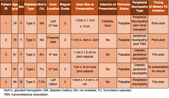
All patients included received the described medical food for diabetic peripheral neuropathy prior to and/or at the time of ulcer presentation. Ulcer occurred within 1–4 weeks of therapy initiation in most cases. One patient presented with an existing ulcer and received the LMF-MC-PLP formulation at the time of initial wound evaluation. Although all patients initiated the medical food prior to ulcer presentation, DFUs arise from chronic neuropathic and microvascular risk, and the timing of clinical presentation does not necessarily reflect the timing of ulcer pathogenesis.
This case series is reported in accordance with the CARE guidelines where applicable.
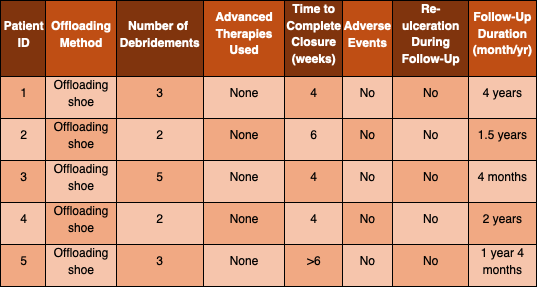
Wound management and exposure. All included patients in this review received standard wound care consistent with clinical practice guidelines. This included regular wound evaluation, sharp debridement as indicated, maintenance of moisture balance, decolonization when clinically appropriate, and offloading using postoperative shoes, crutches, or rolling knee walkers. Patients were evaluated weekly until wound closure was achieved. They did not receive advanced wound therapies (including negative pressure wound therapy, cellular or tissue-based products, hyperbaric oxygen therapy, or growth factors), allowing evaluation within a standard of care context. Details of wound management, debridement frequency, time to complete closure, and adverse events are summarized in Table 2.
The prescription LMF-MC-PLP formulation was administered orally twice daily. The medical food was prescribed for the dietary management of diabetic peripheral neuropathy and continued throughout the wound healing period.
Outcomes and follow-up. The primary clinical outcome was time to complete wound closure, defined as full epithelialization of the ulcer with absence of drainage, confirmed at clinical evaluation. All 5 patients with Wagner grade 2 DFUs achieved complete wound closure within 6 weeks of presentation. Three patients achieved closure by week 4, and 2 closed by week 6. No adverse events were reported.
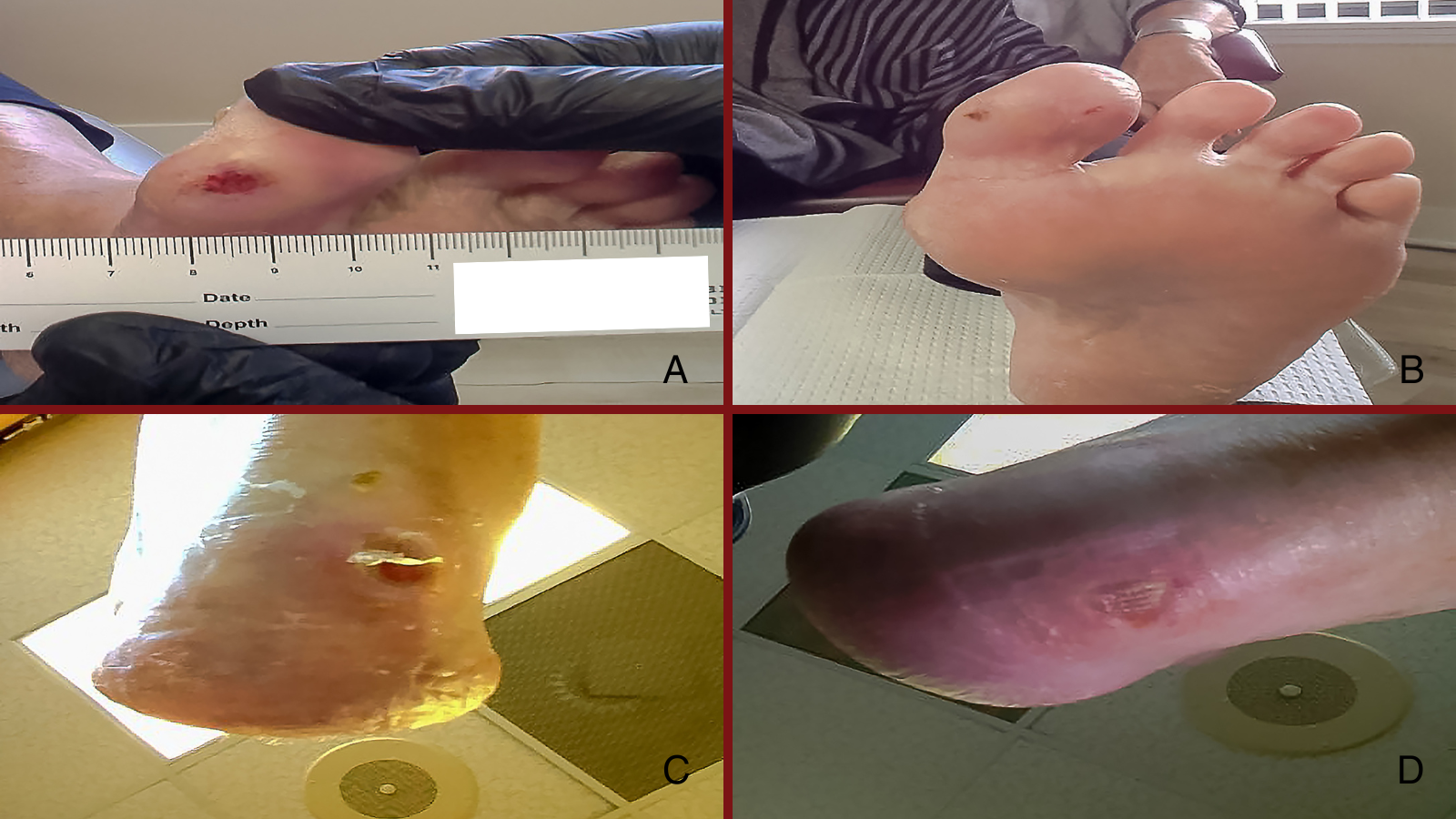
Representative clinical images demonstrated progressive wound reduction and re-epithelialization over time, with visible decreases in ulcer size and tissue improvement between initial presentation and follow-up at weeks 4 and 6, consistent with the observed timelines of wound closure (Figure 1).
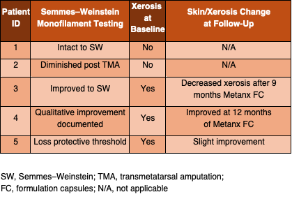
Follow-up periods ranged from 6 to 20 months after wound closure. No patient experienced reulceration during follow-up. Additional clinical observations included qualitative changes in peripheral neuropathy and skin condition. Over time, qualitative improvement in peripheral neuropathy was documented on Semmes–Weinstein monofilament testing in patients who continued the medical food. All patients initially presented with xerotic changes of the lower extremities characterized by dry, scaly skin, consistent with autonomic peripheral neuropathy. In 3 of 5 patients who continued the LMF-MC-PLP formulation for 9–12 months, xerosis decreased and overall skin appearance improved, suggesting possible improvement in autonomic function affecting sweat and oil gland activity. In addition to reduced xerosis, patients demonstrated improvement in skin hydration and natural oil production, with resolution of fissured and dry skin appearance in affected areas. Secondary qualitative observations regarding Semmes–Weinstein monofilament testing, baseline xerosis, and changes in skin condition during follow-up are summarized in Table 3. Patients also reported subjective improvement in neuropathic symptoms and proprioception of the lower extremities over the course of treatment.
This retrospective chart review involved analysis of existing medical records and did not include any patient identifiers. The study was conducted in accordance with institutional standards for clinical record review and complied with HIPAA regulations. Formal institutional review board approval was not required for this descriptive case series.
Discussion
In this retrospective case series, patients with Wagner grade 2 DFUs managed with standard wound care while receiving a prescription medical food formulation (pyridoxal 5′-phosphate 35 mg, L-methylfolate 3 mg, and methylcobalamin 2 mg, administered orally twice daily) achieved complete wound closure within 4–6 weeks. In a prospective study of DFUs, Sheehan and colleagues reported that the percent change in wound area at 4 weeks in ulcers that ultimately healed was 82% (95% CI 70–94), whereas in ulcers that failed to heal the percent change was 25% (95% CI 15–35; P < 0.001).21 These findings support the use of early percent change in wound area as a predictor of healing and provide a benchmark against which the relatively rapid closure observed in the present case series can be qualitatively considered.21 No adverse events were observed. Serial clinical images documenting wound progression further supported these observations, demonstrating visible reduction in ulcer size and progressive tissue repair over the treatment period.
The observed wound-healing course occurred in the context of a medical food designed to support endothelial function, methylation-dependent pathways, and neuronal integrity, which are biologically relevant to microvascular perfusion, tissue repair conditions, and nerve function. While causality cannot be inferred, the convergence of relatively rapid healing, sustained closure, and qualitative improvements in neuropathic findings align with the hypothesized role of neurovascular and metabolic support in DFU management. These observations are consistent with prior studies of the LMF-MC-PLP formulation demonstrating reductions in homocysteine, improvements in epidermal nerve fiber density and monofilament sensation, and preclinical evidence of improved endothelial nitric oxide synthase coupling and oxidative stress modulation, all of which may be relevant to microvascular function and tissue repair conditions.17,22,23 In addition, patient-reported improvements in proprioception suggest a potential functional dimension to the observed neuropathic changes, which may be relevant to pressure redistribution, gait stability, and ulcer risk.
No patient experienced reulceration during follow-up periods ranging from 6 to 20 months after wound closure, and qualitative improvement in sensory testing and skin condition was documented in patients who continued Metanx. These additional observations were not systematically measured but are clinically relevant given the established role of sensory and autonomic neuropathy in ulcer development, skin integrity, and recurrence risk. Improvement in skin hydration and reduction in fissuring observed in this cohort may reflect changes in autonomic nerve function influencing sweat and oil gland activity, which are important for maintaining skin integrity and barrier function in patients with diabetes.
Taken together, sustained ulcer closure, absence of reulceration, qualitative improvement in sensory testing, and changes in skin condition suggest that adjunctive neurovascular and metabolic support during the wound-healing period may influence longer-term tissue resilience. The absence of reulceration during follow-up, in conjunction with improvements in skin condition and neuropathic findings, may suggest a more durable tissue environment, though this observation requires confirmation in larger, controlled studies.
Limitations
This report is limited by its retrospective design, small sample size, and absence of a control group, with outcomes interpreted relative to historical benchmarks rather than contemporaneous comparators. Observed healing times appear shorter than historical benchmarks for similar ulcers treated with standard care alone; interpretation remains limited by the uncontrolled design. While causality cannot be inferred, findings are discussed in the context of biologically plausible mechanisms relevant to microvascular function and tissue repair. These findings do not establish a direct effect on wound healing but support a biologic and clinical framework for investigation of wound-healing outcomes in patients receiving this prescription LMF-MC-PLP formulation. Confounding from other aspects of standard wound care or patient characteristics cannot be excluded, and findings may not be generalizable beyond similar clinical contexts. Although all patients initiated the medical food prior to ulcer presentation, the timing of clinical presentation does not necessarily reflect the timing of ulcer pathogenesis; given the chronic pathogenesis of DFUs, this temporal relationship does not imply causation. Secondary observations regarding neuropathy and skin changes were qualitative and not systematically measured, extracted from routine clinical documentation rather than collected using standardized instruments. These findings should be viewed as preliminary and hypothesis-generating, and confirmation in larger, controlled studies is needed.
Conclusions
In this retrospective case series of 5 patients with Wagner grade 2 diabetic foot ulcers, wound closure was achieved within 4–6 weeks while patients received standard wound care and the medical food LMF-MC-PLP formulation. These observations, together with qualitative improvements in neuropathic signs and skin condition and the absence of reulceration during follow-up, suggest a possible favorable association between targeted neurovascular and metabolic support and wound healing trajectories and longer-term tissue resilience.
Dr. Trathen practices in Fort Myers, FL. He discloses that he serves on the speakers bureau for the medical food discussed in this article, and receives honoraria from Alfasigma USA, Inc. William Nikolic and Eric Kassel are employees of Alfasigma USA, Inc.
Dr. Nikolic is an Associate Director Medical Science Liaison, Medical Affairs with Alfasigma, USA. Dr. Kassel is the Senior Director of Clinical Affairs and Scientific Operations at Alfasigma, USA.
Acknowledgments
The authors thank Olivia Edwards, PharmD, for her editorial assistance.
References
1. McDermott K, Fang M, Boulton AJM, Selvin E, Hicks CW. Etiology, epidemiology, and disparities in the burden of diabetic foot ulcers. Diabetes Care. 2023;46(1):209-221.
2. Collins N. Medicare spending on wound care: the first comprehensive study. WCEI.net. Published October 2017. Accessed [insert access date].
3. Boulton AJM. The pathway to foot ulceration in diabetes. Med Clin North Am. 2013;97(5):775-790.
4. Jeffcoate WJ, Harding KG. Diabetic foot ulcers. Lancet. 2003;361(9368):1545-1551.
5. Armstrong DG, Boulton AJM, Bus SA. Diabetic foot ulcers and their recurrence. N Engl J Med. 2017;376(24):2367-2375.
6. Falanga V. Wound healing and its impairment in the diabetic foot. Lancet. 2005;366(9498):1736-1743.
7. Schäfer M, Werner S. Oxidative stress in normal and impaired wound repair. Pharmacol Res. 2008;58(2):165-171.
8. Brem H, Tomic-Canic M. Cellular and molecular basis of wound healing in diabetes. J Clin Invest. 2007;117(5):1219-1222.
9. Metanx [package insert]. Shreveport, LA: Alfasigma USA Inc; 2024.
10. Julian T, Syeed R, Glascow N, Angelopoulou E, Zis P. B12 as a treatment for peripheral neuropathic pain: a systematic review. Nutrients. 2020;12(8):2221. doi:10.3390/nu12082221.
11. Okada K, Tanaka H, Temporin K, et al. Methylcobalamin increases Erk1/2 and Akt activities through the methylation cycle and promotes nerve regeneration in a rat sciatic nerve injury model. Exp Neurol. 2010;222(2):191-203. doi:10.1016/j.expneurol.2009.12.017.
12. Nakamura S, Li H, Adijiang A, Pischetsrieder M, Niwa T. Pyridoxal phosphate prevents progression of diabetic nephropathy. Nephrol Dial Transplant. 2007;22(8):2165-2174.
13. Mascola E, Verni F. Vitamin B6 and diabetes: relationship and molecular mechanisms. BioMed Research International. Int J Mol Sci. 2020 May 23;21(10):3669. doi: 10.3390/ijms21103669.
14. Raghubeer S, Matsha TE. Methylenetetrahydrofolate (MTHFR), the one-carbon cycle, and cardiovascular risks. Nutrients. 2021;13(12):4562. doi:10.3390/nu13124562.
15. Kaye AD, Jeha GM, Pham AD, et al. Folic acid supplementation in patients with elevated homocysteine levels. Adv Ther. 2020;37(10):4149-4164. doi:10.1007/s12325-020-01474-z.
16. Schwartzfarb EM, Romanelli P. Hyperhomocysteinemia and lower extremity wounds. Int J Low Extrem Wounds. 2008;7(3):126-136. doi:10.1177/1534734608322490.
17. Fonseca VA, Lavery LA, Thethi TK, et al. Metanx in type 2 diabetes with peripheral neuropathy: a randomized trial. Am J Med. 2013;126(2):141-149. doi:10.1016/j.amjmed.2012.06.022.
18. Jacobs AM, Cheng D. Management of diabetic small-fiber neuropathy with combination L-methylfolate, methylcobalamin, and pyridoxal 5′-phosphate. Rev Neurol Dis. 2011;8(1-2):39-47.
19. McNamara VF, Vinik AI, Barrentine LW, De Vol EB. Effectiveness of Metanx prescription medical food on small nerve fibers and monofilament sensation in patients with diabetic peripheral neuropathy. J Diabetes Mellitus. 2016;6(2):166-174. doi: 10.4236/jdm.2016.62018.
20. Shevalye H, Watcho P, Stavniichuk R, et al. Metanx alleviates multiple manifestations of peripheral neuropathy and increases intraepidermal nerve fiber density in Zucker diabetic fatty rats. Diabetes. 2012;61(8):2126-2133. doi:10.2337/db11-1524.
21. Sheehan P, Jones P, Caselli A, Giurini JM, Veves A. Percent change in wound area of diabetic foot ulcers over a 4-week period is a robust predictor of complete healing in a 12-week prospective trial. Diabetes Care. 2003;26(6):1879-1882.[GU8]
22. Trippe BS, Ciaramitaro P, et al. Improvement in loss of protective sensation with L-methylfolate, methylcobalamin, and pyridoxal-5′-phosphate in patients with diabetic peripheral neuropathy. J Am Podiatr Med Assoc. 2016;106(5):336-343.
23. Smolek MK, Notaroberto NF, Jaramillo AG, Pradillo LR. Intervention with L-methylfolate, methylcobalamin, and pyridoxal-5′-phosphate improves vascular and neural biomarkers in experimental diabetic neuropathy. Nutr Res. 2013.
© 2026 HMP Global. All Rights Reserved.
Any views and opinions expressed are those of the author(s) and/or participants and do not necessarily reflect the views, policy, or position of Podiatry Today or HMP Global, their employees, and affiliates.