Minimally Invasive Surgical Options for Haglund's Deformity and Retrocalcaneal Heel Pain
Advances in minimally invasive surgery have expanded treatment options for patients with Haglund's deformity, insertional Achilles tendinopathy, and retrocalcaneal heel pain. This review outlines imaging-based decision-making, procedure selection, and surgical pearls to help match the appropriate technique to the underlying pathology while minimizing soft tissue disruption.
Key Takeaways
- Identifying proper etiology of retrocalcaneal heel pain is paramount for proper procedure selection and maximizing patient outcomes. Clinical examination, radiographs, and advanced imaging all help distinguish tendon-driven, osseous-driven, and mixed pathology.
- Procedure selection is best guided by pathology rather than a one-size-fits-all approach. For instance, patients with isolated osseous deformity, but without soft tissue pathology may benefit from a dorsal calcaneal closing wedge osteotomy.
- Minimally invasive techniques may reduce soft tissue morbidity without compromising outcomes. Smaller incisions, limited tendon detachment, and modern fixation constructs may contribute to decreasing wound complications and support an earlier return to activity in appropriately selected patients.
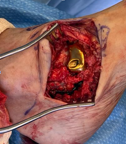
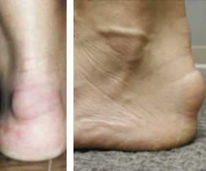
There are descriptions in existence of open and minimally invasive techniques for treating retrocalcaneal pain resulting from conditions such as Haglund’s deformity, insertional Achilles tendinopathy, and retrocalcaneal exostosis (Figure 1). Conservative treatment may include stretching (night splint), using heel lifts, and physical therapy. If pain is severe enough, temporary immobilization may be necessary to reduce the pain (using a removable boot or cast). Once one exhausts conservative therapy, further imaging is appropriate, as is consideration of surgical options. Open resection with or without Achilles tendon detachment provides reliable pain relief and functional improvement, but carries risks of wound complications and nerve irritation.1-3
Emerging evidence challenges the traditional paradigm that direct resection of the posterosuperior calcaneal prominence is necessary in all cases. Improved radiographic assessment of calcaneal morphology and its relationship to insertional Achilles pathology has facilitated more individualized treatment algorithms and surgical decision-making.4 Recently, investigations of minimally invasive techniques have shown reduction of soft tissue morbidity while achieving similar outcomes in appropriately selected patients.5 These techniques allow for posterior calcaneoplasty of the Haglund’s deformity and/or retrocalcaneal exostosis and reinforcement of the Achilles tendon insertion with bone anchors. Additional modifications to open posterior calcaneoplasty have focused on addressing concomitant biomechanical contributors. Adding gastrocnemius recession to Haglund’s deformity surgery has shown improved outcomes in appropriately selected patients by reducing tension across the Achilles tendon insertion.3 These findings further emphasize the importance of protecting the Achilles repair construct and minimizing postoperative stress at the tendon-bone interface.
Zadek originally described the dorsal calcaneal closing wedge osteotomy to reduce tension on the Achilles tendon by altering the calcaneal geometry and the reducing posterior superior calcaneal exostosis irritating the anterior Achilles tendon.6 Recent studies have shown favorable biomechanical and clinical outcomes from this approach in chronic Achilles insertional disease.7,8
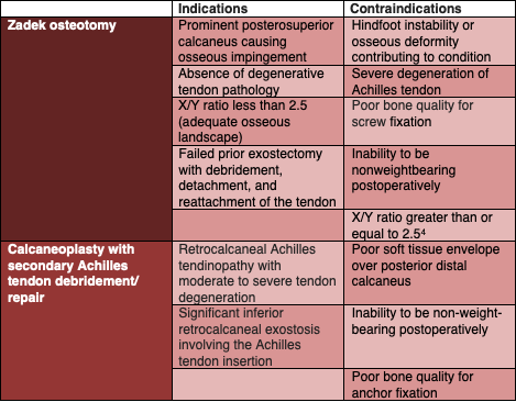
Table 1 summarizes the indications and contraindications for each procedure. These procedures are not new concepts, but the techniques have now shifted to favor minimally invasive methods. The learning curves are steep and require understanding of the anatomy and surgical principles to optimize outcomes. The significant advantages to these minimally invasive techniques include smaller incisions, less soft tissue injury, and limiting Achilles tendon detachment.
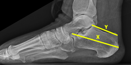
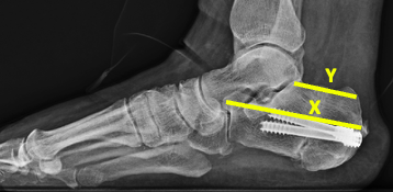
Insertional Achilles tendinopathy and Haglund’s deformity are challenging causes of posterior heel pain with multifactorial pathophysiology involving mechanical impingement of the posterosuperior calcaneus, degenerative changes at the Achilles insertion, retrocalcaneal bursitis, prior trauma, and altered calcaneal morphology. The most significant radiographic parameter to assess for Haglund’s syndrome is the X/Y ratio being less than 2.5 to help guide surgical decision-making (Figure 2).4 One measures the calcaneal length (X) from the most anterior aspect of the calcaneus to the most posterior aspect of the calcaneus (excluding tendon calcifications). One then calculates the length of the greater tuberosity (Y) by measuring from the posterior aspect of the posterior facet to the posterior superior calcaneus.
Advances in surgical techniques, specifically minimally invasive ones using double-row constructs with bone anchors, aim to optimize tendon security while minimizing soft tissue injury. By evenly distributing the contractile force of the gastrocnemius-soleus complex across the calcaneal footprint, in our experience, double-row fixation eliminates micromotion and prevents tension gaps at the healing interface. This construct maximizes surface area contact between the distal Achilles tendon stump and the resected bone, effectively eliminating dead space or uneven pressure across the insertion site. Furthermore, modern, low-profile bone anchors may reduce the incidence of local skin breakdown and prominent hardware. Ultimately, the rigidity of this construct allows for accelerated rehabilitation, potentially translating to a faster return to activity.
An alternative procedure is the dorsal closing wedge calcaneal osteotomy, which reduces Achilles tendon strain by reducing the mechanical lever arm of the tendon on the calcaneus. Historically, the dissection needed for the osteotomy has been associated with wound complications. Recent minimally invasive techniques can help reduce soft tissue injury while protecting the sural nerve by incision placement planning.9,10
Despite increasing interest in both tendon-focused and structural procedures, there is no current clear consensus or decision-making algorithm for surgical management of retrocalcaneal Achilles tendinopathy and Haglund’s syndrome.
Radiographic considerations
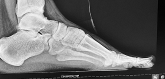
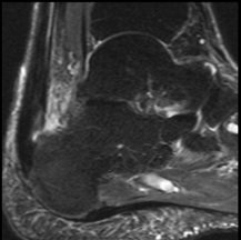
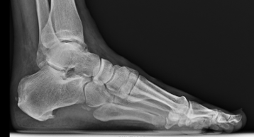
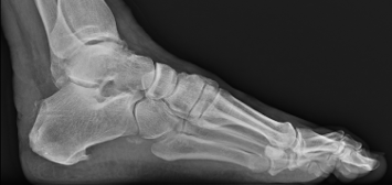
Evaluate the lateral foot view for retrocalcaneal exostoses (Figure 3). Correlate the location(s) of clinical pain to the radiographic landmarks. Pain at the posterior superior aspect of the calcaneus indicates a Haglund’s condition, as the Achilles tendon is irritated by the bony prominence. Presence of radiographic exostosis at the inferior attachment of the Achilles tendon may correlate to clinical pain at this location. Magnetic resonance imaging (MRI) is essential to evaluate for any degeneration or partial tears of the Achilles tendon (Figure 4).
Surgical Pearls to Consider
Below are some technique tips and pearls that our team finds helpful in these types of procedures.
Zadek osteotomy
- Position the patient prone for better posterior calcaneal access with a minimally invasive technique, easier fluoroscopic imaging, and ergonomic use of instrumentation.
- The incision is on the lateral body of the calcaneus (approximating the central point of the osteotomy). Care must be taken to avoid the sural nerve, the peroneal tendons anteriorly, and the Achilles tendon posteriorly.
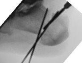
- Utilizing fluoroscopy, place percutaneous wires through the inferior aspect of the calcaneus with an apex plantar and base dorsal to mimic the borders of the wedge to use as boundaries for the osteotomy (Figure 5). The width of the wedge resection should be calculated such that the X/Y ratio is greater than or equal to 2.5. The authors highly recommend planning out preoperatively for the desired size of the dorsal wedge to remove to achieve proper correction.
- Using the minimally invasive burr, follow the outline of one of the wires to create a through-and-through osteotomy. Starting superiorly, resect the remaining bone with a scooping action. Irrigate constantly and pause the burr periodically to lessen risk of thermal necrosis. An angiocath can be used to irrigate the bone slurry periodically. Periodic use of fluoroscopy can assist in ensuring proper alignment.
- Preserve the plantar hinge and feather at the end while applying pressure to close the osteotomy site.
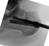
- Temporarily fixate the osteotomy with guidewires placed from posterior to anterior. Two headless cannulated screws can provide compression and stabilization (Figure 6).
Calcaneoplasty with secondary Achilles tendon debridement/repair
- Mark the medial and lateral borders of the Achilles tendon distally on the posterior calcaneus. The 2 proximal incisions will be just inferior to the superior corners of the calcaneus where the Achilles tendon insertion begins. The 2 distal incisions will be distal to the other incisions at the most posterior prominence of the calcaneus. Surgeons should confirm these sites radiographically before making the incisions.
- It is very important to create space between the Achilles tendon and the skin using a blunt elevator so that later in the procedure, one can pass sutures through this pathway.
- Using one of the proximal incisions, the surgeon introduces the minimally invasive burr to begin resecting the posterior superior prominence of the calcaneus after radiographic confirmation. We then recommend periodically switching portals on the superior incisions until satisfied with the bone resection. The burr can also be angled distally to resect any inferior retrocalcaneal exostosis. Irrigate constantly and pause the burr periodically to lessen risk of thermal necrosis. An angiocath can help irrigate the bone slurry periodically. Using fluoroscopy periodically can help ensure proper placement.
- Arthroscopy may be used to inspect Achilles tendon integrity.
- Drill and insert 2 bone anchors into the calcaneus at the insertion of the tendon and pass the suture tapes up through the lower end of the detached Achilles tendon.
- Go 2 cm distal to the previous anchors and drill 2 pilot holes for the distal anchors.
- Take the suture tapes coming out of the tendon and cross them over each other to form an "X." Bring the top-left sutures down to the bottom-right hole, and the top-right sutures down to the bottom-left hole.
- Thread the crossed sutures into 2 knotless anchors. Hold the foot in slight plantarflexion, pull the sutures tight until the tendon is compressed flat against the bone, and drive the anchors into the bottom holes to lock the construct in place.
Algorithms From the Lead Author
The treatment algorithm for insertional Achilles tendinopathy begins with careful identification of the patient's pain through clinical examination, radiographs, and MRI evaluation. We then correlate the location of pain with imaging findings to determine the primary driver of the patient’s symptoms. As a result, we can classify the pathology into one of three categories:
- tendon-driven;
- mixed; or
- osseous-driven disease.
Tendon-driven pathology is characterized by diffuse insertional tendinosis, partial-thickness tearing, or intra-tendinous calcification confirmed with MRI. In these cases, the tendon itself represents the primary source of pain and dysfunction.1,2 Mixed pathology is defined by focal insertional Achilles changes occurring in conjunction with a prominent Haglund's deformity, suggesting both intrinsic tendon disease and extrinsic mechanical compression.11 Osseous-driven pathology is identified when a large Haglund's prominence and abnormal calcaneal angles are present in the setting of a relatively healthy Achilles tendon, indicating that biomechanical impingement is the predominant pain generator.4,12
Procedure selection stems from the identified pathology. For tendon-driven disease, treatment consists of direct debridement of diseased tendon through either a central-splitting or partial-detachment approach, followed by reattachment using a double-row suture bridge construct. In these patients, we do not expect a Zadek osteotomy alone to provide adequate symptom relief because the underlying pathology resides within the tendon. For mixed pathology, the preferred approach is exostectomy combined with limited Achilles tendon debridement while preserving as much of the tendon as possible. A Zadek osteotomy may be added when a prominent posterosuperior calcaneal prominence contributes substantially to tendon compression. For patients with purely osseous, biomechanically driven pathology and no significant Achilles tendon degeneration, performing a Zadek osteotomy can decompress the Achilles insertion and reduce pathologic loading of the posterior calcaneus. In these cases, one should avoid Achilles tendon detachment because the tendon itself is not the primary source of pathology.
Complications and Concerns
These procedures do have their share of potential postoperative complications. With the tissue anchors, we have experiences instances where we had to remove the anchors due to sinus tract infections. Initial treatment with intravenous antibiotics and subsequent removal after 6 weeks allowed for sufficient tendon reattachment such that anchor removal did not compromise tendon healing. Use of absorbable antibiotic pellets into the anchor holes augmented antibiotic delivery to the involved site(s). Immobilization in a removable boot with short initial course of non-weight-bearing can support optimal healing; the length of non-weight-bearing depends on factors such as tendon integrity, bone quality, and infection clearance. For the osteotomy, a similar complication can occur with sinus tract development into the screw site. The treatment is identical to the infected anchor removal process. If possible, delaying removal until 6 weeks postop will allow for enough osteotomy stability. One can use percutaneous wires if there is osteotomy stability and later remove them in the office. In the event of calcaneal osteotomy displacement, open reduction and internal fixation will be necessary.
Highlights of Representative Cases
Case 1. A 61-year-old female presented with a 15-month history of posterior heel pain. On physical examination, she had no pain on palpation along the Achilles tendon above the insertion site, but related pain at the superior posterior calcaneus. Radiographs are shown in Figure 2. Radiographic calculation shows an X/Y ratio of 2.11 (X=84.7mm, Y=40.23mm) and MRI of the ankle did not show any tendon pathology. The patient underwent a successful Zadek osteotomy (Figure 7) where the X/Y ratio was improved to 2.77 (X=78.24mm, Y=28.28mm). Preoperative planning targeted an approximately 10 mm wedge resection to achieve proper correction.
Case 2. A 71-year-old male presented with a more than 3-year history of posterior heel pain. On physical examination, he has pain at the posterior calcaneus at the Achilles insertion site where there is a bony prominence, and pain at posterior superior calcaneus. His radiograph is shown in Figure 8 and MRI is shown in Figure 4.
Conclusion
Heel pain primarily caused by osseous pathology, such as retrocalcaneal exostosis or Haglund’s deformity without much Achilles tendon pathology, can be successfully treated by addressing the bony deformity through retrocalcaneal exostectomy or dorsal closing-wedge osteotomy using minimally invasive methods. When the exostectomy may disrupt the Achilles tendon insertion or MRI confirms Achilles tendon pathology, concomitant soft tissue repair is needed. Both pathologies now have minimally invasive technique options that can provide less soft tissue injury and potentially a faster return to activity.
Dr. Husain is the program director of the McLaren Oakland Hospital Podiatric Medicine and Surgery Residency Program and is in private practice in the metro-Detroit area..
Drs. Panchal (PGY1) and Cohen (PGY2) are residents at the McLaren Oakland Hospital Podiatric Medicine and Surgery Residency Program.
The authors have no non-financial or commercial, proprietary, or financial interest in the products or companies described in the manuscript. The author(s) did not receive grants or a consultant honorarium to conduct the study, write the manuscript or otherwise assist in the development of the above-mentioned manuscript.
References
1. Xia Z, Yew AKS, Zhang TK, Su HCD, Ng YCS, Rikhraj IS. Surgical correction of Haglund’s triad using a central tendon-splitting approach: a retrospective outcomes study. J Foot Ankle Surg. 2017;56(6):1132-8.
2. Sammarco GJ, Taylor AL. Operative management of Haglund’s deformity in the nonathlete: a retrospective study. Foot Ankle Int. 1998;19(11):724-9.
3. Vesely BD, Reardon BK, Scott AT, Medda AW. Retrospective comparison of isolated Haglund’s deformity surgery versus combined with gastrocnemius recession. J Foot Ankle Surg. 2023;62(4):719-22.
4. Tourné Y, Baray AL, Barthélémy R, Moroney P. Contribution of a new radiologic calcaneal measurement to the treatment decision tree in Haglund syndrome. Orthop Traumatol Surg Res. 2018;104(8):1215-9.
5. Musikachart P, Tharmviboonsri T, Chuckpaiwong B, Lertwattanachai P, Harnroongroj T. Does central-splitting or complete detachment provide better clinical results in surgical treatment of Haglund’s disease? a 2 to 6 years retrospective comparative follow-up study. J Foot Ankle Surg. 2025;54(5):608-12.
6. Zadek I. An operation for the cure of achillobursitis. Am J Surg. 1939;43(2):542-6.
7. Hall SR, Schipper ON, Kaplan JRM, Johnson AH, Gonzalez TA, Vulcano E. Outcomes after percutaneous Zadek osteotomy for insertional Achilles tendinopathy. Foot Ankle Int. 2024;45(9):931-9.
8. Rutishauser T, Stephan A, Stadelmann VA. Open dorsal closing-wedge calcaneal osteotomy for Haglund exostosis-related heel pain. Foot Ankle Int. 2024;45(12):1319-29.
9. Kiriluk SH, Vulcano E, Schipper ON, Kaplan JRM, Johnson AH, Ventresca H, Gauthier C, Davis HT, Harrison P, Lewis T, Lam P, Jackson JB, Gonzalez TA. Percutaneous Zadek osteotomy vs open Haglund resection for insertional Achilles tendinopathy: early outcomes and complication rates. Foot Ankle Int. 2025;46(10):1103-14.
10. Talusan PG, Cata E, Tan EW, Parks BG, Guyton GP. Safe zone for neural structures in medial displacement calcaneal osteotomy: a cadaveric and radiographic investigation. Foot Ankle Int. 2015;36(12):1493-8.
11. Cusumano A, Martinelli N, Bianchi A. Bertelli A, Marangon A, Sansone V. Transtendinous approach calcaneoplasty versus endoscopic calcaneoplasty for Haglund’s disease. Int Orthop. 2021;45(1):225-31.
12. Pfeffer G, Gonzalez T, Zapf M, Nelson TJ, Metzger MF. Achilles pullout strength after open calcaneoplasty for Haglund’s syndrome. Foot Ankle Int. 2018;39(8):966-9.
© 2026 HMP Global. All Rights Reserved.
Any views and opinions expressed are those of the author(s) and/or participants and do not necessarily reflect the views, policy, or position of Podiatry Today or HMP Global, their employees, and affiliates.