Recurrent Inflammatory Plaques on Upper Extremities in a Patient With Breast Cancer Surgery History
A 49-year-old woman was seen by the dermatology consult service with inflammatory, tender, indurated, pink plaques on the upper extremities, as well as an edematous right upper extremity (Figure 1). Her past medical history included invasive ductal carcinoma of the right breast with metastasis to lymph nodes, which underwent surgical intervention in 2019 with bilateral mastectomy and right axillary lymph node dissection. One year prior, she presented similarly, which was clinically favored to be erysipelas, and showed subsequent improvement with antibiotics. A punch biopsy of the patient’s current rash demonstrated dilated dermal vessels with intravascular histiocytes and neutrophils with surrounding perivascular lymphocytic inflammation (Figure 2). Keratin staining was negative. Associated symptoms included pain, fever, edema, and chills. Pertinent laboratory results included negative antistreptolysin O (ASO) titer and sterile tissue cultures. The presentation and history of this case in conjunction with hallmark histopathology confi rmed the diagnosis of intravascular histiocytosis (IVH).
Discussion
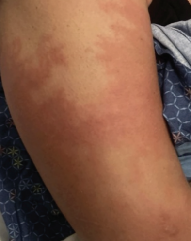
IVH is a rare condition characterized by a clinical presentation of poorly demarcated and irregularly shaped erythematous plaques.1 Although this patient’s presentation was consistent with the erythematous plaques that often accompany IVH, there can be marked variation in clinical appearance. Despite this varied clinical appearance, the histologic pattern is pathognomonic for diagnosis of IVH. Thus, taking a biopsy is necessary for definitive diagnosis.1
Immunohistochemical (IHC) stain is not necessary for diagnosis; however, obtaining an IHC stain can further describe the histopathologic findings as including aggregates of CD68+ histiocytes within dilated lymphatic vessels in the papillary and reticular dermis, an irregular shape of thin walls, and a single discontinuous layer of endothelial cells without nuclear pleomorphism.1 An IHC stain could not be performed in this case due to cells disappearing on deeper levels.
Although also unnecessary for diagnosis, a negative keratin stain would be expected. A keratin stain was performed in this case, and its negative result was interpreted to exclude carcinoma as a possible differential diagnosis.
IVH may be idiopathic or secondary to inflammatory conditions, including rheumatoid arthritis, Crohn’s disease, dermatomyositis, monoclonal gammopathy, metallic prosthesis, chronic infections, and malignancy—notably breast cancer.1 Secondary presentations of IVH are localized to the lesion of the primary condition. In cases associated with rheumatoid arthritis, osteoarthrosis, and metallic prosthesis, the cutaneous reaction localizes near the affected joint; cases associated with mastectomy present with cutaneous lesions localized to the surgical site.1 This localization pattern is hypothesized to be due to localized immune dysregulation.1 In this patient’s case, the cutaneous presentation localized to the upper extremities, further confirming the correlation with her previous breast surgery and axillary lymph node removal.
IVH commonly presents as reticular erythematous or violaceous plaques due to an inflammatory state.1 It is indeed plausible that this presentation without the diagnostic biopsy result of intravascular histiocytes could be attributed to infectious causes, including cellulitis or erysipelas. The cutaneous presentation of erysipelas is characterized as a well-demarcated raised plaque of erythema.2 Further, the most common organism that causes erysipelas is group A streptococcus.2 This patient’s current clinical presentation was poorly demarcated and a negative ASO titer was obtained, allowing erysipelas to be excluded as a potential diagnosis. In addition, the recurrent nature of her rash would be unusual with a diagnosis of erysipelas. With the patient’s previous presentation having been attributed to erysipelas, the antibiotics used for treatment likely provided transient improvement due to the immunomodulatory and anti-inflammatory properties of the drug class, rather than a bacterial cause.3 However, the cutaneous reaction recurred likely due to insufficient immunomodulating treatment. This is further supported by the patient’s current presentation sufficiently improving upon treatment with prednisone and topical triamcinolone.
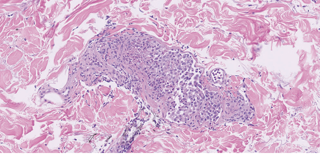
Other differential diagnoses in this case can be excluded due to histopathologic findings. Sweet’s syndrome, which may also present with fever and tender, erythematous plaques, would be characterized by neutrophilic infiltration of the upper dermis on histopathology,4 in contrast to the histiocytes within the dermal vessels seen in IVH. Additionally, given this patient’s history of breast cancer, inflammatory breast cancer could be considered a differential diagnosis. Inflammatory breast cancer can similarly have an erysipeloid appearance with rapid onset and no underlying mass. However, the histopathologic findings in this case did not show invasive carcinoma cells, which would be expected in inflammatory breast cancer.5 Further, the patient’s negative keratin stain argues against a diagnosis of inflammatory breast cancer, as a keratin stain would be expected to be positive.
When considering autoimmune connective tissue disease as a possible differential diagnosis, it is relevant that the cutaneous presentations of cutaneous lupus erythematosus, dermatomyositis, and scleroderma can vary widely. Further, the acute, subacute, and chronic presentations of these autoimmune connective tissue diseases can vary, introducing an incredibly wide array of potential clinical findings.6 While the clinical presentation in this patient’s case does not exclude autoimmune connective tissue diseases, this diagnosis can be excluded as a possibility because they are typically characterized by interface dermatitis on histopathology,6 which was not seen.
Careful diagnosis of IVH is reliant on not only pathologic findings, but also in recognizing high-risk settings of presentations, including the breast cancer history with bilateral mastectomy and ipsilateral lymph node dissection noted in the past medical history of the presented patient. Alternative high-risk clues in the medical history that might lead a physician to consider a diagnosis of IVH may include rheumatoid arthritis, which is implicated in nearly 40% of cases.1
It is also notable that this patient’s cutaneous reaction secondary to mastectomy and axillary lymph node removal manifested approximately 6 years later. This extended timeline is not unprecedented, as a case of IVH reported in association with breast carcinoma excision 9 years prior has been well characterized.3 Therefore, a remote history of breast cancer should not remove the diagnosis of IVH from the differential in a patient with edematous upper extremity plaques. The delayed presentation in this patient was likely due to the pathogenic correlation of IVH with inflammatory conditions. Lymph node removal is commonly associated with lymph stasis, which can cause diminished clearance of antigens and precipitate local immune dysfunction and persistent inflammation.3
Conclusion
This patient was managed with a prednisone taper and topical triamcinolone 0.1% ointment. She subsequently improved, with decreased pain, swelling, and redness and increased mobility. Ultimately, she was lost to follow up. Her case demonstrates how IVH can manifest in high-risk situations, including patients undergoing breast cancer surgery, years after intervention.
© 2026 HMP Global. All Rights Reserved.
Any views and opinions expressed are those of the author(s) and/or participants and do not necessarily reflect the views, policy, or position of the Dermatology Learning Network or HMP Global, their employees, and affiliates.
Shannon C. Lyden is a medical student at The Ohio State University College of Medicine in Columbus, OH. Dr Plaza is a pathologist and Dr Korman is a dermatologist in the department of dermatology at The Ohio State University Wexner Medical Center in Columbus, OH.
Disclosure: The authors report no relevant fi nancial relationships.
References
1. Barba E, Colato C, Girolomoni G. Intralymphatic histiocytosis: a case report and review of literature. J Cutan Pathol. 2015;42(9):593-599. doi:10.1111/cup.12613
2. Michael Y, Shaukat NM. Erysipelas. In: StatPearls [Internet]. StatPearls Publishing; 2025.
3. Requena L, El-Shabrawi-Caelen L, Walsh SN, et al. Intralymphatic histiocytosis. A clinicopathologic study of 16 cases. Am J Dermatopathol. 2009;31(2):140-151. doi:10.1097/DAD.0b013e3181986cc2
4. Vashisht P, Goyal A, Hearth Holmes MP. Sweet syndrome. In: StatPearls [Internet]. StatPearls Publishing; 2025.
5. Chippa V, Barazi H. Inflammatory breast cancer. In: StatPearls [Internet]. StatPearls Publishing; 2025.
6. Reddy BY, Hantash BM. Cutaneous connective tissue diseases: epidemiology, diagnosis, and treatment. Open Dermatol J. 2009;3(1):22-31. doi:10.2174/1874372200903010022