Reed Nevus
Reed nevus, also referred to as pigmented spindle cell nevus or, in some contexts, pigmented spindle cell nevus of Reed, is a benign melanocytic lesion characterized by spindle-shaped melanocytes and prominent pigmentation first described by Dr Richard J. Reed in 1975.1-4 It is considered part of the Spitz nevus spectrum and is most often encountered in adolescents and young adults.2,3 In the fifth edition of the World Health Organization Classification of Skin Tumours, spitzoid melanocytic lesions are no longer conceptualized as a single morphologic category but are instead classified according to their underlying molecular drivers, with morphology understood as a phenotypic consequence of specific oncogenic alterations.5 This modern framework acknowledges that lesions with overlapping histologic features, including Spitz nevus and Reed nevus, may represent biologically distinct entities defined by different kinase fusions rather than mere morphologic variants of the same lesion.5
Case Report
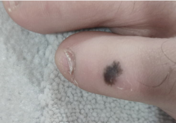
slightly irregular peripheral borders.
A 15-year-old male patient presented with a slightly raised pigmented lesion on the left dorsal aspect of the fifth toe, measuring approximately 6 mm × 4 mm (Figure 1). The lesion developed over the past 2 years, so was not congenital, and the patient reported no recent rapid growth or interval changes. On examination, it demonstrated homogeneous dark-brown central pigmentation with slightly lighter, irregular peripheral borders. The patient was otherwise healthy, and the lesion was asymptomatic, without bleeding or ulceration. He was subsequently referred to a plastic surgeon, who performed an excision of the lesion with a 4-mm margin.
Clinical Presentation and Comparison With Spitz Nevus
Reed nevi typically present as small, darkly pigmented macules or papules, most often in adolescents and young adults and frequently on the lower extremities.1,6 Lesions are usually less than 7 mm in diameter and may exhibit rapid growth, which can raise concern for melanoma due to their striking appearance. A slight female predominance has been reported in the literature.1,2,6
Reed nevus shares many clinical and biologic features with Spitz nevus, reflecting their close relationship within the Spitz nevus spectrum. Both lesions commonly occur in younger patients and may exhibit rapid growth, symmetry, and well-defined borders.2,3 As a result, many dermatopathologists consider Reed and Spitz nevi to represent closely related variants within the same biologic category.
Large-scale genomic studies have demonstrated that Spitz tumors are genetically heterogeneous and are most commonly driven by mutually exclusive kinase fusions involving ROS1, ALK, NTRK1, BRAF, or RET, which together account for the majority of oncogenic events in spitzoid neoplasms.7 These kinase fusions occur across the full biologic spectrum of spitzoid lesions, including benign Spitz nevi, atypical Spitz tumors, and spitzoid melanomas, supporting the concept that molecular alterations precede and shape morphologic appearance rather than determine biologic behavior in isolation.7
Clinically, Reed nevi are characteristically darkly pigmented, ranging from dark brown to black, and are composed almost exclusively of spindle-shaped melanocytes. In contrast, classic Spitz nevi are often pink, red, or lightly pigmented and may contain a mixture of spindle and epithelioid cells.1-3,8 Reed nevi also tend to be smaller and more frequently localized to the lower extremities, whereas Spitz nevi may arise on any body site and are more common in children.1,8
Reed nevi have been shown to exhibit a strong association with NTRK3 fusions, particularly in lesions demonstrating classic pigmented spindle cell morphology, providing molecular melanocytic neoplasia, which emphasizes molecular alterations as primary classifiers and morphology as a secondary expression of tumor biology.10
From a molecular taxonomy perspective, benign Spitz and Reed nevi typically harbor single initiating oncogenic events, such as support for their distinction from other Spitz variants.9 These findings support the concept that Reed nevus represents a molecularly enriched subset within the broader Spitz tumor family rather than simply a heavily pigmented Spitz nevus.9 This distinction aligns with the modern integrated taxonomy of melanocytic neoplasia, which emphasizes molecular alterations as primary classifiers and morphology as a secondary expression of tumor biology.10
From a molecular taxonomy perspective, benign Spitz and Reed nevi typically harbor single initiating oncogenic events, such as kinase fusions, without accompanying secondary alterations in tumor suppressor genes, explaining their limited growth potential and benign clinical behavior.10 Progression to atypical Spitz tumor or spitzoid melanoma requires additional secondary genetic alterations, including involvement of CDKN2A, TP53, or TERT, which are absent in conventional Reed nevi.10
Dermoscopy
The characteristic dermoscopic feature of Reed nevus is the starburst pattern, consisting of regular, symmetric peripheral streaks radiating from a central dark area. This pattern is highly suggestive of Reed nevus and most commonly observed in larger or more mature lesions.11 However, Reed nevi can display a range of dermoscopic patterns depending on their size and stage of evolution. In small or early-stage lesions (< 6 mm), the most frequent patterns are reticular (40%), starburst (20%), globular (6.5%), homogeneous (6.5%), and atypical (27%); these patterns may be present from the outset and are not strictly dependent on lesion age.6 Additional dermoscopic clues include a superficial black network, which corresponds histologically to pigmented parakeratosis and is seen in a minority of cases.12 Reed nevi may also show rapid changes in dermoscopic appearance over time, including loss of the starburst pattern with involution.13
Histology and Immunohistochemistry
In the presented patient case, histologic analysis shows a symmetric, sharply circumscribed proliferation of pigmented spindleshaped melanocytes arranged in confluent junctional nests on the dermoepidermal junction (Figure 2A). In acral and other specialsite nevi, upward scatter of melanocytes into the granular layer and focal melanin deposition within the stratum corneum may be observed, particularly in irritated lesions. These features should not be overinterpreted as malignant in the setting of preserved symmetry and bland cytology. In the presented patient case, pagetoid spread is confined to the central portion of the lesion and lacks pleomorphism, mitotic activity, or asymmetry concerning for melanoma14 (Figure 2B).
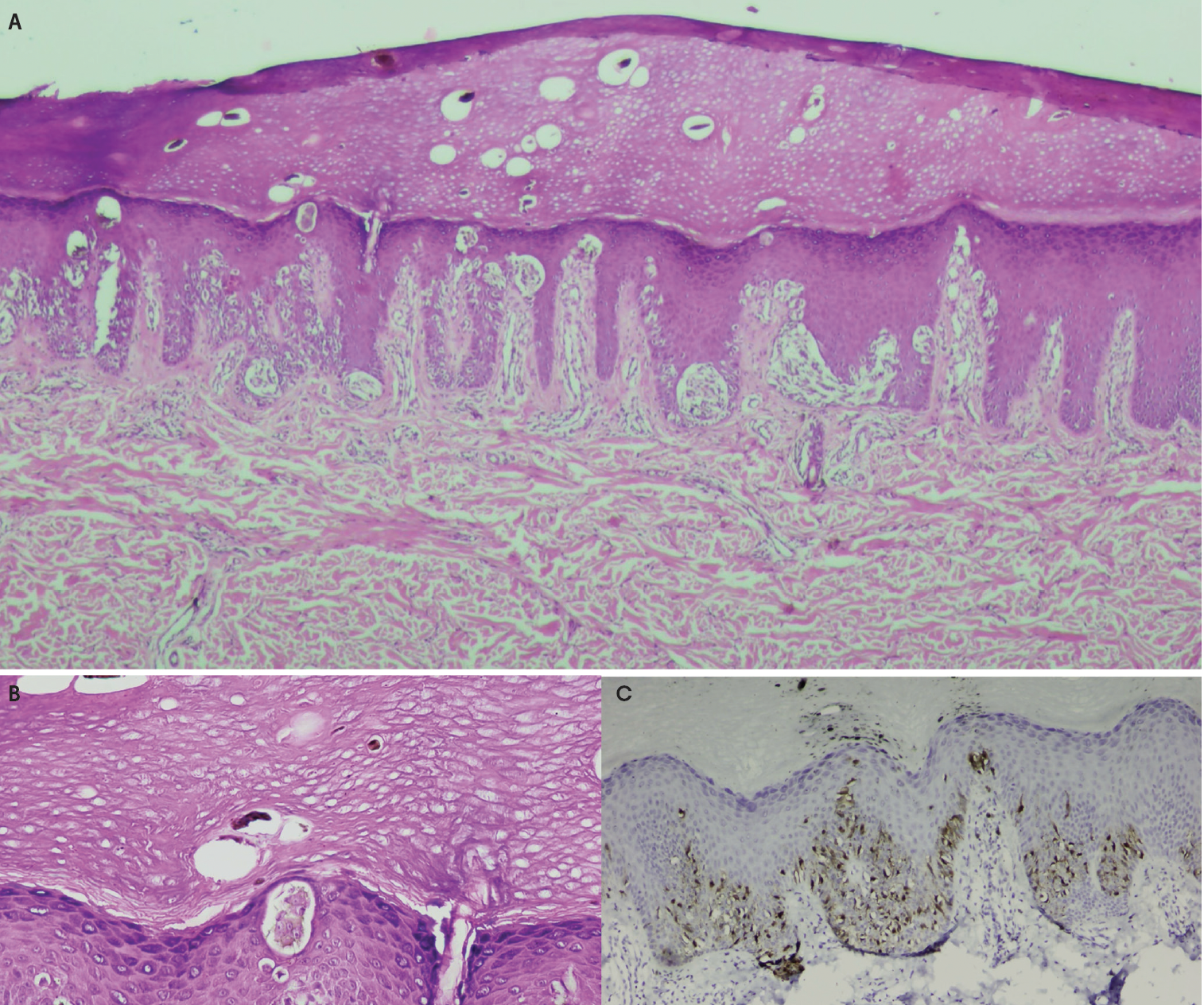
Immunohistochemical (IHC) studies further support the benign nature of this proliferation in the presented patient case. Reed nevi are PRAME negative with retained BAP1 expression.15,16 P16 is also diffusely preserved, consistent with a Spitz or Reed nevus lineage17 (Figure 2C). Ki-67 staining shows increased proliferative activity in the epidermal component but is low in dermal nests, which is typical of acral or irritated Spitz or Reed nevi.18 Melan-A staining shows junctional nests and pagetoid cells without atypia. Taken together, the architecture, cytologic findings, and IHC findings support a diagnosis of a benign acral Reed nevus.
Differential Diagnoses
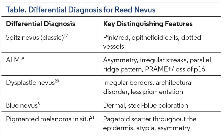
Reed nevus may mimic several benign and malignant melanocytic lesions, particularly when arising on acral sites in adolescents (Table). It must first be distinguished from an early acral lentiginous melanoma (ALM), which may show pagetoid scatter with irregular pigmentation on histologic and clinical examination.19 However, Reed nevi remain symmetrical, have uniform cytology, and have reassuring IHC patterns. Classic Spitz nevi are in the differential due to their shared lineage within the Spitz/Reed spectrum, particularly in less heavily pigmented lesions, but will typically contain a mixture of spindle cells and epithelioid cells, have a pink to red coloration, and be distinguished on dermoscopy by dotted vessels in place of the starburst pattern typical of Reed nevi.17 Dysplastic nevi may be considered, but will display cytologic atypia and architectural disorder while lacking the typical starburst pattern of Reed nevi.20 Blue nevi are heavily pigmented but are dermal and demonstrate a steel-blue coloration.8 Melanoma in situ, particularly the acral lentiginous type, can be excluded as it presents with asymmetry, atypia, and concerning IHC staining.21 Recognizing the characteristic symmetry, starburst pattern, uniform spindle cell morphology, and reassuring IHC patterns prevents overdiagnosis and unnecessary re-excision of this benign pigmented lesion.
Management
Complete excision is considered curative for Reed nevi. When the lesion is removed with clear margins, as in the presented patient case, no additional treatment, re-excision, or routine follow up is recommended. Recurrence is rare. In cases where the diagnosis is uncertain, such as when the lesion arises in an acral site, excisional biopsy with narrow margins is the preferred approach. If margins are involved or if histopathologic features are concerning for an atypical or malignant lesion, re-excision may be considered according to the degree of atypia. For definitively diagnosed Reed nevi, patient reassurance is appropriate, emphasizing the benign nature of this lesion and that there is no need for further intervention after removal.14,22
Conclusion
Reed nevi are benign melanocytic lesions that most often present in adolescents and young adults as small, heavily pigmented macules or papules. Dermoscopically, the starburst pattern may raise concern for melanoma and prompt excisional biopsy. Histologically, Reed nevi demonstrate junctional nests of pigmented, spindle-shaped melanocytes and may show limited pagetoid scatter in acral lesions. IHC reveals reassuring benign features, including PRAME negativity, preserved p16 expression, and low dermal proliferative activity. Complete excision is curative.
References
1. Díaz A, Valera A, Carrera C, et al. Pigmented spindle cell nevus: clues for differentiating it from spindle cell malignant melanoma. A comprehensive survey including clinicopathologic, immunohistochemical, and FISH studies. Am J Surg Pathol. 2011;35(11):1733-1742. doi:10.1097/PAS.0b013e318229cf66
2. VandenBoom T, Quan VL, Zhang B, et al. Genomic fusions in pigmented spindle cell nevus of Reed. Am J Surg Pathol. 2018;42(8):1042-1051. doi:10.1097/ PAS.0000000000001074
3. Requena C, Requena L, Sánchez-Yus E, et al. Hypopigmented Reed nevus. J Cutan Pathol. 2008;35(Suppl 1):87-89. doi:10.1111/j.1600-0560.2007.00957.x
4. Mohan LS, Khan AU, Zhang B, et al. Retrospective cohort: genomic differences between pigmented spindle cell nevi of Reed and Reed-like melanomas. Am J Dermatopathol. 2020;42(9):641-647. doi:10.1097/DAD.0000000000001603
5. Goldman-Lévy G, Barnhill R, Bastian BC, et al. WHO classification of skin tumours: key updates in the fifth edition. Histopathology. 2026;88(3):555-568. doi: 10.1111/his.15562
6. de Giorgi V, Savarese I, Rossari S, et al. Clinical and dermoscopic features of small Reed nevus (<6 mm). J Eur Acad Dermatol Venereol. 2013;27(7):919-921. doi:10.1111/ j.1468-3083.2012.04457.x
7. Wiesner T, He J, Yelensky R, et al. Kinase fusions are frequent in Spitz tumours and spitzoid melanomas. Nat Commun. 2014;5(1):3116. doi:10.1038/ncomms4116
8. Moscarella E, Ronchi A, Brancaccio G, et al. Spectrum of blue nevus-like lesions, including blue nevus, pigmented epithelioid melanocytoma, and animal-type melanoma. Clin Dermatol. 2025;43(3):323-333. doi:10.1016/j.clindermatol.2024.09.003
9. Yeh I, Tee MK, Botton T, et al. NTRK3 kinase fusions in Spitz tumours. J Pathol. 2016;240(3):282-290. doi:10.1002/path.4775
10. Bastian BC. The molecular pathology of melanoma: an integrated taxonomy of melanocytic neoplasia. Annu Rev Pathol. 2014;9:239-271. doi:10.1146/annurevpathol- 012513-104658
11. Zalaudek I, Docimo G, Argenziano G. Using Dermoscopic criteria and patientrelated factors for the management of pigmented melanocytic nevi. Arch Dermatol. 2009;145(7):816-826. doi:10.1001/archdermatol.2009.115
12. Argenziano G, Soyer HP, Ferrara G, et al. Superficial black network: an additional dermoscopic clue for the diagnosis of pigmented spindle and/or epithelioid cell nevus. Dermatology. 2001;203(4):333-335. doi:10.1159/000051784
13. Fikrle T, Pizinger K. Rapid growth and slow involution of Reed nevus in a long-term dermoscopic follow-up. Pediatr Dermatol. 2022;39(6):1000-1001. doi:10.1111/pde.15085
14. Barnhill RL, Barnhill MA, Berwick M, Mihm MC. The histologic spectrum of pigmented spindle cell nevus: a review of 120 cases with emphasis on atypical variants. Human Pathology. 1991;22(1):52-58. doi:10.1016/0046-8177(91)90061-S
15. Warbasse E, Mehregan D, Utz S, Stansfield RB, Abrams J. PRAME immunohistochemistry compared to traditional FISH testing in spitzoid neoplasms and other difficult to diagnose melanocytic neoplasms. Front Med (Lausanne). 2023;10:1265827. doi:10.3389/ fmed.2023.1265827
16. Wiesner T, Obenauf AC, Murali R, et al. Germline mutations in BAP1 predispose to melanocytic tumors. Nat Genet. 2011;43(10):1018-1021. doi:10.1038/ng.910
17. Luo S, Sepehr A, Tsao H. Spitz nevi and other Spitzoid lesions part I. Background and diagnoses. J Am Acad Dermatol. 2011;65(6):1073-1084. doi:10.1016/j.jaad.2011.04.040
18. Díaz A, Valera A, Carrera C, et al. Pigmented spindle cell nevus: clues for differentiating it from spindle cell malignant melanoma. A comprehensive survey including clinicopathologic, immunohistochemical, and FISH studies. Am J Surg Pathol. 2011;35(11):1733-1742. doi:10.1097/PAS.0b013e318229cf66
19. Warren MP, Harvey VM. Acral lentiginous melanoma. N Engl J Med. 2015;373(19):1864-1864. doi:10.1056/NEJMicm1500906
20.Drozdowski R, Spaccarelli N, Peters MS, Grant-Kels JM. Dysplastic nevus part I: historical perspective, classification, and epidemiology. J Am Acad Dermatol. 2023;88(1):1- 10. doi:10.1016/j.jaad.2022.04.068
21. Elbendary A, Xue R, Valdebran M, et al. Diagnostic criteria in intraepithelial pagetoid neoplasms: a histopathologic study and evaluation of select features in Paget disease, Bowen disease, and melanoma in situ. Am J Dermatopathol. 2017;39(6):419-427. doi:10.1097/DAD.0000000000000704
22. Beijnen UEA, Walsh LR, Nuzzi LC, Schmidt BAR, Labow BI, Taghinia AH. Management of residual Spitz nevus in surgical specimens following biopsy and excision. Plast Reconstr Surg Glob Open. 2020;8(12):e3244. doi:10.1097/GOX.0000000000003244