

Superficial Femoral Artery Aneurysm as a Cause of Deep Vein Thrombosis Treated With a Covered Stent
Adrian Mercado-Alamo, MD; Anwar Zaitoun, MD; Saroj Neupane, MD; Thomas Davis, MD
J INVASIVE CARDIOL 2018;30(11):E124-E125.
Key words: cardiac imaging, computed tomography angiography, deep vein thrombosis
Superficial femoral artery (SFA) aneurysm occurs in approximately 5 per 100, 000 patients. Deep vein thrombosis due to SFA aneurysm is an extremely rare condition that develops due to direct compression from the aneurysm on a segment of the venous system.
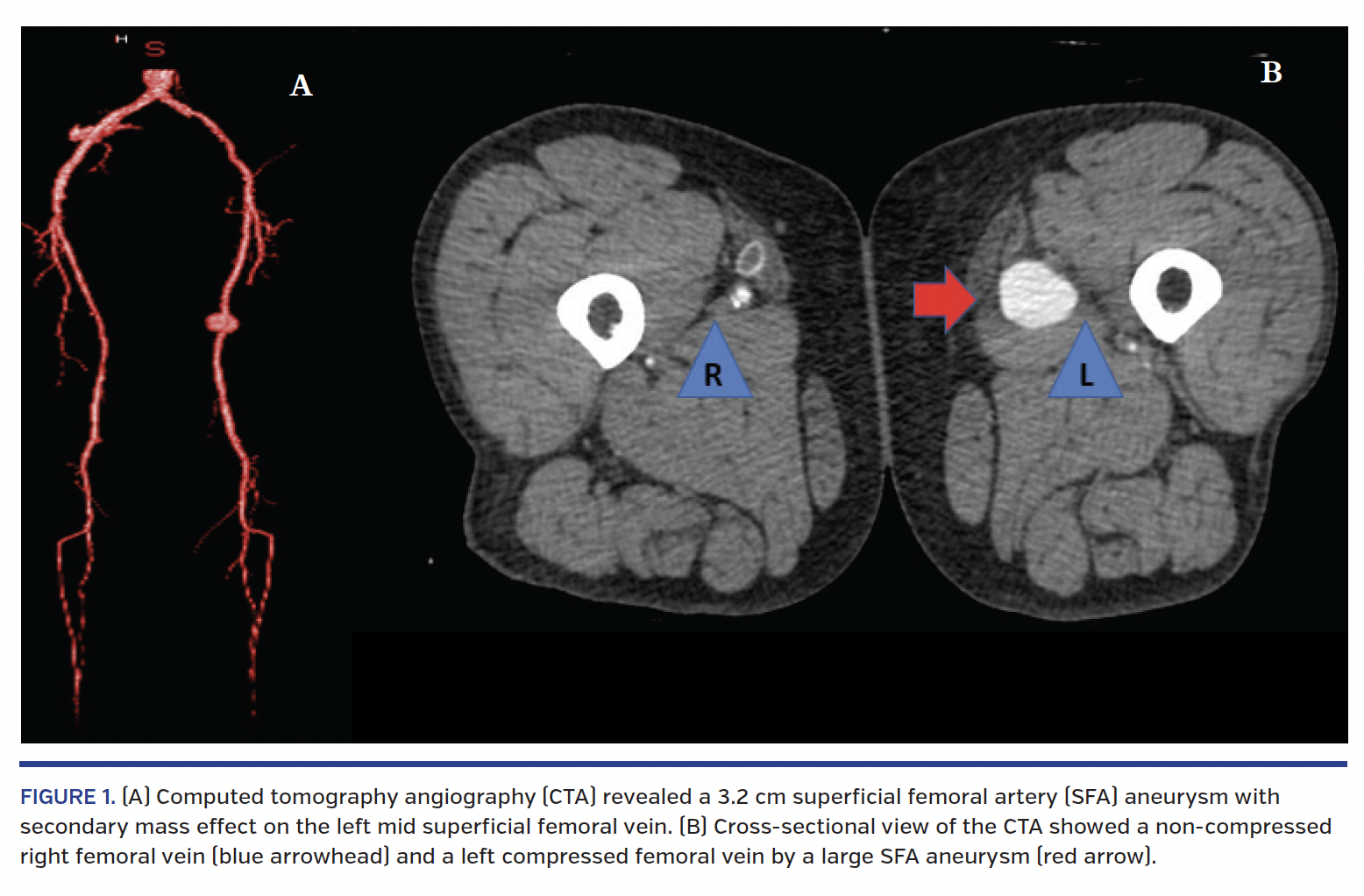
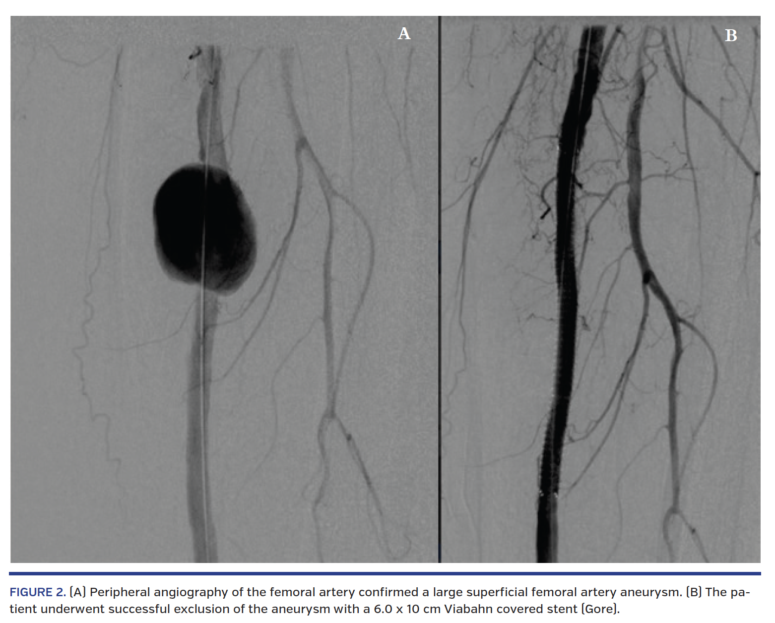
We present a 57-year-old female who came to the emergency department complaining of left thigh pain and swelling for a 2-week duration. The patient had recently undergone a left SFA intervention at an outside facility due to significant peripheral artery disease. Physical exam revealed a non-cyanotic extremity, warm with adequate capillary refill, and palpable pulses throughout bilaterally. Venous duplex ultrasonography revealed acute thrombosis of the superficial femoral vein. Computed tomography angiography (CTA) revealed a 3.2 cm SFA aneurysm (Figure 1) with secondary mass effect on the left mid superficial femoral vein. Peripheral angiography of the femoral artery confirmed a large SFA aneurysm (Figure 2A). The patient underwent successful exclusion of the aneurysm with a 6.0 x 10 cm Viabahn covered stent (Gore) (Figure 2B) and was discharged home afterward on oral anticoagulation regimen for 3 months.
From the Department of Cardiology, St. John Providence Health System, Detroit, Michigan.
Disclosure: The authors have completed and returned the ICMJE Form for Disclosure of Potential Conflicts of Interest. The authors report no conflicts of interest regarding the content herein.
Manuscript accepted May 18, 2018.
Address for correspondence: Anwar Zaitoun, MD, St. John Hospital and Medical Center, 22101 Moross Rd, Detroit, MI 48236. Email: anzaitoun@gmail.com


