
Advertisement

ADVERTISEMENT

Clinical Images

The Use of a Homemade "BAG" Repositioning System to Salvage Complete Coronary Stent Dislodgement

Siu-Fung Wong, MBChB; Tak-Shun Chung, MBBS

March 2024
1557-2501
J INVASIVE CARDIOL 2024;36(3). doi:10.25270/jic/23.00153. Epub February 26, 2024.


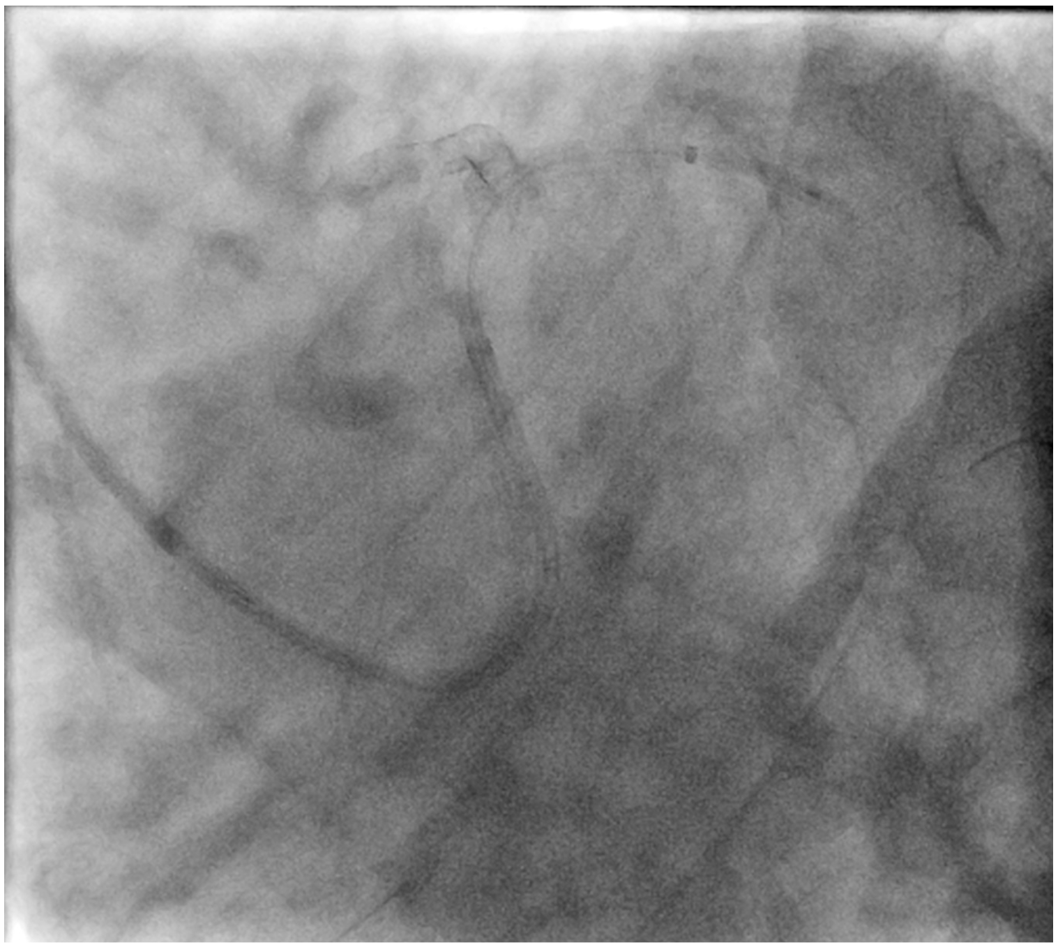
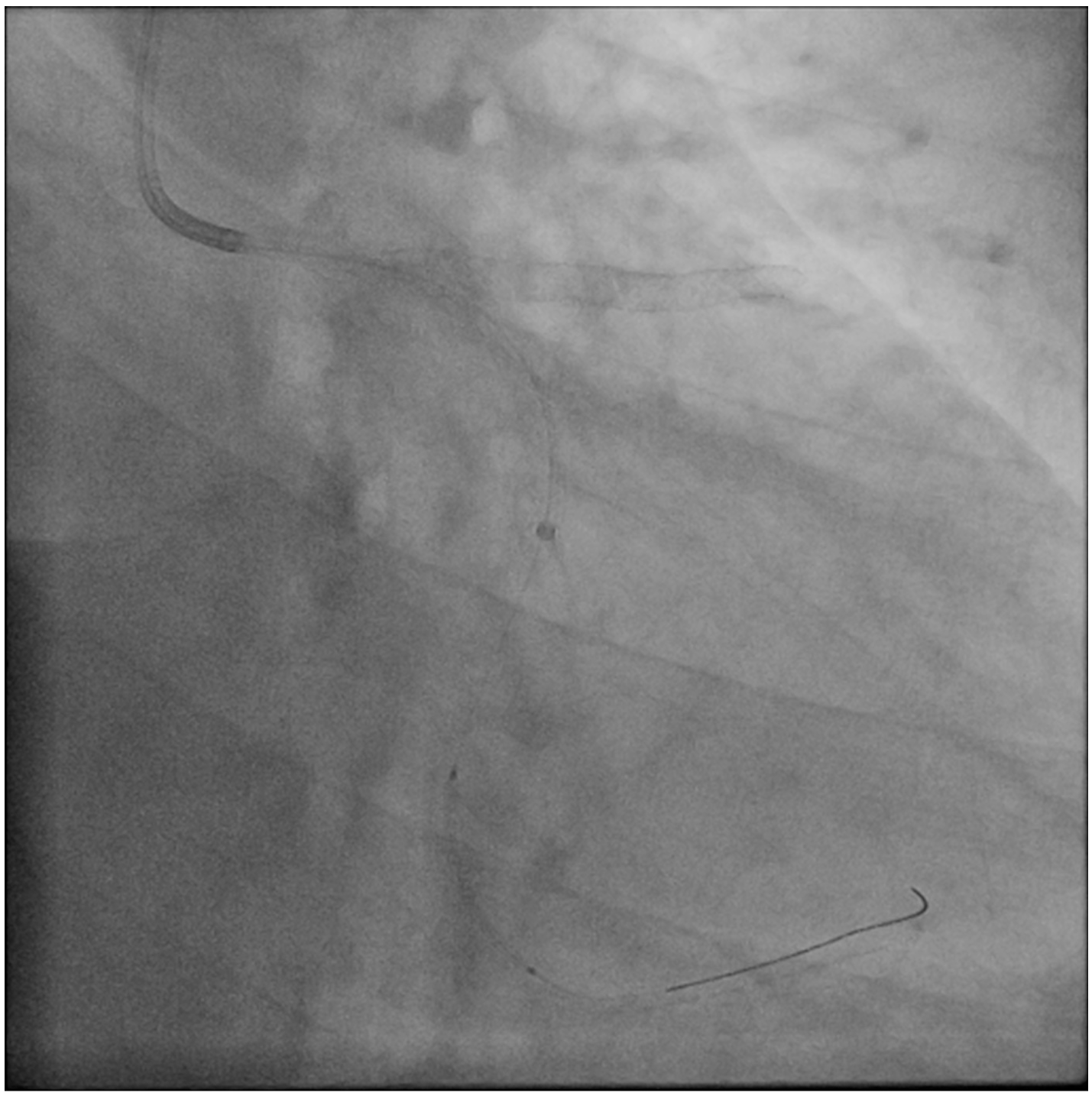
A 71-year-old man who had undergone percutaneous transluminal coronary angioplasty (PTCA) in 2013 was admitted for unstable angina. Coronary angiogram showed 2 de novo lesions at the proximal and distal left circumflex artery (LCX) (Figure 1A). PTCA was performed transradially via a 6-French (Fr) sheath. With a 6-Fr extra-backup 3.5 guide catheter engaging to the left main coronary artery (LM), a 2.5 x 18-mm drug-eluting stent (DES) was delivered to the distal LCX lesion but was inadequate for complete lesion coverage (Figure 1B). During retrieval, the stent was found dislodged at the distal LM bifurcation (Figure 1C).

Figure 1. (A) Preprocedural angiogram.
Figure 1. Coronary stent dislodgement salvaged by the Balloon-Assisted Guide-extension system. (A) Preprocedural angiogram.
Figure 1. (B) Inadequate stent length for distal LCX lesion.
Figure 1. Coronary stent dislodgement salvaged by the Balloon-Assisted Guide-extension system. (B) Inadequate stent length for the distal left circumflex artery lesion.
Figure 1. (C) Stent dislodged during retrieval (arrow); asterisk (*) indicates stent balloon.
Figure 1. Coronary stent dislodgement salvaged by the Balloon-Assisted Guide-extension system. (C) Stent dislodged during retrieval (arrow); asterisk (*) indicates stent balloon.


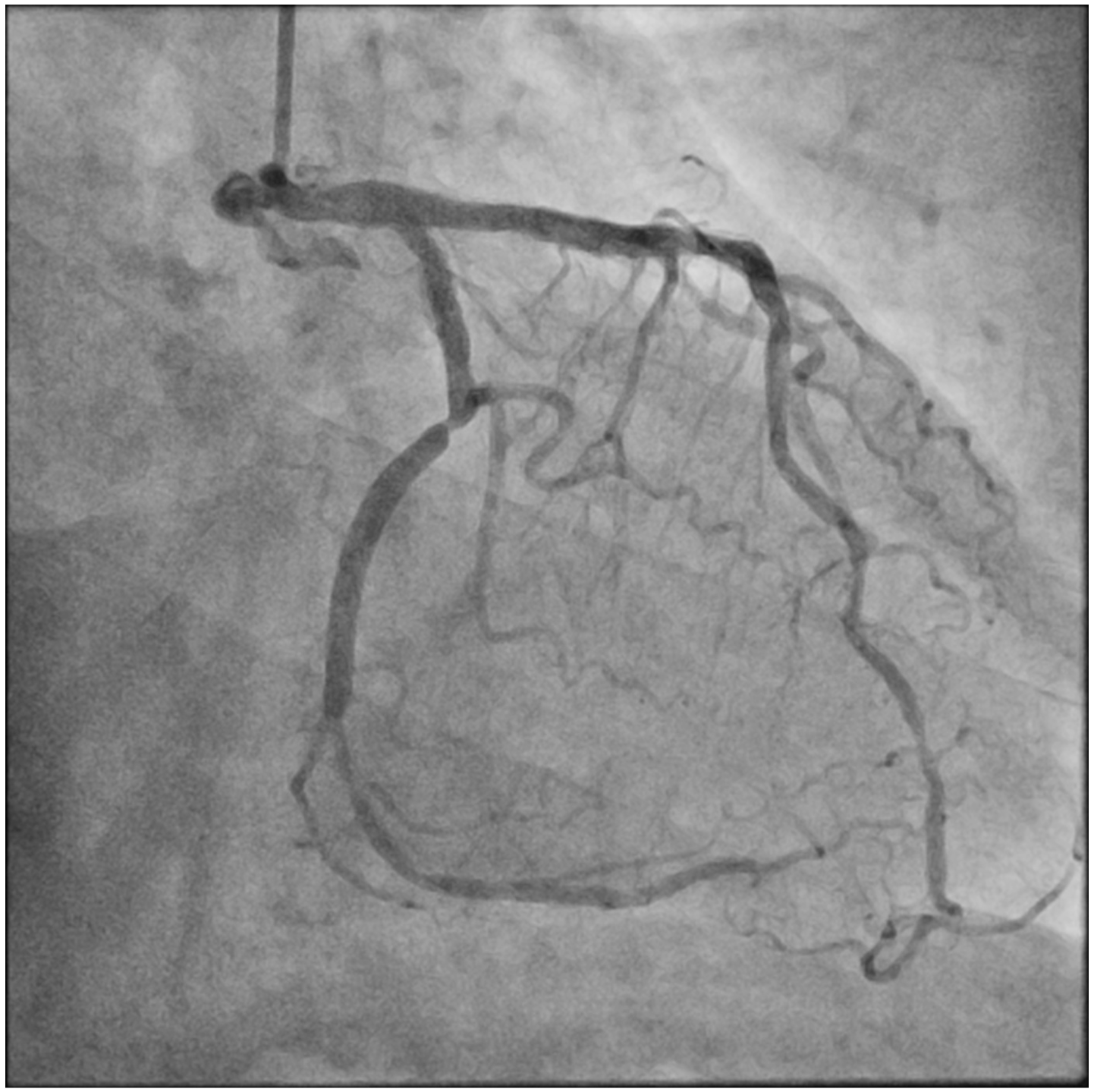
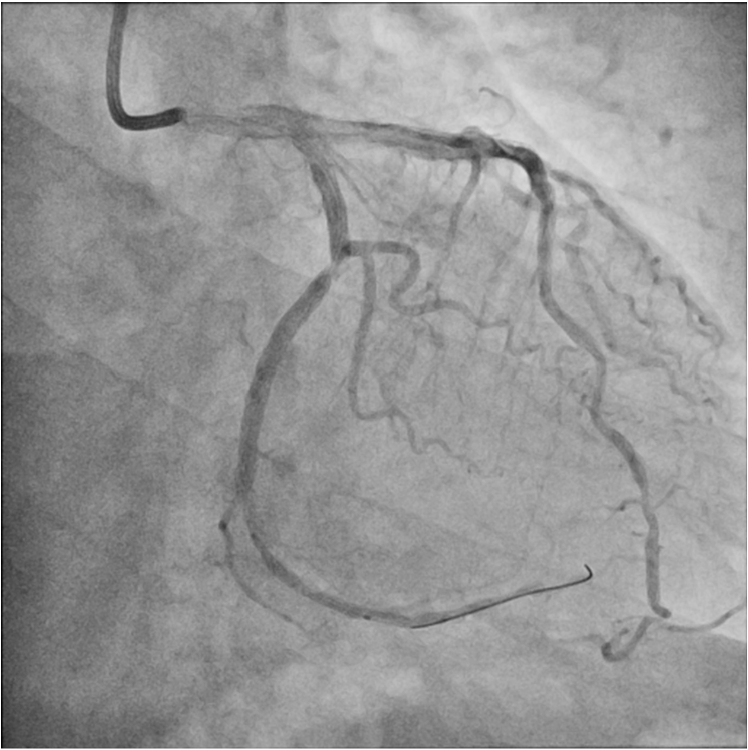
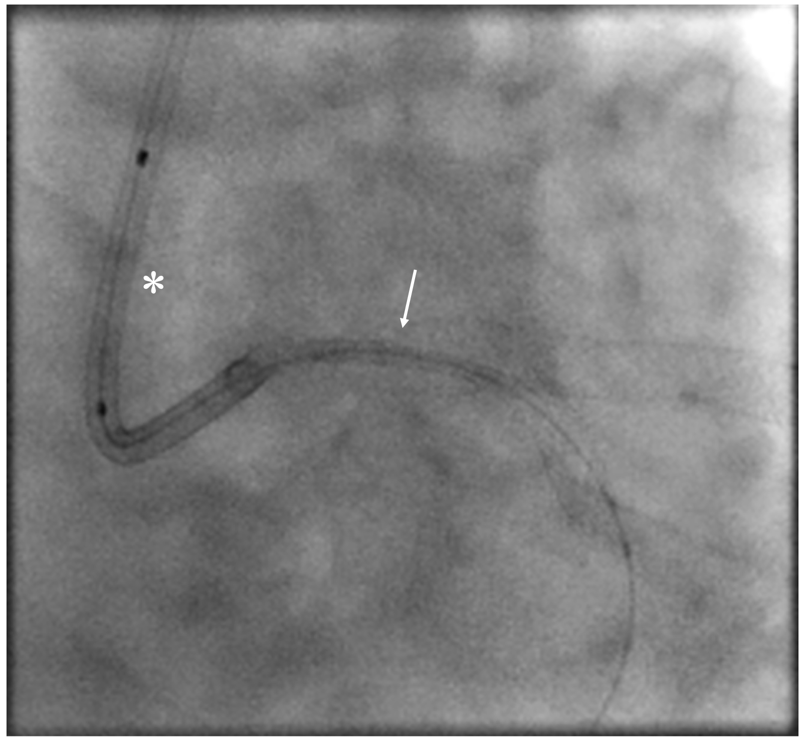
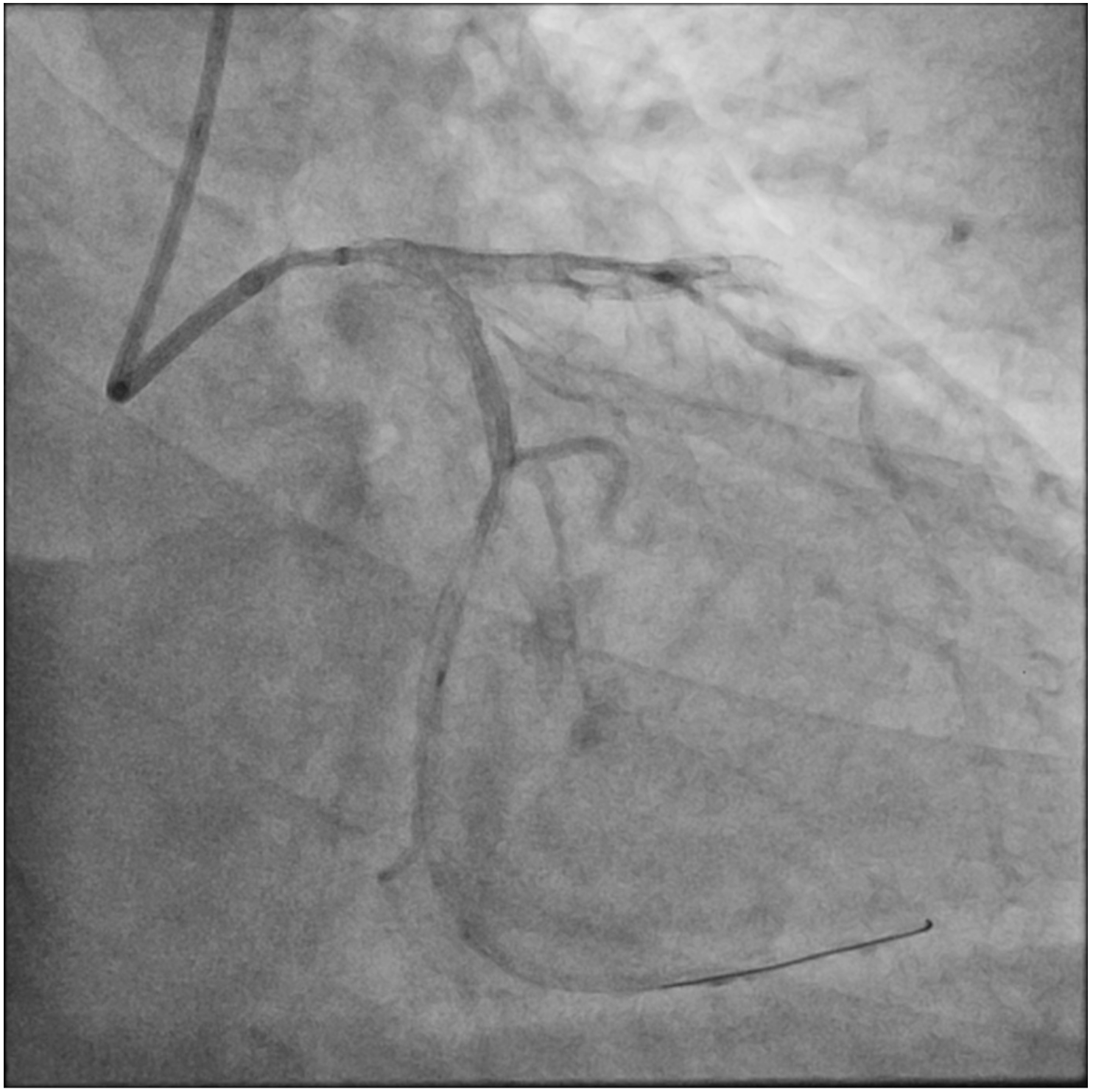
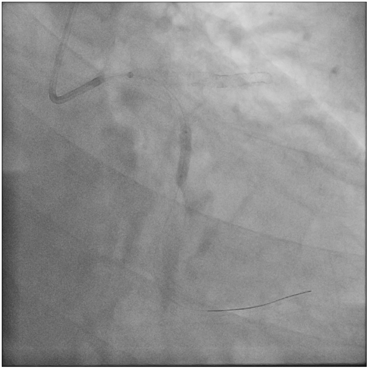
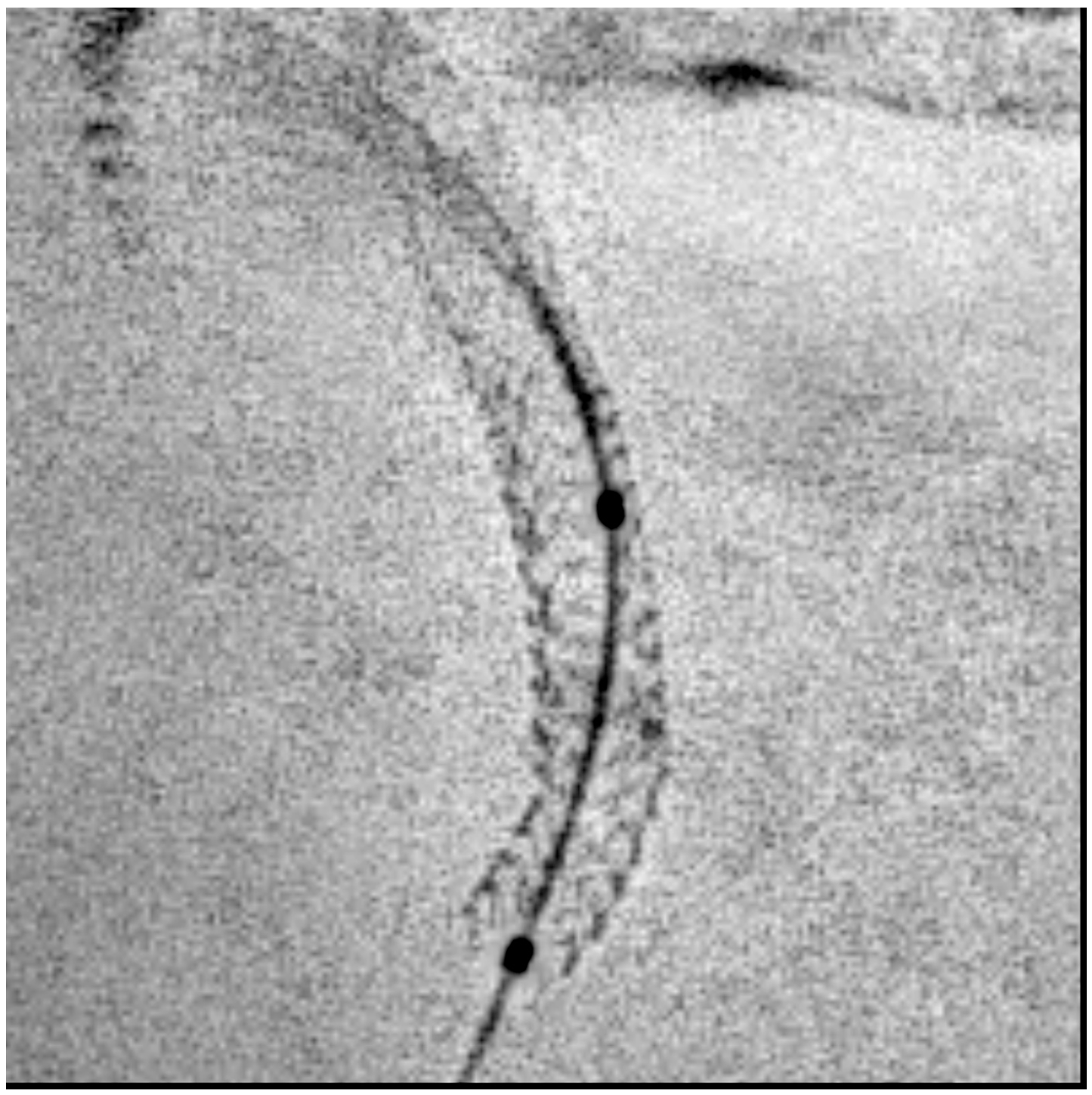
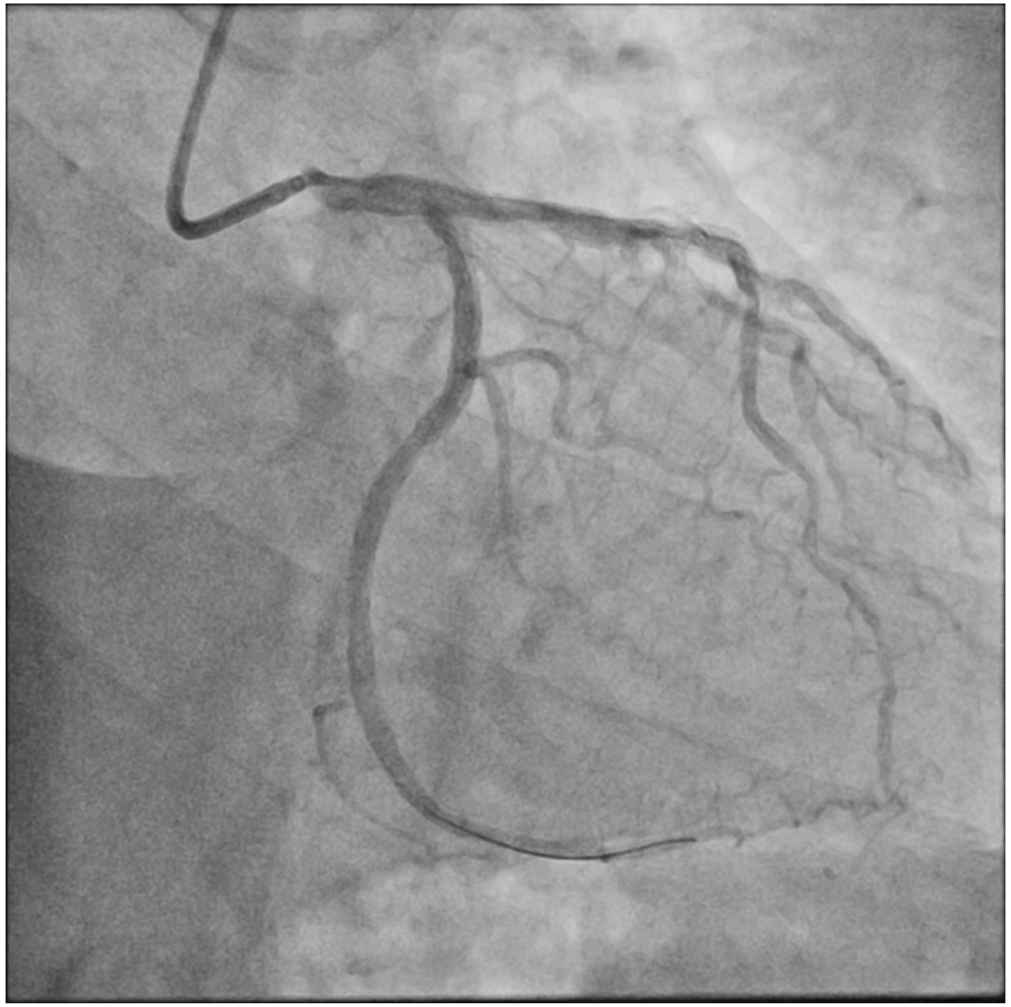
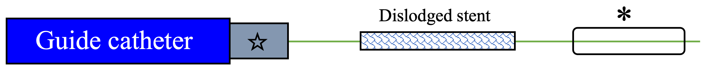
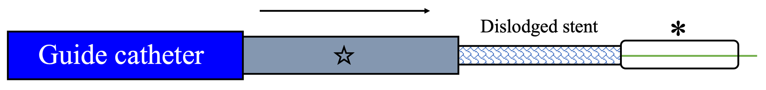
Distal small balloon retrieval was unsuccessful (Figure 1D). With the help of the Balloon-Assisted Guide-extension (BAG) system, the stent could be optimally positioned at the proximal LCX lesion by forward pushing with a 6-Fr Guideliner V3 catheter (Teleflex) and backward pulling by the small balloon (Figure 1E and F; Video). It was then deployed by sequential balloon dilatation with good expansion (Figure 1G and H). Another 2.5 x 24-mm DES was deployed at the distal LCX, which gave an excellent final angiographic result (Figure 1I and J).


Figure 1. (D) Failed retrieval of dislodged stent
Figure 1. Coronary stent dislodgement salvaged by the Balloon-Assisted Guide-extension system. (D) Failed retrieval of dislodged stent into the Guideliner (Teleflex); asterisk (*) indicates 1.5-mm semi-compliant balloon.
Figure 1. Coronary stent dislodgement salvaged by the Balloon-Assisted Guide-extension (BAG) system. (E) Repositioning of dislodged stent by the BAG system.
Figure 1.  (F) Angiography
Figure 1. Coronary stent dislodgement salvaged by the Balloon-Assisted Guide-extension system. (F) Angiography confirming optimal position of stent at the proximal left circumflex artery.
Figure 1. (G) Sequential balloon dilatation.
Figure 1. Coronary stent dislodgement salvaged by the Balloon-Assisted Guide-extension system. (G) Sequential balloon dilatation.
Figure 1. (H) Good stent expansion.
Figure 1. Coronary stent dislodgement salvaged by the Balloon-Assisted Guide-extension system. (H) Good stent expansion.
Figure 1. (I) Second DES to distal LCX.
Figure 1. Coronary stent dislodgement salvaged by the Balloon-Assisted Guide-extension system. (I) Second drug-eluting stent to distal left circumflex artery.
Figure 1. (J) Final angiography.
Figure 1. Coronary stent dislodgement salvaged by the Balloon-Assisted Guide-extension system. (J) Final angiography.

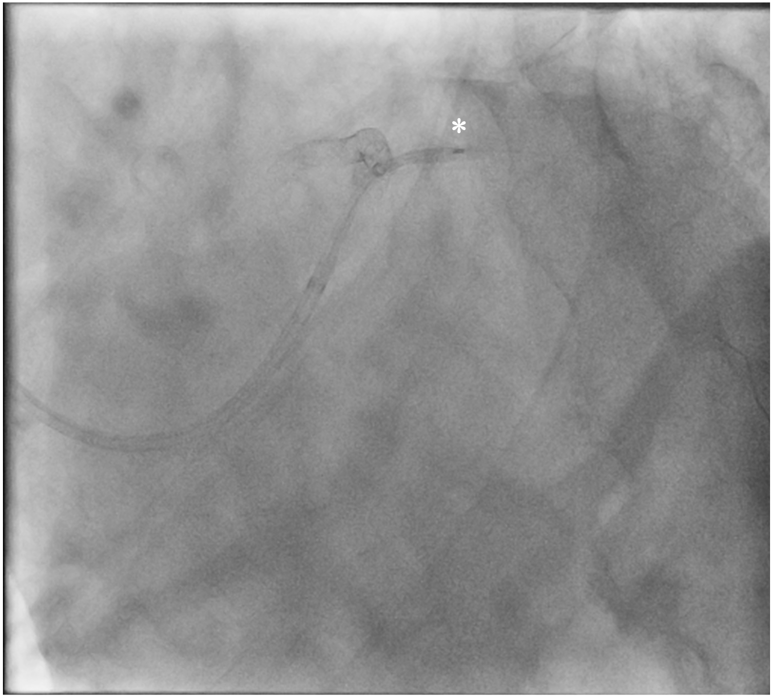
Our case illustrates a novel repositioning system that can relocate and deploy a lost stent at an optimal site even in the absence of its original stent platform (Figure 2A-C). This can avoid wasting stents and serve as a viable bailout when conventional retrieval methods are unsuccessful.


Figure 2. (A) system setup
Figure 2. Schematic illustration of the Balloon-Assisted Guide-extension system: (A) system setup. Asterisk (*), small semi-compliant balloon; star, guide extension.
Figure 2. (B) distal small balloon retraction
Figure 2. Schematic illustration of the Balloon-Assisted Guide-extension system: (B) distal small balloon retraction. Asterisk (*), small semi-compliant balloon; star, guide extension.
Figure 2. (C) advancement by guide extension.
Figure 2. Schematic illustration of the Balloon-Assisted Guide-extension system: (C) advancement by guide extension. Asterisk (*), small semi-compliant balloon; star, guide extension.


Affiliations and Disclosures

From the Division of Cardiology, Department of Medicine & Geriatrics, United Christian Hospital, Hong Kong SAR, China

Disclosures: The authors report no financial relationships or conflicts of interest regarding

the content herein.

Address for correspondence: Siu-Fung Wong, MBChB, Department of Medicine &

Geriatrics, United Christian Hospital, Hong Kong SAR, China. Email: anthonywaaf1992@hotmail.com


Advertisement

Advertisement

Advertisement