Access to Atrial Flow Regulators for Implantation in Critically Ill Children Via the US Food and Drug Administration’s Emergency Use Pathway
© 2026 HMP Global. All Rights Reserved.
Any views and opinions expressed are those of the author(s) and/or participants and do not necessarily reflect the views, policy, or position of the Journal of Invasive Cardiology or HMP Global, their employees, and affiliates.
J INVASIVE CARDIOL 2026. doi:10.25270/jic/26.00133. Epub July 28, 2026.
Key Clinical Summary
- Occlutech Atrial Flow Regulator (AFR) implantation through the FDA’s emergency use authorization (EUA) pathway was successfully performed in 8 critically ill pediatric patients with complex congenital heart disease after approved treatment options were exhausted.
- All patients progressed clinically after AFR implantation; 2 required repeat catheterization for device thrombosis, demonstrating overall feasibility with acceptable procedural safety in emergent pediatric cardiac care.
- The EUA pathway may provide timely access to investigational pediatric cardiac devices when no approved alternatives exist, supporting lifesaving intervention and future regulatory evidence generation.
Abstract
Objectives. The authors conducted a review of the US Food and Drug Administration’s (FDA) emergency use authorization (EUA) pathway as a mechanism to use the Occlutech Atrial Flow Regulator (AFR) (Occlutech International AB) for critically ill pediatric patients with congenital heart disease.
Methods. The authors describe 8 pediatric patients in whom the EUA pathway was activated to permit AFR implantation and provide potentially lifesaving therapy.
Results. Emergency authorization was granted for each of the 8 cases with successful implantation of the AFR. There were 2 cases in which the patient required a repeat catheterization for thrombus on the device, but all 8 patients were able to progress their care. These cases demonstrate the feasibility of the EUA pathway to access emerging technologies for pediatric patients.
Conclusions. The Occlutech AFR may provide an important therapeutic option for critically ill children with congenital heart disease. The EUA pathway can facilitate timely access to unapproved but potentially beneficial technologies when no satisfactory approved alternatives exist.
Introduction
The Occlutech Atrial Flow Regulator (AFR) (Occlutech International AB) is a self-expanding nitinol device designed to provide a controlled atrial septal communication in patients with heart failure. Available in multiple sizes (Figures 1 and 2), the introduction of this device has encouraged congenital cardiologists to consider its use in patients with hypoplastic left heart syndrome (HLHS) with restrictive atrial septums and those with failing Fontan physiology.
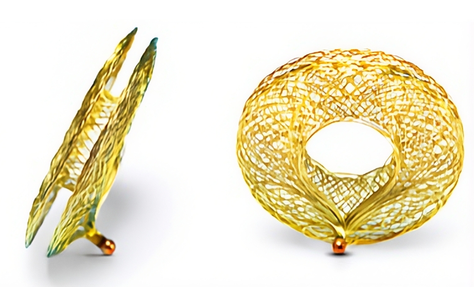
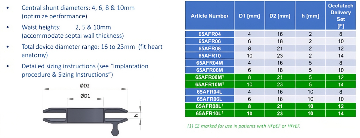
Although the device has been used off label in Europe and in the United States for over 6 years, it remains an investigational product and is available only under expanded access pathways, such as compassionate or emergency use authorization by the US Food and Drug Administration (FDA).1
In adult cardiology, the AFR has been shown to be safe, easy to implant, and offer symptom relief in both heart failure with reduced (HFrEF) and preserved ejection fraction (HFpEF).2-4 Pediatric experience has increased in recent years with early data from Europe and select US centers supporting its utility in complex congenital heart disease and pulmonary hypertension.5-7
Congenital heart disease often presents unique anatomical and physiological challenges that require innovative, patient-specific interventions. In urgent and life-threatening scenarios where no FDA-approved alternative exists, physicians can request special permission from the FDA to use unapproved devices under the emergency use provisions.8 This case series details our institution’s experience using the FDA’s emergency use authorization (EUA) pathway to obtain AFR devices for critically ill pediatric patients with limited viable therapeutic alternatives.
Methods
We conducted a retrospective, descriptive review of 8 pediatric cases in which the Occlutech AFR was implanted under the FDA emergency use mechanism. All cases were discussed in a multidisciplinary forum involving interventional cardiology, cardiothoracic surgery, and critical care teams. Informed consent for the procedure and for inclusion in publication was obtained from the patients' parents or guardians prior to each procedure. Emergency use authorization was obtained through direct communication with the FDA and in collaboration with Occlutech. Institutional review board (IRB) notification and FDA compliance oversight were obtained for each procedure.
All interventions were performed by experienced congenital interventional cardiologists. Device sizing and implantation strategies were individualized based on each patient’s anatomy and hemodynamic status. The waist height was determined based on the measurement of thickness of the septum or Fontan conduit on echocardiogram. The determination of the fenestration size was based on clinical judgement of the interventionalist regarding the degree of shunting needed based on the patient’s physiology and clinical status.
CLINICAL CASES
Case 1: Failing Fontan requiring venoarterial extracorporeal membrane oxygenation (VA-ECMO)
A 5-year-old girl with hypoplastic left heart syndrome (HLHS) underwent an extracardiac Fontan with a 4-mm fenestration. Upon arrival in the cardiac intensive care unit (CICU), she demonstrated high oxygen saturation and low cardiac output with escalating inotropic and vasopressor support. She was emergently returned to the operating room for cannulation onto VA-ECMO. By postoperative day (POD) 2, she had not made any progress clinically and was unable to tolerate any weaning of mechanical support. She was taken to the cardiac catheterization laboratory urgently for hemodynamic evaluation. Baseline hemodynamics revealed elevated Fontan pressure (16 mm Hg) and right ventricular (RV) end-diastolic pressure (9 mm Hg) with a saturation of 93%, while on full ECMO flow at 90 cc/kg/min giving a cardiac index of 2.2 L/min/m2. Angiography of the inferior vena cava revealed a tiny fenestration with minimal right to left shunting.
A rapid conference was convened, and we agreed that a large reliable fenestration may allow progress from mechanical support. An 8 mm x 2-cm Conquest balloon (BD) was used to dilate the fenestration and prepare it for AFR implantation. Under transesophageal echocardiogram (TEE) and fluoroscopic guidance, a 6-mm fenestration size x 5-mm height AFR was successfully deployed across the fenestration with satisfactory angiographic and echocardiographic result. A Fontan pressure post-AFR was recorded (12 mm Hg) and saturations were stable around 85%.
The patient was able to wean off inotropes and vasopressors and was weaned and decannulated from ECMO 3 days after AFR placement. She was discharged 10 days later. A repeat elective catheterization was performed 1 month later, during which her Fontan circuit pressures remained low at 11 mm Hg and saturations were still in the mid 80s.
Case 2: Failed Fontan with single ventricle assist device (sVAD)
A 2-year-old boy with HLHS and a history of extracardiac fenestrated Fontan suffered from recalcitrant chylothorax, elevated pulmonary vascular resistance, and low cardiac output despite surgical pleurodesis and thoracic duct ligation. Initial catheterization revealed elevated Fontan circuit pressures, for which he underwent serial balloon dilations of the fenestration with up to an 8-mm noncompliant balloon to intentionally significantly decompress the Fontan circuit. Several months later he presented with moderate to severely reduced RV function. He was admitted to the heart failure service and was treated with standard heart failure therapy before ultimately going to the operating room for placement of an sVAD. The combination of the sVAD and a large fenestration resulted in profound desaturation, hindering the optimization of his sVAD flows.
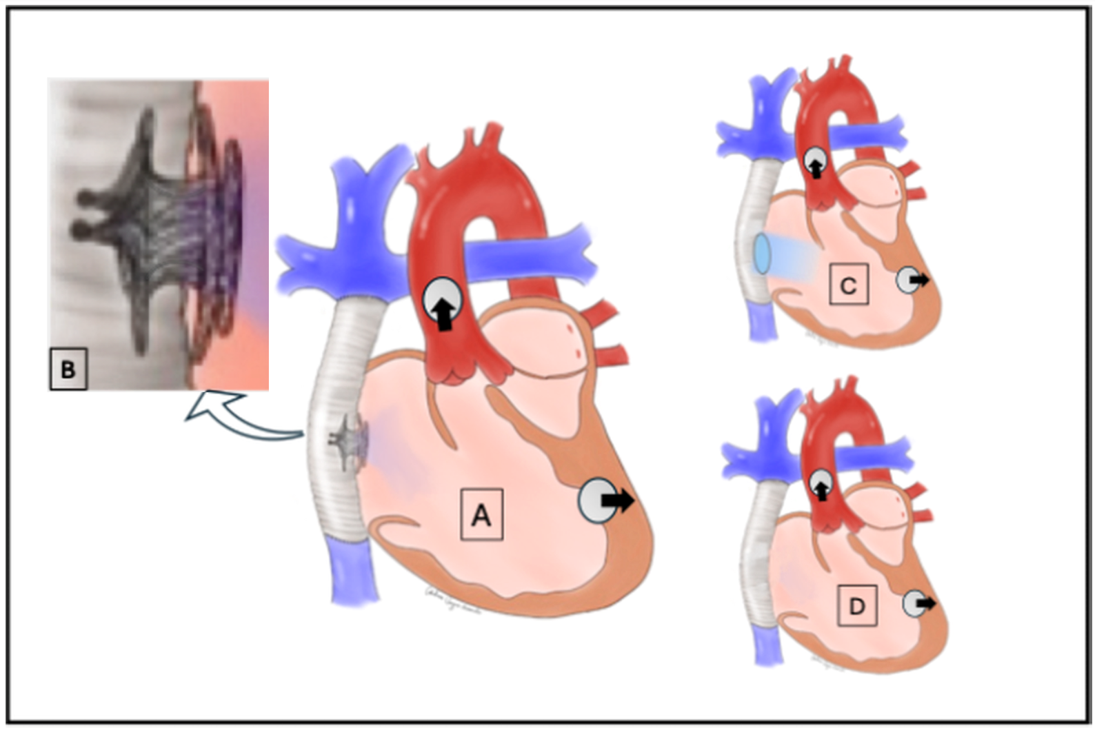
Two weeks later, he was taken to the catheterization laboratory where a 4-mm fenestration x 5-mm height AFR was deployed into the fenestration. When the sVAD flow was optimized, there was still significant hypoxia; hence, we implanted a second 4 x 5-mm device nested within the first (Figure 3). This combination provided enough restriction to the flow being drawn across the fenestration, and the saturations improved to the high 70s while still in the catheterization laboratory. The patient stabilized and remained admitted on heart failure treatment and sVAD with saturations in the low 80’s until cardiac transplant 13 months later. He was discharged home 2 weeks following the heart transplant.
Case 3: Pulmonary hypertension leading to cardiac arrest
A 15-year-old girl with pulmonary arterial hypertension (PAH) secondary to interstitial lung disease and prematurity (25-week gestation twin) experienced recurrent syncope and RV dysfunction. The patient had previously been maintained on dual oral pulmonary antihypertensive therapy, but over time developed worsening PAH with multiple episodes of syncope and evidence of moderately depressed RV function, which prompted admission to the CICU. While in the CICU, the patient suffered another pulmonary hypertensive crisis associated with cardiac arrest. She was successfully resuscitated, and after a short period of stability, the decision was made to bring her to the cardiac catheterization laboratory for intervention. The patient underwent radiofrequency perforation of the atrial septum followed by implantation of a 10 x 2-mm Occlutech AFR within the newly created atrial communication.
Two-month follow-up echocardiogram demonstrated persistent moderate RV dilation with improving RV systolic function. It is important to recognize that the AFR was not the source of this RV recovery so much as the triple PAH therapy. The right heart offloading provided by the device allowed time and stability for the pulmonary antihypertensives to take effect. The patient has not had any recurrence of syncopal or pre-syncopal episodes at follow-up, most recently 2 years post-procedure.
Case 4: Fontan with persistent chylothorax
A 2-year-old boy with HLHS status post fenestrated, extracardiac Fontan was evaluated for persistent high-output chylothorax 1 month after Fontan completion. Upon referral to the catheterization laboratory, angiography revealed thrombotic occlusion of his Fontan fenestration and significant thrombus burden in the inferior limb of the Fontan. After successful thrombectomy, the fenestration was ballooned with a 6 mm x 1-cm NanoCross Elite balloon (Medtronic) but re-occluded acutely during the case. A 6 x 5-mm AFR was placed and post-dilated with an 8-mm Mustang balloon (Boston Scientific).
The echocardiogram the following day revealed repeat occlusion of the fenestration. The patient was brought back to the catheterization laboratory for repeat thrombectomy and balloon dilation (7 mm x 2-cm Mustang balloon) in an attempt to minimize the risk of re-occlusion. Restoration of the shunt was achieved without having to remove the AFR. He was treated with a bivalirudin infusion for 48 hours as a bridge to enoxaparin treatment. The patient was able to discharge home after resolution of his chylous effusions 4 weeks later.
Case 5: Infant HLHS with restrictive atrial septum
A 2-month-old infant with HLHS and a restrictive atrial septum initially underwent static dilation and balloon atrial septostomy emergently following birth before septectomy at the time of a Norwood-Sano procedure. The patient developed worsening pulmonary edema and respiratory distress in the setting of a restrictive atrial septum on echocardiogram. The patient was referred for catheterization, which confirmed a 5-mm Hg gradient across the atrial septum. Repeat balloon septoplasty was unsatisfactory, with elastic compliant septal tissue and a very small left atrium (LA) making septal stenting a difficult option. Hence, a 6 x 5-mm Occlutech AFR device was placed with no residual gradient across the septum (right atrium = LA 5 mm Hg).
Post-procedure imaging confirmed sustained flow with no residual gradient. The patient continued to have a very complex course; however, he was not burdened by restriction of the atrial septum. He is now status post Glenn procedure, but remains admitted for non-cardiac issues.
Case 6: Fontan with persistent chylothorax
A 3-year-old boy with history of HLHS with left ventricle to coronary fistulous connections was initially palliated with a Norwood-Sano procedure followed by a bidirectional Glenn shunt with neo-left pulmonary artery MatraCell patch (LifeNet Health) augmentation. He subsequently underwent a Fontan procedure with an 18-mm extracardiac Fontan conduit with a 4-mm fenestration. An echocardiogram was performed on POD 3 and revealed spontaneous closure of the fenestration. His postoperative course was complicated by high volume non-chylous chest tube output for which he was started on bivalirudin, extensive diuretics, and pulmonary antihypertensive therapy in addition to a fat restricted diet with partial parental nutrition and intralipid (IL) supplementation. His chest tube output continued to be robust despite these efforts, so he was referred to the cardiac catheterization laboratory to re-establish his fenestration flow.
Hemodynamic assessment revealed a Fontan pressure of 19 mm Hg. While in the cardiac catheterization laboratory, his fenestration was crossed and a 6 x 5-mm AFR was placed. His saturations initially decreased from the low 90s to the low 80s, but over the next 5 minutes the saturations increased and the degree of shunting appreciated on transesophageal echocardiogram (TEE) dwindled. The device was post-dilated with an 8 mm x 2-cm Sterling balloon (Boston Scientific) and then again with a 12 mm x 2-cm Mustang balloon. Repeat TEE evaluation confirmed robust flow across the device and saturations were decreased to the high 70s to low 80s.
Following the procedure, he was continued on with dual pulmonary antihypertensive therapy and diuretics. His chylous output reduced over the coming week and ultimately stopped within 2 weeks of the procedure. He was discharged home on POD 19 and his fenestration remains patent.
Case 7: Fontan with persistent chylothorax
A 3-year-old boy with history of HLHS initially palliated with Norwood and subsequently Glenn procedures underwent a Fontan procedure with an 18-mm extracardiac Fontan conduit and a 4-mm fenestration. Postoperative TEE was notable for open fenestration, mild tricuspid regurgitation, and normal function. His postoperative course was complicated by high output chylothorax, for which he was made nil per os (NPO) on total parenteral nutrition and IL. Computed tomography angiography was completed and reassuring against thrombosis or obstruction as a driver of chylothorax. His bilateral chest tubes were dislodged with rapid re-accumulation of effusion and therefore replaced. His chylous output was refractory despite maximal medical management, including octreotide, midodrine, and methylprednisolone. Repeat echocardiogram confirmed that his fenestration was not patent.
He was referred to the cardiac catheterization laboratory where his fenestration was crossed and a 6 x 5-mm AFR was placed. TEE showed decreasing flow through the AFR device over time, so the device was post-dilated with 6 mm x 2-cm and 8 mm x 2-cm Sterling balloons. The level of chylous output decreased over the next 3 days before starting to increase again up to greater than 20mL/kg. The patient was ultimately referred back to the cardiac catheterization laboratory for a lymphatic intervention with selective embolization of lymphatic channels. The patient’s course was complicated by continued left-sided effusion, though at significantly lower output. He was treated with a 14-day NPO course after which his effusions improved, and he was able to wean off octreotide and midodrine before discharging home. His fenestration remained patent as of his 1-month follow-up appointment post discharge.
Case 8: Fontan with persistent Chylothorax
A 2-year-old boy with history of double outlet right ventricle with transposed great vessels, mitral stenosis, and coarctation of the aorta had undergone a Fontan procedure with a 16-mm extracardiac Fontan conduit with a 4-mm fenestration. On POD 4, his saturations increased to the high 90s. Echocardiography failed to demonstrate fenestration flow. He developed a large right-sided effusion, necessitating right chest tube placement, and was initiated on bivalirudin at that time. He continued with high-volume right chest tube output (>30 mL/kg/day) despite being made NPO. He was referred for cardiac catheterization, during which a hemodynamic evaluation exhibited a 16-mm Hg Fontan circuit pressure. A 4 x 5-mm AFR was placed, which was post-dilated with an 8 mm x 2-cm Sterling balloon. Follow up echocardiograms on POD 1, 2, and 3 revealed no flow across the device and his saturations were increased to the low to mid 90s, despite treatment with bivalirudin.
He was brought back to the cardiac catheterization laboratory on POD 4 for mechanical thrombectomy directed at the occluded lumen of the AFR and balloon dilation (8 mm x 2-cm Mustang balloon) in an attempt to minimize the risk of re-occlusion. Restoration of the shunt was achieved without having to remove the AFR. He was treated with a bivalirudin infusion for 48 hours as a bridge to enoxaparin treatment. The patient was discharged home 14 days after his procedure. His chylous effusions were resolved, and his device was patent on echocardiography at 3-month follow-up. He has been transitioned to rivaroxaban anticoagulation.
Discussion
This case series illustrates the practical application of the FDA’s EUA pathway to facilitate access to life-saving technology in critically ill pediatric patients. In each of the 8 cases, standard treatment options were either exhausted, contraindicated, or deemed to be invalid options. No FDA-approved alternative existed. Through collaboration with the FDA and industry, our team was able to rapidly obtain and implant the Occlutech AFR device under emergency authorization.
From a clinical standpoint, AFR placement offers a distinct safety and hemodynamic benefit. In the case of a restrictive atrial septum, the AFR offers a precisely calibrated orifice that remains reliably patent, reducing the unpredictability of shunt size and long-term patency that is often seen with stents or balloon septoplasty. This is likely accompanied by a lower risk profile than stenting of the atrial septum, which is prone to many complexities and complications.9 The patient subgroups described, particularly those with acutely “failing Fontan” physiology, provide some of the most challenging in the medium term to our specialty. Our interventions here provided a path to acute improvement in desperate circumstances. The longer-term prognosis for all of them is fraught, and they remain subject to the complications associated with Fontan circulation, cyanosis, and anticoagulation. This report is purely focused on the use of the AFR to rescue an emergent situation.
The traditional FDA approval process takes between 3 to 7 years for medical devices, a timeline shorter than that for drug approval, but of no benefit to children who are acutely unwell.10 The expanded access pathway is a process that allows for the use of unapproved medical devices when the traditional regulatory pathway would not accommodate an acute patient need. This pathway is subdivided into 2 categories: emergency expanded use and nonemergency expanded use (commonly referred to as “compassionate use”). Both processes exist to solve the central problem: a clinical team, aware of a potential benefit from a non-approved device, needs a pathway to allow use in a patient in urgent or emergent need.
The compassionate use pathway requires the following criteria: the patient has a serious disease or condition that requires treatment, and there is no suitable alternative approved device or therapy. FDA approval is required upfront and the application process varies to some degree depending on whether or not the device is already under an investigational device exemption (IDE) protocol (already in a clinical trial).1 In both instances, the clinical team must provide the FDA with an explanation of the clinical circumstances, why alternative treatments or therapies are inadequate, and describe measures taken to protect the patient from cavalier practices. IRB approval is also required. Each compassionate use application is seen as an individual patient IDE, hence the involvement of research infrastructure. After the procedure, the physician must have a defined plan for monitoring to identify any complications or sequelae. Furthermore, they must submit letters to the FDA and IRB compliance office to document patient follow-up data and outcomes, usually 40 days after the device implant date.
The emergency expanded use process is centered upon focused criteria that include the patient having a “life-threatening or serious disease or condition that needs immediate treatment”, pathology that lacks a reasonable alternative approved treatment or device, and a patient’s status that is too critical to await prospective FDA approval by the aforementioned compassionate use pathway.10 Effective utilization of this pathway requires centers to develop an open line of communication with the device manufacturer in order to effectively communicate the urgency of the patient condition. Following device implantation, the onus is on the clinical team to retrospectively contact the FDA and IRB to provide the appropriate information that lead to the decision to use the non-approved device, as well as follow-up data on patient diagnosis, procedure, device use, and follow-up outcomes when available.
Notably, the terminology used by the FDA does not perfectly align with clinical language: “emergency use” applies to situations requiring urgent patient care, whereas “urgent use” describes scenarios that, in practice, are closer to elective interventions. Understanding this distinction is critical for appropriate pathway selection.
This experience underscores the importance of maintaining clear procedural workflows and strong working relationships with regulatory agencies and device companies—especially when access to off-label or investigational technology may be required. Devices such as the AFR are stored in small quantities by manufacturers at international locations, designed with availability for urgent cases across the United States via these pathways. This can present a significant logistical challenge, especially in urgent clinical scenarios, where approval and a clinical decision may be determined with only an aspiration of receiving the device on time. In at least two of the clinical scenarios described above, the devices were available only because a return shipment of AFR devices from a prior compassionate use case was still in the hospital inventory.
All 8 of these patients benefited from AFR implantation in urgent or emergent clinical scenarios, suggesting that, in select cases, this device and this process can provide critical support and improve outcomes. Use of expanded access pathways provides 2 additional benefits: they serve as a source of background data for the device in question, which while not necessarily part of a study, provides valuable insight to governing agencies, clinical investigators, and the manufacturer. Furthermore, increased use can augment regulatory momentum toward full device approval. In light of these opportunities, physicians should be thoughtful in their application and follow-up data provision to preserve the integrity of the process.
Limitations
This study is limited by the small sample size, which restricts the power and prevents any meaningful statistical analysis of the cohort. The single-center nature of this study is also limiting, in that it possibly introduces a selection bias because these were patients at a high-volume, specialized academic institution with resources that may not be available at other centers. This also reduces the generalizability of the work, as small centers may not have access to the administrative resources to utilize the FDA emergency use pathway as nimbly.
Conclusions
The FDA EUA pathway enabled our team to deploy the Occlutech AFR in 8 pediatric patients with complex cardiovascular disease and no viable approved alternatives. These cases demonstrate that with appropriate planning, collaboration, and regulatory compliance, investigational devices can be safely and effectively used to manage emergent hemodynamic instability in children. As pediatric applications of the AFR continue to expand, we advocate for continued refinement of access pathways to facilitate timely and life-saving intervention and provide data to be used towards eventual regulatory approval.
Affiliations and Disclosures
Christopher Iskander, MD, MPH; Mehmet Kucuk, MD; Anne Taylor, MD; Ryan Leahy, MD, MA; Jenny E. Zablah, MD; Gareth J. Morgan, MB, BaO, BCh, MPhil
From The Heart Institute, Children's Hospital Colorado, Aurora, Colorado; and the University of Colorado, School of Medicine, Anschutz Medical Campus, Colorado.
Disclosure Statement: Dr Zablah and Dr Morgan serve as proctors for Occlutech. The remaining authors report no financial relationships or conflicts of interest regarding the content herein.
Address for correspondence: Gareth J. Morgan, MB, BaO, BCh, MPhil, 13123 E 16th Ave, Aurora, CO 80045, USA. Email: drgarethjmorgan@gmail.com
References
- U.S. Food and Drug Administration. Expanded access for medical devices. January 1, 2018. Accessed August 8, 2025. https://www.fda.gov/medical-devices/premarket-submissions-selecting-and-preparing-correct-submission/investigational-device-exemption-ide
- Paitazoglou C, Bergmann MW. The atrial flow regulator: current overview on technique and first experience. Ther Adv Cardiovasc Dis. 2020;14:1753944720919577. doi:10.1177/1753944720919577
- Paitazoglou C, Bergmann MW, Özdemir R, et al; AFR-PRELIEVE Investigators. One-year results of the first-in-man study investigating the Atrial Flow Regulator for left atrial shunting in symptomatic heart failure patients: the PRELIEVE study. Eur J Heart Fail. 2021;23(5):800-810. doi:10.1002/ejhf.2119
- Atrial flow regulator in heart failure (PROLONGER). Clinicaltrials.gov identifier: NCT04334694. Updated April 6, 2020. Accessed January 10, 2023. https://clinicaltrials.gov/study/NCT04334694
- Bautista-Rodriguez C, Hascoët S, Fraisse A. Feasibility, safety, and efficacy of Atrial Flow Regulator in children under 10 kg. Catheter Cardiovasc Interv. 2023. doi:10.1002/ccd.30536
- Castaldi B, Cuppini E, Sirico D, et al. feasibility, safety, and efficacy of the Atrial Flow Regulator in pediatric patients: a single-center experience. J Soc Cardiovasc Angiogr Interv. 2023;2(6Part B):101209. doi:10.1016/j.jscai.2023.101209
- O’Callaghan B, Zablah J, Vettukattil J, et al. Multi-institutional US experience of the Occlutech© AFR device in congenital and acquired heart disease. Congenit Heart Dis. 2022;17(1):107-116. doi:10.32604/CHD.2022.018590
- Nightingale SL, Prasher JM, Simonson S. Emergency Use Authorization (EUA) to enable use of needed products in civilian and military emergencies, United States. Emerg Infect Dis. 2007;13(7):1046-1051. doi:10.3201/eid1307.061188
- Mainzer G, Goreczny S, Morgan GJ, et al. Stenting of the inter-atrial septum in infants and small children: indications, techniques and outcomes. Catheter Cardiovasc Interv. 2018;91(7):1294-1300. doi:10.1002/ccd.27462
- Van Norman GA. Expanded patient access to investigational new devices: review of emergency and nonemergency expanded use, custom, and 3D-printed devices. JACC Basic Transl Sci. 2018;3(4):533-544. doi:10.1016/j.jacbts.2018.06.006