Floating Atrial Septal Defect Occluder in the Left Atrium
© 2026 HMP Global. All Rights Reserved.
Any views and opinions expressed are those of the author(s) and/or participants and do not necessarily reflect the views, policy, or position of the Journal of Invasive Cardiology or HMP Global, their employees, and affiliates.
J INVASIVE CARDIOL 2026. doi:10.25270/jic/26.00290. Epub August 28, 2026.
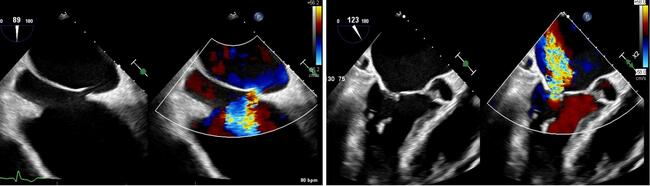
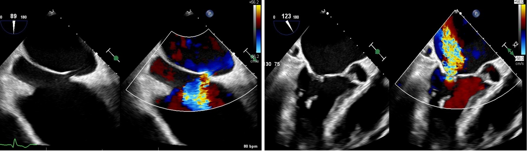
An 83-year-old man with diastolic heart failure, chronic atrial fibrillation on warfarin, severe atrial-functional mitral regurgitation, and a secundum atrial septal defect (ASD) in an anterior/superior location (baseline Qp:Qs 1.5:1) (Figure 1) underwent transcatheter edge-to-edge repair with the MitraClip (Abbott). Final imaging demonstrated enlargement of the ASD with fragile septal tissue and suspected multifenestrated anatomy due to the proximity of the transseptal puncture (TSP) to the pre-existing ASD (Video 1, Figure 2).
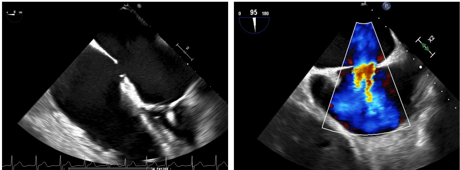
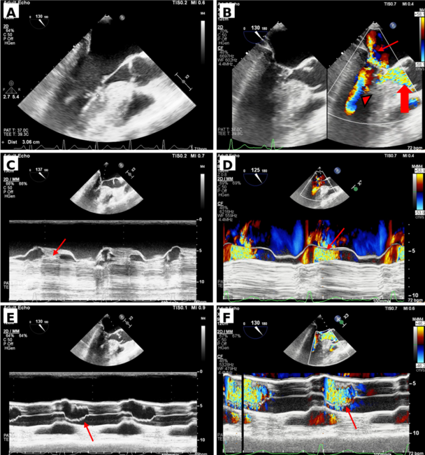
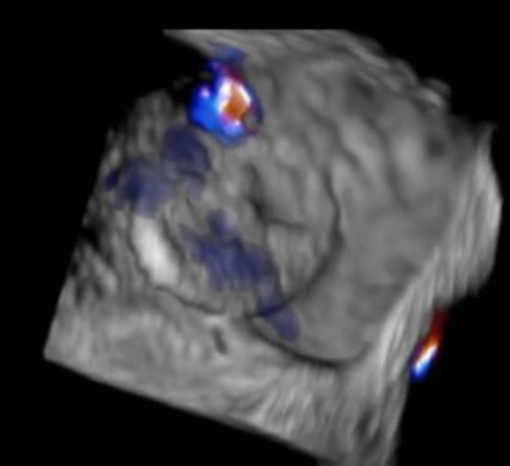
Given recent gastrointestinal bleeding, left atrial appendage occlusion (LAAO) was performed 3 months later, followed by ASD closure using a 30-mm Amplatzer Cribriform Occluder (Abbott). Residual shunting posterior/inferior to the device was attributed to the LAAO TSP (Figure 3). However, transthoracic echocardiography (TTE) the following morning revealed embolization of the Cribriform device into the left atrium (LA) (Video 2).
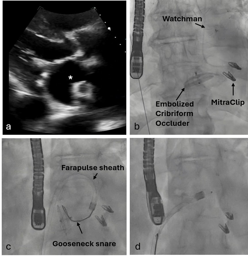
The ASD was crossed with a multipurpose catheter and a J-wire. A 13F FARADRIVE steerable sheath (part of FARAPULSE pulse-field ablation platform) (Boston Scientific) was advanced and looped along the LA wall toward the mitral valve, limiting embolized device mobility. A 25-mm Gooseneck snare was introduced through a 6F Judkins Right-4 guide within the steerable sheath; the snare grasped and cinched the device’s right atrial pin, allowing successful recapture and removal (Video 3, Figure 4). After careful reassessment, the ASD was closed with a 24-mm Amplatzer Septal Occluder (ASO) (Abbott) (Video 4). At 10-month follow-up, TTE confirmed intact septal closure.
The left-sided embolization likely resulted from elevated right atrial pressure due to pulmonary hypertension and incomplete capture of the right atrial tissue at the inferior margin of the Cribriform device. Fortunately, the MitraClip confined the device to the LA, preventing potentially catastrophic embolization into the left ventricle or aorta.1,2 Large-bore, steerable electrophysiology sheaths may facilitate retrieval of foreign bodies from the LA. For ASDs greater than 10 mm, the ASO may be preferable to Cribriform devices, even with multifenestrated septal anatomy.
Affiliations and Disclosures
Neeraj Shah, MD, MPH1; Raveen Bazaz, MD2; Bassel Sayegh, MD2; Mitsugu Ogawa, MD3; Dennis Phillips, MD4; Benjamin Kristobak, MD4
From the Departments of 1Interventional Cardiology, 2Cardiac Electrophysiology, 3Cardiothoracic Surgery, and 4Cardiac Anesthesia, Independence Health Westmoreland Hospital, Greensburg, Pennsylvania.
Disclosures: The authors report no financial relationships or conflicts of interest regarding the content herein.
Consent statement: The authors confirm that informed consent was obtained from the patient for the interventions described in the manuscript and for the publication thereof, including any and all images.
Address for correspondence: Neeraj Shah, MD, Independence Health Westmoreland Hospital, 532 W Pittsburgh St, Greensburg, PA 15601, USA. Email: neerajshah86@gmail.com; X: @NeerajShahMD
References
1. Garre S, Gadhinglajkar S, Sreedhar R, Krishnamoorthy KM, Pillai VV. Atrial septal defect occluder device embolization: experience of a tertiary care cardiac center. Ann Card Anaesth. 2023;26(2):149-154. doi:10.4103/aca.aca_28_22
2. Hierlmeier BJ, Ostrovsky G, Zarth M. Embolization of an atrial septal defect occluder device into the left ventricle. Cureus. 2020;12(11):e11417. doi:10.7759/cureus.11417