Totally Percutaneous Transfemoral Transcatheter Aortic Valve Replacement Via Synthetic Vascular Grafts: A Single-Center Case Series
© 2026 HMP Global. All Rights Reserved.
Any views and opinions expressed are those of the author(s) and/or participants and do not necessarily reflect the views, policy, or position of the Journal of Invasive Cardiology or HMP Global, their employees, and affiliates.
J INVASIVE CARDIOL 2026. doi:10.25270/jic/26.00134. Epub September 14, 2026.
Key Clinical Summary
- Totally percutaneous transfemoral transcatheter aortic valve replacement (TAVR) through synthetic aortobifemoral vascular grafts achieved 100% procedural success and no 30-day vascular complications in 3 high-risk patients with severe aortic stenosis.
- Preprocedural CT angiography, image-guided graft puncture, prophylactic safety wires, and vascular closure devices enabled safe access-site management and effective hemostasis.
- Transfemoral TAVR through synthetic vascular grafts may provide a less invasive alternative to nonfemoral access in carefully selected patients, warranting validation in larger studies.
Abstract
Objective. Transfemoral (TF) vascular access is the preferred route for transcatheter aortic valve replacement (TAVR) in patients with severe aortic stenosis; however, patients with prior peripheral vascular surgery are often treated via alternative, more invasive access sites. The authors aimed to assess the feasibility of valve delivery via synthetic vascular grafts in patients with aortobifemoral bypass surgery.
Methods. The authors report 3 consecutive patients with peripheral vascular disease and a history of aortobifemoral bypass graft surgery who underwent TF-TAVR via direct puncture of the synthetic vascular grafts. Preprocedural computed tomography angiography (CTA) guided access site selection and vascular closure strategy in all cases.
Results. Prophylactic 0.014-inch safety wires were delivered to the primary access sites via a secondary access site in order to facilitate hemostasis and enable percutaneous vascular repair if necessary. Self-expanding and balloon-expandable valves were successfully delivered via synthetic grafts in all 3 procedures. A prophylactic occlusive balloon was inflated within the abdominal aorta in 1 case in order to prevent potential hemorrhage during sheath removal. Successful hemostasis was achieved in all patients, using a plug-based vascular closure device (VCD) in 1 patient and combined suture-mediated VCDs and plug-based closure devices in 2 patients.
Conclusions. Percutaneous TF-TAVR with valve delivery via synthetic vascular grafts appears feasible in select patients. VCDs achieved access site hemostasis in all cases. Procedural planning based on CTA appears essential for selecting the optimal vascular access strategy.
Introduction
Minimally invasive transcatheter aortic valve replacement (TAVR) has supplanted surgical aortic valve replacement as the preferred mode of treatment in the majority of patients with severe aortic stenosis (AS).1,2 The transfemoral (TF) approach is the preferred route for valve delivery as it is less invasive than alternative vascular access sites, enables the procedure to be performed under conscious sedation, and is associated with shorter procedure time, reduced periprocedural morbidity, and earlier mobilization
The presence of peripheral artery disease in patients undergoing TAVR is associated with adverse clinical outcomes and increased risk of vascular complications.6-8 At least 5% of patients with AS who are referred for TAVR have severe peripheral vascular disease, which is considered incompatible with TF valve delivery.9 Some of these patients have a history of prior peripheral vascular bypass graft surgery. Totally percutaneous delivery of large-bore sheaths and devices via synthetic vascular grafts10 may be challenging because the inelastic graft may undergo structural deterioration, vascular closure devices (VCDs) may not seal the puncture-site within the graft wall, and there is a risk of graft infection. Clinicians therefore often select alternative access routes for valve delivery in such patients, which are more invasive and may require specialized technical expertise.11-14
We aimed to assess the feasibility, technical considerations, and procedural safety of totally percutaneous TF-TAVR via synthetic aortobifemoral bypass grafts. We hypothesized that percutaneous TF valve delivery may be feasible in select patients with patent vascular grafts, providing appropriate measures are taken to facilitate hemostasis and vascular repair if VCD failure occurs.
Methods
Study population
All TAVR procedures performed at the Carmel Medical Center in Haifa, Israel, since initiation of the program in March 2010 were prospectively documented in an institutional registry using a standardized data collection form. We analyzed our prospective institutional TAVR registry and identified all patients who had undergone TF-TAVR via a synthetic vascular graft. Clinical, demographic, and procedural data, and clinical outcomes were analyzed. The study was approved by the hospital’s institutional review board, and informed consent was obtained from the patients for the interventions described in the manuscript and to the publication of their data, including any and all images.
TAVR procedure
Prior to the procedure, patients were assessed by the multidisciplinary heart team and underwent computed tomography angiography (CTA) to assess potential vascular access sites. TF access was used as the default approach. Puncture of the bypass graft was guided by combination of ultrasound and fluoroscopic imaging. Large-bore sheaths were inserted over a stiff 0.035-inch guidewire under fluoroscopic surveillance. Access site hemostasis was achieved using VCDs with aim to perform a totally percutaneous procedure.
We initially used a plug-based MANTA VCD (Teleflex); however, we subsequently switched to a combined strategy of the suture-based Prostyle VCD (Abbott) and Angio-Seal (Terumo). In all cases, hemostasis was confirmed by control angiography which was performed via the secondary access site. In cases considered to be at risk for vascular complication, an 0.014-inch safety wire was delivered to the primary access site via a secondary access site (contralateral femoral artery, distal ipsilateral femoral artery, or brachial artery).15,16 The safety wire was deployed in order to enable rapid vascular repair in case of vessel injury, using balloon inflation or stent graft implantation.17 All patients received unfractionated heparin, aiming for an activated clotting time of greater than 250 seconds. All patients also received antibiotic prophylaxis with intravenous amoxicillin/clavulanate potassium unless they were allergic to penicillin, in which case they received clindamycin.
Follow-up
Access site hemostasis was confirmed in all cases by control angiography, which was performed via the secondary access site. Postprocedural follow-up was based on review of the patients’ medical records and analysis of the national electronic database that captures all medical events and hospitalizations. Routine postprocedural vascular imaging was not performed. In view of the patients’ history of recurrent vascular interventions in the period preceding the TF-TAVR procedure, we focused on procedural outcomes during 30 days of follow-up. Clinical events during hospitalization and 30-day follow-up (mortality, hospitalization, bleeding, transfusions, and vascular and access site complications) were defined according to the Valve Academic Research Consortium (VARC)-3 updated endpoint definitions.
Results
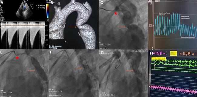
Between 2010 and 2025, 1502 patients with severe AS underwent TAVR procedures at our institute. We identified 4 patients with a history of previous aortofemoral bypass surgery who underwent TAVR at our institution. The procedure was performed via TF vascular access in all patients. In one of these patients, we were able to puncture the femoral artery below the anastomosis with the synthetic graft. The present study describes procedural outcomes in the remaining 3 patients in whom TF-TAVR required direct puncture of a synthetic vascular graft with percutaneous hemostasis using VCDs (Tables 1 and 2). Due to difference in diameter between the synthetic graft and the diseased native femoral artery to which it was anastomosed, we aimed to puncture the graft above the anastomosis in order to leave a distal margin that would accommodate a stent graft if vascular repair was necessary. Puncture of the bypass graft was guided by combination of ultrasound and fluoroscopic imaging.
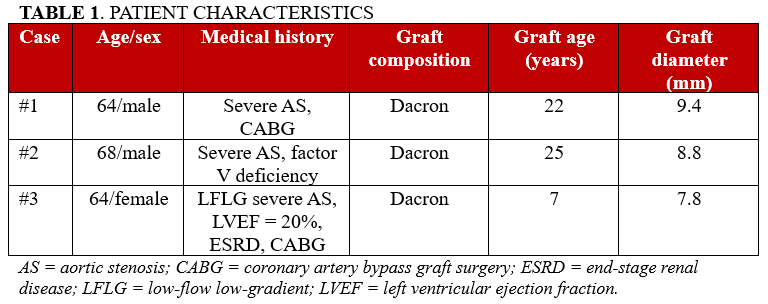
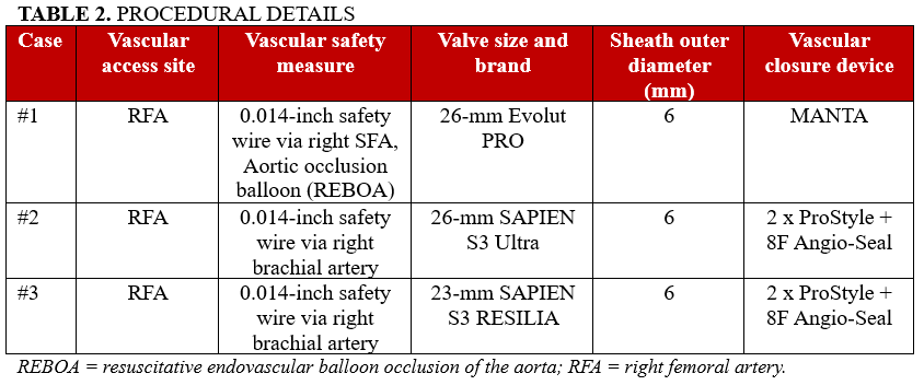
No cases of mortality, rehospitalization, VARC-3 bleeding, blood transfusion, vascular or access-site complication, graft infection, or vascular reintervention occurred during hospitalization or within 30 days.
Case 1
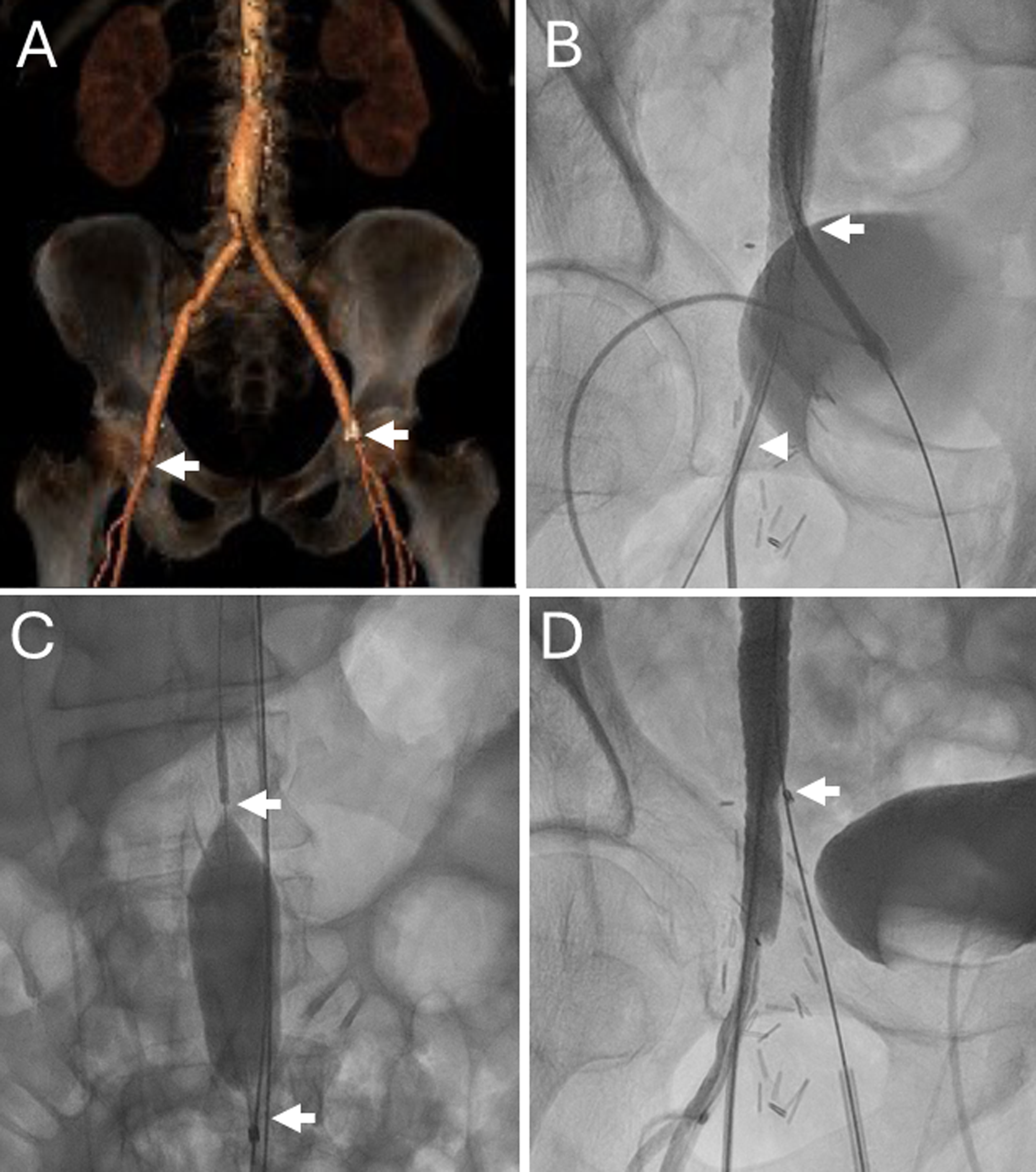
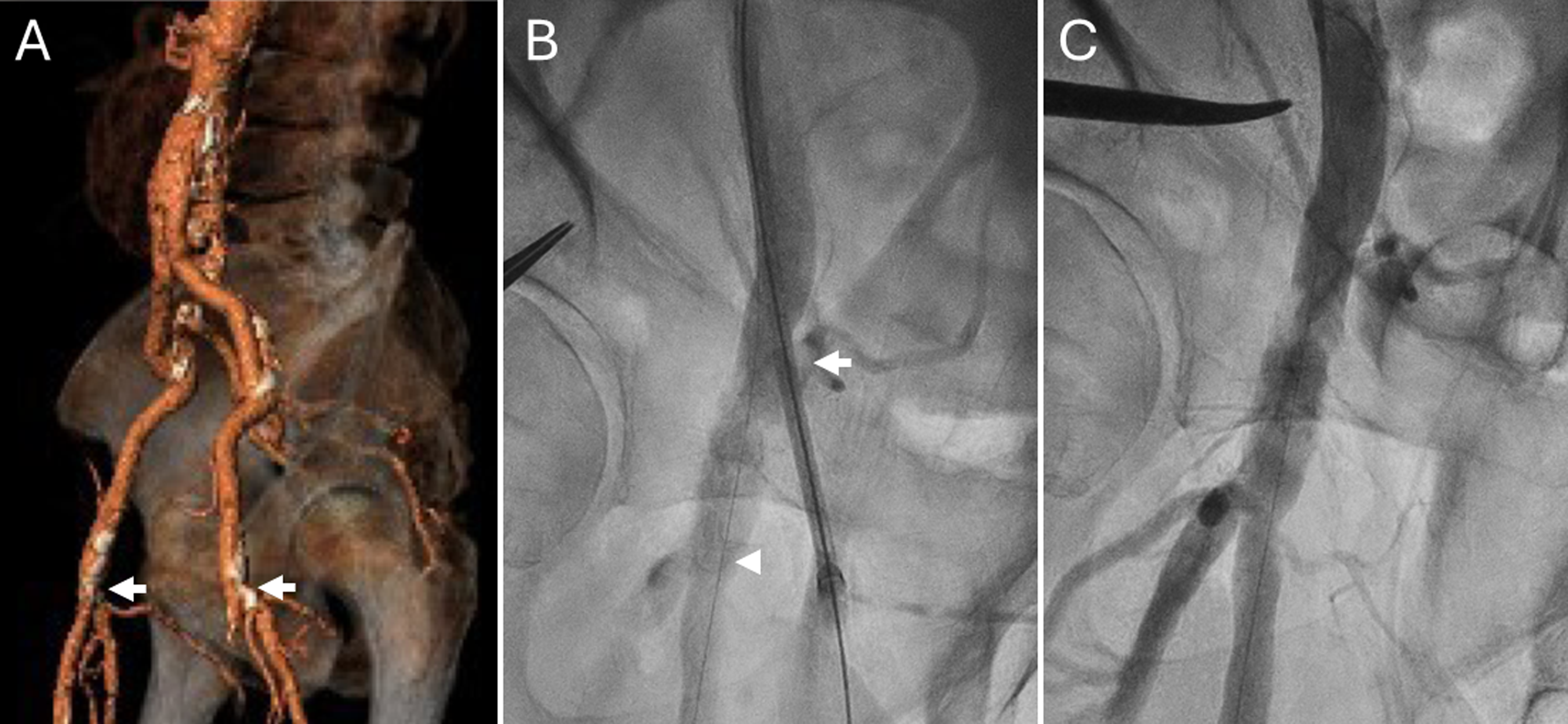
A 64-year-old man with severe AS, coronary artery disease, and a history of coronary artery bypass grafting and aortobifemoral bypass surgery was referred for TAVR (Figure 1, Videos 1-5). A 0.014-inch Grand Slam safety wire (Asahi Intecc) was placed within the abdominal aorta via the right superficial femoral artery. The right femoral artery graft was punctured under combined ultrasound and fluoroscopic guidance. Direct implantation of a 26-mm Evolut PRO valve (Medtronic) was performed. Prior to removal of the sheath, a balloon for resuscitative endovascular balloon occlusion of the aorta (REBOA, Tokai Medical) was positioned within the abdominal aorta via the left deep femoral artery to enable immediate hemorrhage control if necessary. A MANTA VCD achieved successful hemostasis at the graft puncture site.
Case 2
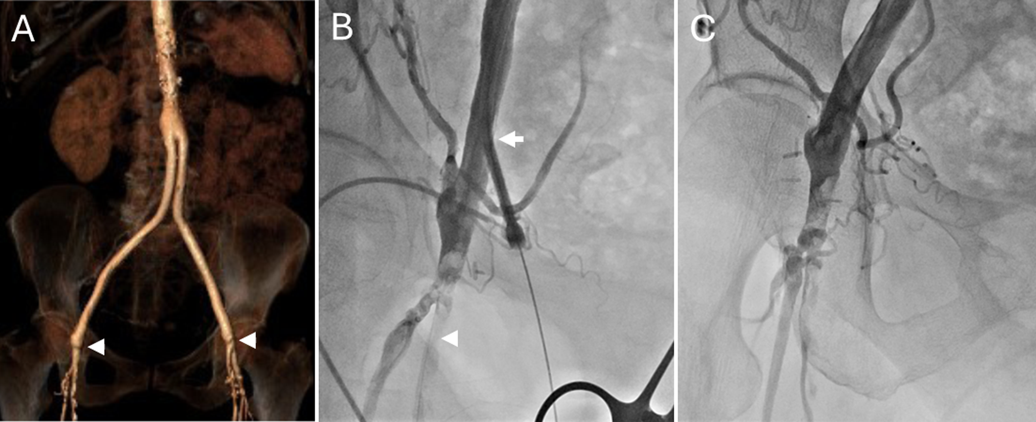
A 68-year-old man with severe AS, aortobifemoral bypass grafts and hereditary factor V deficiency presented a dual challenge of complex vascular anatomy and high bleeding risk (Figure 2, Videos 6-9). A 6F brachial sheath was used to deliver a 300-cm long, 0.014-inch Grand Slam safety wire to the right femoral artery via the graft.15 The right femoral artery graft was punctured under combined ultrasound and fluoroscopic guidance. A 26-mm SAPIEN 3 Ultra valve (Edwards Lifesciences) was implanted successfully. Hemostasis at the graft puncture site was achieved using the combination of 2 ProStyle VCDs and an 8F Angio-Seal VCD. Distal perfusion was confirmed post-closure and no hemorrhagic complications occurred despite the underlying coagulopathy.
Case 3
A 64-year-old woman with end-stage renal disease and prior coronary bypass surgery and aortobifemoral bypass surgery presented with low-flow, low-gradient severe AS and severely reduced left ventricular ejection fraction (20%) (Figure 3, Videos 10-13). A 300-cm long, 0.014-inch Grand Slam safety wire was advanced via the right brachial artery to the right femoral artery in order to facilitate immediate bleeding control if necessary. The right femoral bypass graft was punctured under ultrasound and fluoroscopic guidance. A 23-mm SAPIEN 3 RESILIA valve (Edwards Lifesciences) was implanted successfully. Hemostasis was achieved with 2 ProStyle suture-mediated VCDs and an 8F Angio-Seal, resulting in immediate and complete hemostasis without vascular compromise or hemodynamic deterioration.
Discussion
We report 3 cases in which totally percutaneous TF-TAVR was performed in patients with previous aortobifemoral bypass surgery, using the synthetic graft as the primary access site. Preprocedural CTA was performed in all cases in order to confirm patency of the bypass grafts and to select appropriate puncture sites and closure strategy. Ultrasound and fluoroscopy were used in a complementary fashion for guiding vessel puncture. In all 3 cases, VCDs achieved immediate angiographic hemostasis, and no clinically apparent vascular complications or vascular reinterventions were identified during 30-day follow-up. Our findings corroborate previous case reports18,19 and extend the evidence base to a more heterogeneous population, including patients with increased bleeding risk and severely reduced ejection fraction.
A major concern in these procedures was whether currently available VCDs would reliably achieve hemostasis at the access site within the vascular graft. We used a plug-based MANTA VCD in the first case; however, we switched to a combined strategy of a suture-based Prostyle VCD and Angio-Seal in the subsequent 2 cases following reports that this approach was safer and more effective.20 We decided to deploy 2 suture-based VCDs in order to increase the likelihood of successful hemostasis because of uncertainty regarding the efficacy of these devices when used in synthetic grafts.
In this limited series, VCDs achieved immediate angiographic hemostasis in all 3 patients without clinically apparent access site complications during 30-day follow-up. Based on our previous experience,16,17 prophylactic 0.014-inch safety wires were placed within the primary access site in all cases in order to facilitate delivery of balloons for bleeding control and stent grafts for vascular repair if necessary. An occlusive aortic balloon (REBOA) was used during sheath removal in 1 patient who was considered at high risk for VCD failure. These adjunctive safety measures may be considered when performing percutaneous large-bore access through a synthetic graft, particularly when failure of the planned closure strategy would pose a substantial bleeding risk.
The decision to perform TF-TAVR via prosthetic grafts was based on our institutional familiarity with performing such procedures in patients with hostile vascular anatomy. Alternative vascular access may have been considered a reasonable strategy in these patients; however, there is currently no consensus available to help operators select among different access options, and the decision is based on the local operator’s personal experience and expertise.13,14
Based on our limited experience, the following technical considerations may be useful: (1) procedural planning based on CTA, (2) positioning a prophylactic 0.014-inch safety wire within the primary access site in order to facilitate bleeding control and vascular repair if necessary, (3) image-guided puncture away from vascular anastomoses, (4) use of an extra-support stiff 0.035-inch guidewire for sheath advancement, and (5) planned hybrid or plug-based closure with a rescue balloon on standby.
Limitations
This is a small case series of selected patients who had documentation of vascular graft patency before the procedure. Follow-up was limited to 30 days following the procedure, was based on review of electronic patient records, and did not include systematic postprocedural vascular imaging. Larger cohorts should be studied in order to validate our findings.
Conclusions
Performing totally percutaneous TF-TAVR via synthetic vascular grafts may be feasible in select patients with patent access sites. This approach may enable a less invasive route for valve delivery compared with alternative access sites, which may facilitate early mobilization of these patients who often have severe comorbidities. We recommend that procedural planning should be based on analysis of CTA. Bailout strategies in case of structural failure of the grafts or VCD failure to achieve hemostasis should be planned meticulously prior to initiating the procedure.
Affiliations and Disclosures
Hussein Sliman, MD1,2; Amnon Eitan, MD1,2; Hashem Hayeq, MD3; Avinoam Shiran, MD1,2; Keren Zissman, MD1; Ronen Jaffe, MD1,2
From 1the Department of Cardiology, Carmel Medical Center, Haifa, Israel; 2Technion-Israel Institute of Technology, Haifa, Israel; 3the Department of Vascular Surgery, Carmel Medical Center, Haifa, Israel.
Disclosures: The authors report no financial relationships or conflicts of interest regarding the content herein.
Address for correspondence: Ronen Jaffe, MD, Department of Cardiology, Carmel Medical Center, Haifa 34361, Israel. Email: jafferonen@gmail.com
References
1. Siontis GCM, Overtchouk P, Cahill TJ, et al. Transcatheter aortic valve implantation vs. surgical aortic valve replacement for treatment of symptomatic severe aortic stenosis: an updated meta-analysis. Eur Heart J. 2019;40(38):3143-3153. doi:10.1093/eurheartj/ehz275
2. Nguyen V, Willner N, Eltchaninoff H, et al. Trends in aortic valve replacement for aortic stenosis: a French nationwide study. Eur Heart J. 2022;43(7):666-679. doi:10.1093/eurheartj/ehab773
3. Morello A, Corcione N, Ferraro P, et al. The best way to transcatheter aortic valve implantation: From standard to new approaches. Int J Cardiol. 2021;322:86-94. doi:10.1016/j.ijcard.2020.08.036
4. Beurtheret S, Karam N, Resseguier N, et al. Femoral versus nonfemoral peripheral access for transcatheter aortic valve replacement. J Am Coll Cardiol. 2019;74(22):2728-2739. doi:10.1016/j.jacc.2019.09.054
5. Eugène M, Duchnowski P, Prendergast B, et al; EORP VHD II Registry Investigators Group. Contemporary management of severe symptomatic aortic stenosis. J Am Coll Cardiol. 2021;78(22):2131-2143. doi:10.1016/j.jacc.2021.09.864
6. Bansal K, Soni A, Shah M, et al. Association between polyvascular disease and transcatheter aortic valve replacement outcomes: insights from the STS/ACC TVT registry. Circ Cardiovasc Interv. 2023;16(12):e013578. doi:10.1161/CIRCINTERVENTIONS.123.013578
7. Ueshima D, Barioli A, Nai Fovino L, et al. The impact of pre-existing peripheral artery disease on transcatheter aortic valve implantation outcomes: A systematic review and meta-analysis. Catheter Cardiovasc Interv. 2020;95(5):993-1000. doi:10.1002/ccd.28335
8. Fanaroff AC, Manandhar P, Holmes DR, et al. Peripheral artery disease and transcatheter aortic valve replacement outcomes: a report from the Society of Thoracic Surgeons/American College of Cardiology transcatheter therapy registry. Circ Cardiovasc Interv. 2017;10(10):e005456. doi:10.1161/CIRCINTERVENTIONS.117.005456
9. Carroll JD, Mack MJ, Vemulapalli S, et al. STS-ACC TVT registry of transcatheter aortic valve replacement. J Am Coll Cardiol. 2020;76(21):2492-2516. doi:10.1016/j.jacc.2020.09.595
10. Ciaramella M, LoGerfo F, Liang P. Lower extremity bypass for occlusive disease: a brief history. Ann Vasc Surg. 2024;107:17-30. doi:10.1016/j.avsg.2023.11.053
11. Palmerini T, Saia F, Kim WK, et al. Vascular access in patients with peripheral arterial disease undergoing TAVR: the Hostile tegistry. JACC Cardiovasc Interv. 2023;16(4):396-411. doi:10.1016/j.jcin.2022.12.009
12. Aufazhafarin NT, Saputra PBT, Widiarti W, eet al. Navigating Vascular access strategies for transcatheter aortic valve replacement in patients with peripheral artery disease: a literature review. Catheter Cardiovasc Interv. 2026;107(6):2100-2109. doi:10.1002/ccd.70573
13. Sherwood M, Allen KB, Dahle TG, et al. SCAI expert consensus statement on alternative access for transcatheter aortic valve replacement. J Soc Cardiovasc Angiogr Interv. 2025;4(3Part A):102514. doi:10.1016/j.jscai.2024.102514
14. Feistritzer HJ, Dumpies O, Rosseel L, et al. Alternative access for TAVR: a state-of-the-art review and practical guide. JACC Cardiovasc Interv. 2025;18(19):2309-2325. doi:10.1016/j.jcin.2025.08.029
15. Sliman H, Eitan A, Shiran A, Zafrir B, Jaffe R. Transbrachial secondary vascular access in transcatheter aortic valve replacement procedures: a single-centre retrospective analysis. Heart Lung Circ. 2022;31(7):1023-1028. doi:10.1016/j.hlc.2022.01.013
16. Naoum I, Eitan A, Galili O, et al. Strategy for totally percutaneous management of vascular injury in combined transfemoral transcatheter aortic valve replacement and endovascular aortic aneurysm repair procedures. Am J Cardiol. 2023;207:130-136. doi:10.1016/j.amjcard.2023.08.148
17. Eitan A, Sliman H, Shiran A, Jaffe R. Strategies for facilitating totally percutaneous transfemoral TAVR procedures. J Clin Med. 2022;11(8):2104. doi:10.3390/jcm11082104
18. Salem JE, Paul JF, Caussin C. Transfemoral aortic valve implantation in a renal transplant patient with a Dacron aorto-bi-iliac bypass. J Invasive Cardiol. 2012;24(12):667-70.
19. Gul M, Akgul O, Erturk M, Eksik A, Yildirim A. Transcatheter aortic valve implantation through a left aortoiliac graft. Tex Heart Inst J. 2012;39(6):898-900.
20. Upadhaya S, Upreti A, Gupta K, et al. Vascular closure devices for large bore femoral access after transfemoral transcatheter aortic valve replacement: a network meta-analysis. Catheter Cardiovasc Interv. 2025;106(1):423-432. doi:10.1002/ccd.31565