Congenital Double Orifice Mitral Valve
© 2026 HMP Global. All Rights Reserved.
Any views and opinions expressed are those of the author(s) and/or participants and do not necessarily reflect the views, policy, or position of the Journal of Invasive Cardiology or HMP Global, their employees, and affiliates.
J INVASIVE CARDIOL 2026. doi:10.25270/jic/26.00261. Epub August 11, 2026.
A 1-year-old boy presented to a pediatric cardiology outpatient clinic with failure to thrive, feeding difficulty, and 2 episodes of pneumonia-related hospitalizations in the year prior, suggestive of increased pulmonary blood flow. Perinatal history revealed lower respiratory tract sepsis requiring neonatal intensive care soon after birth. No evidence of cyanosis, hypoxic spells, neurological deficit, or delayed milestones was recorded.
Clinical examination showed left parasternal heave, loud pulmonary component of second heart sound, and a prominent mid-diastolic rumble at apex. Chest x-ray showed moderate cardiomegaly, biatrial enlargement, and multiple end-on vessels in bilateral lung fields with hilar prominence. Transthoracic echocardiogram demonstrated a large non-restrictive perimembranous ventricular septal defect (VSD) with significant left-to-right shunt. Additionally, an accessory papillary muscle was seen in the left ventricle (LV). The pulmonary artery, LV, and left atrium were dilated, suggestive of severe pulmonary arterial hypertension (RVSP = 60 mm Hg).
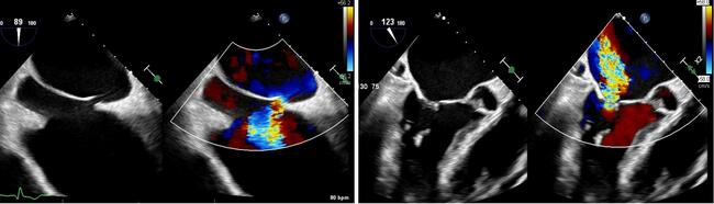
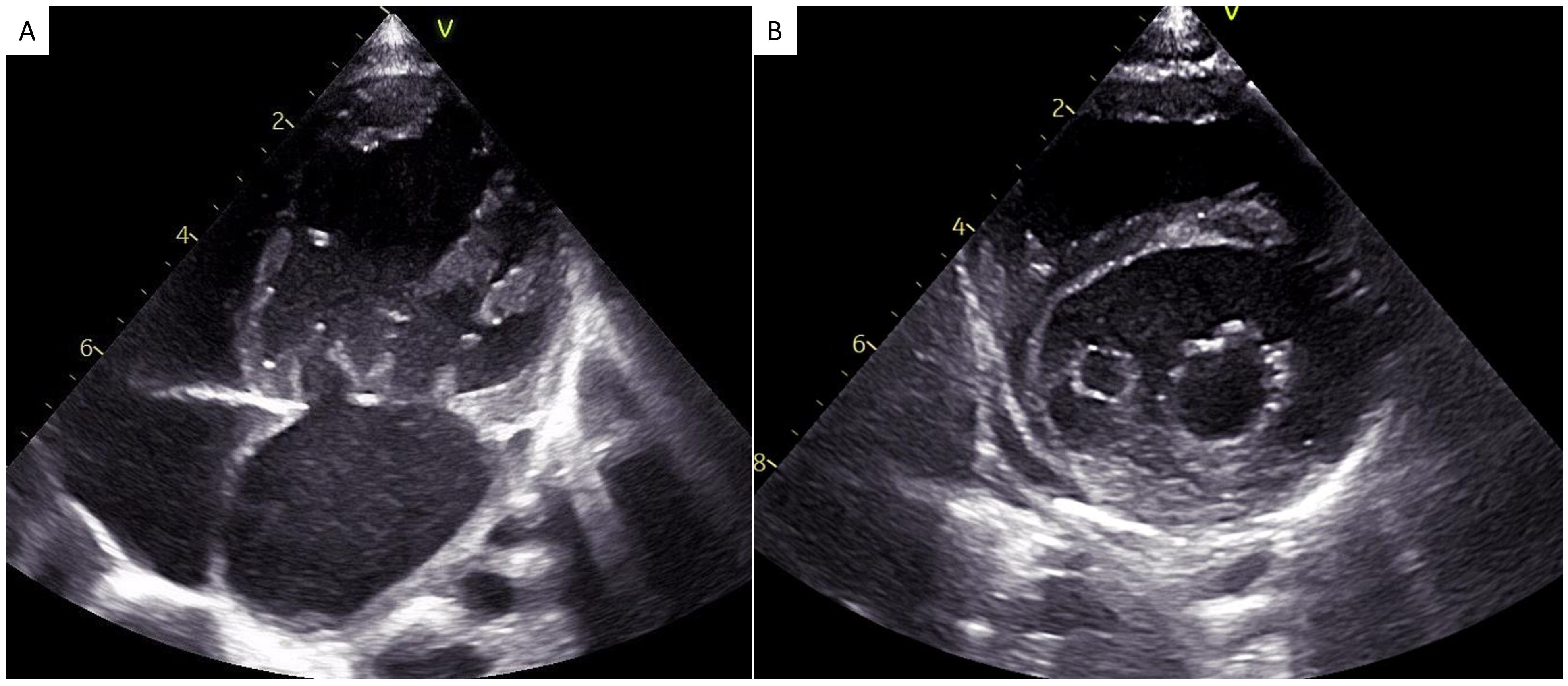
Four-chamber apical view showed an abnormal mitral inflow in the form of 2 separate valvelike structures attached to the papillary muscles in the LV via chordae tendineae; each valvular opening was guarded by 2 leaflets opening simultaneously into the LV during diastole (Figure A), suggestive of congenital eccentric (hole) type double orifice mitral valve (DOMV). Short-axis view at the level of the mitral valve also confirmed 2 unequal circular openings with planimetry-derived areas of 1.7 cm2 and 0.7 cm2 (Figure B). Color Doppler showed flow acceleration across the smaller orifice with peak and mean gradients across the DOMV of 10 and 6 mm Hg, respectively, suggestive of moderate mitral stenosis without any mitral regurgitation (Videos 1-3).
The patient underwent successful surgical VSD closure with mitral valve repair. Post-surgery, the patient was discharged in stable condition with plans to return after 3 months for a follow-up.
Affiliations and Disclosures
Ankit Kumar Sahu, DM; Aditya Srivastava, DM; Arpita Katheria, DM
From the Department of Cardiology, Sanjay Gandhi Postgraduate Institute of Medical Sciences, Lucknow, Uttar Pradesh, India.
Disclosures: The authors report no financial relationships or conflicts of interest regarding the content herein.
Consent statement: The authors confirm that informed consent was obtained from the patient’s parents for the intervention(s) described in the manuscript and for the publication thereof, including any and all images.
Data availability statement: All data generated or analyzed during this study are included in this published article and its supplementary information files. Any other additional data is available from the corresponding author on reasonable request.
Address for correspondence: Ankit Kumar Sahu, Department of Cardiology, Sanjay Gandhi Postgraduate Institute of Medical Sciences, Lucknow, Uttar Pradesh 226014, India. Email: ankitsahu.md@gmail.com; X: @ankitksahu