Halfway Rotablation to Improve Wire Bias in a Severely Calcified and Angulated Proximal Left Anterior Descending Artery Lesion
© 2026 HMP Global. All Rights Reserved.
Any views and opinions expressed are those of the author(s) and/or participants and do not necessarily reflect the views, policy, or position of the Journal of Invasive Cardiology or HMP Global, their employees, and affiliates.
J INVASIVE CARDIOL 2026. doi:10.25270/jic/26.00313. Epub September 22, 2026.
A 78-year-old man was admitted with non-ST-segment elevation myocardial infarction. He had a history of ischemic heart disease with prior percutaneous coronary intervention (PCI) to the right coronary artery (RCA) and mid-left anterior descending artery (mLAD) in 2015, as well as a known chronic total occlusion of the ostial left circumflex artery without demonstrable reversibility.
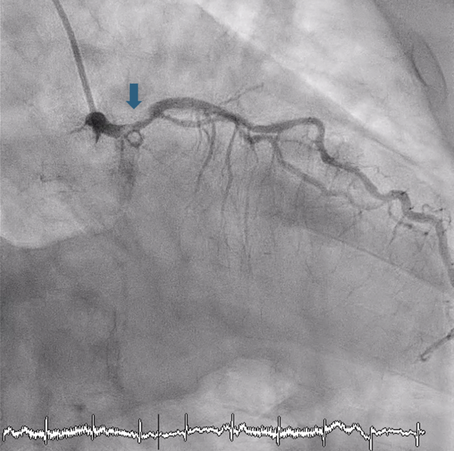
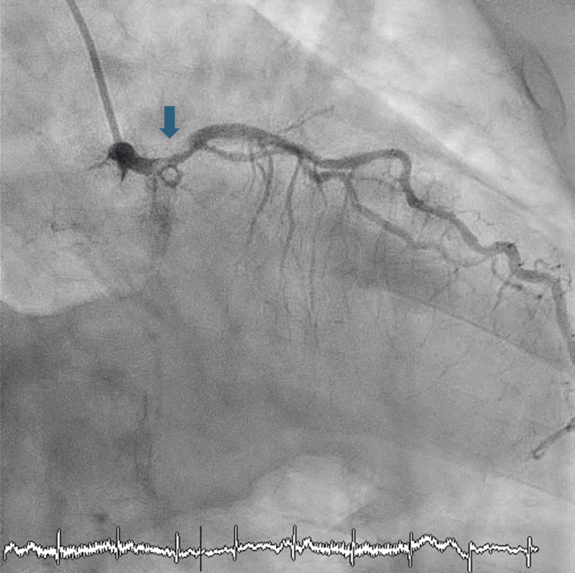
Coronary angiography demonstrated an 80% calcified stenosis extending from the distal left main to the mLAD (Figures 1 and 2). The previous mLAD and RCA stents were patent.
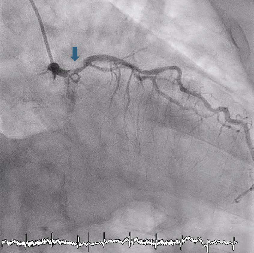
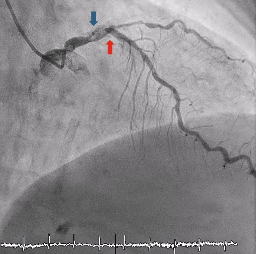
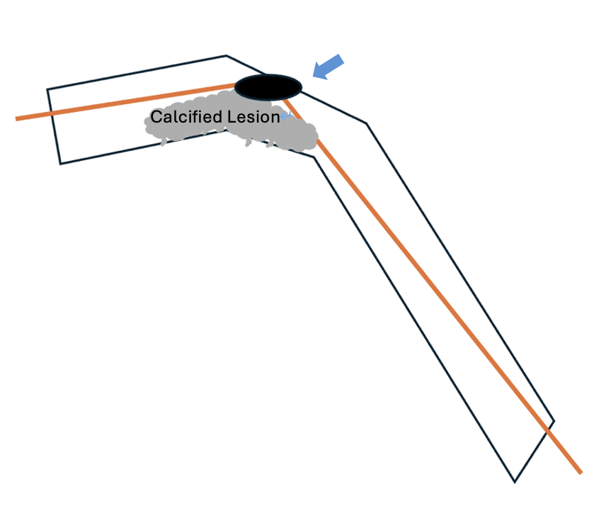
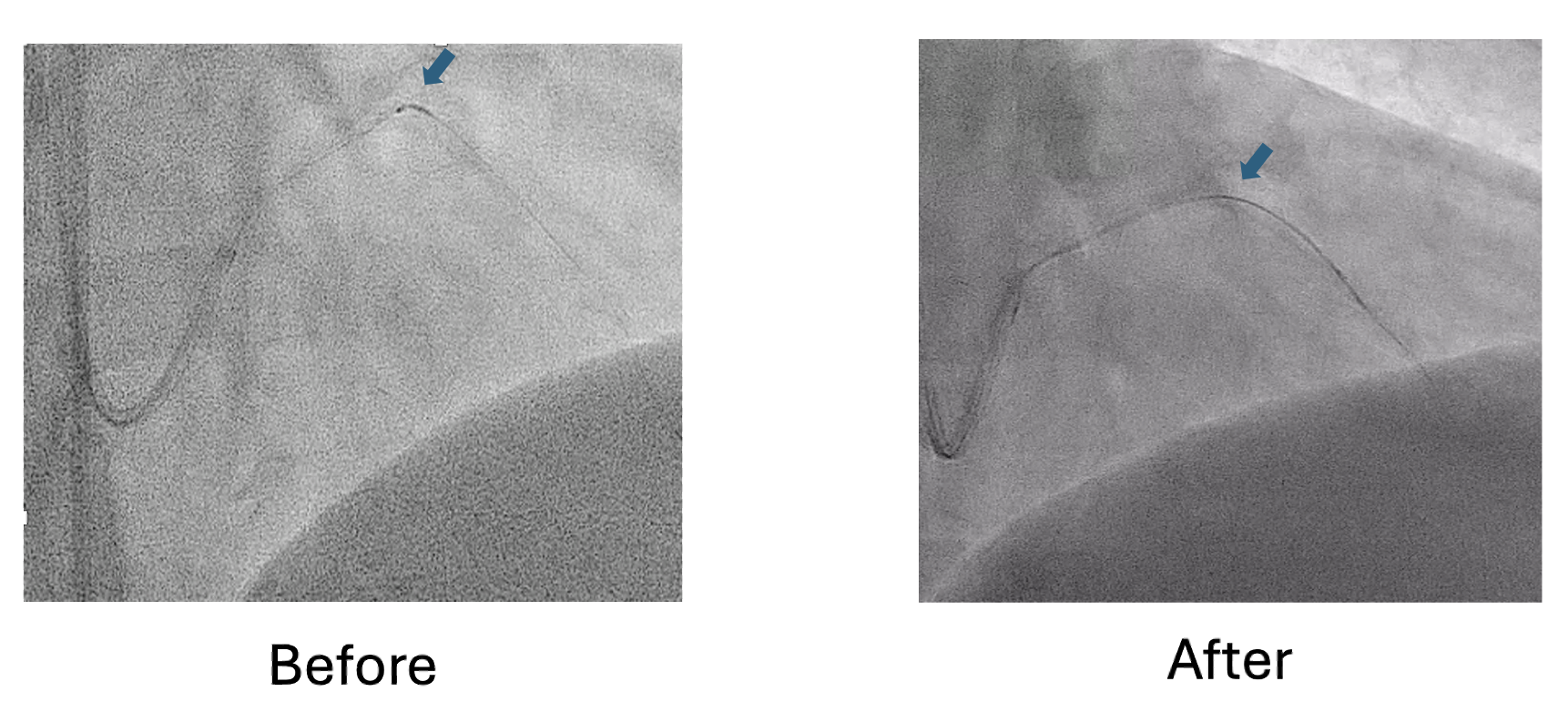
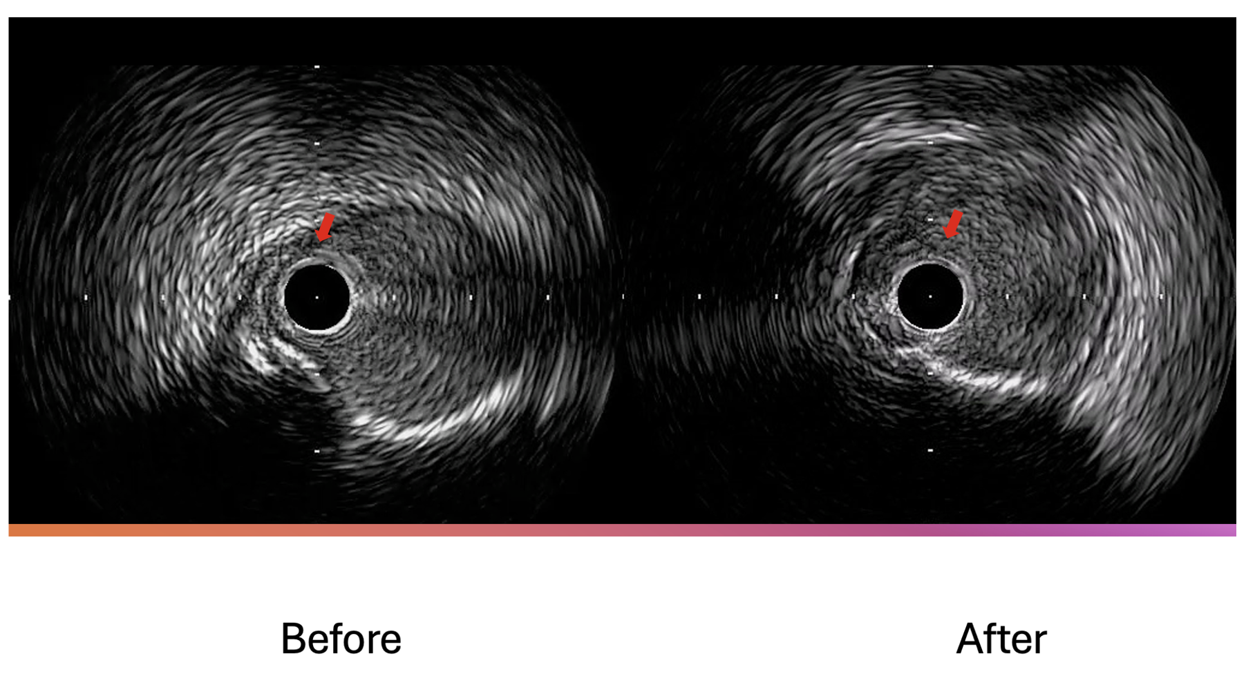
The LAD lesions were successfully wired with a Sion Blue wire (Asahi Intecc) using an Extra Backup 3.5 (Medtronic) guiding catheter. However, an AnteOwl WR intravascular ultrasound (IVUS) catheter (Terumo) could not advance through the proximal LAD because of calcification and vessel angulation (Figure 3). An IVUS pullback from the angulated segment revealed significant calcified plaque from the ostial to the proximal LAD. At the point where the IVUS catheter failed to pass, the wire bias was directed toward the normal vessel wall (Figure 4).
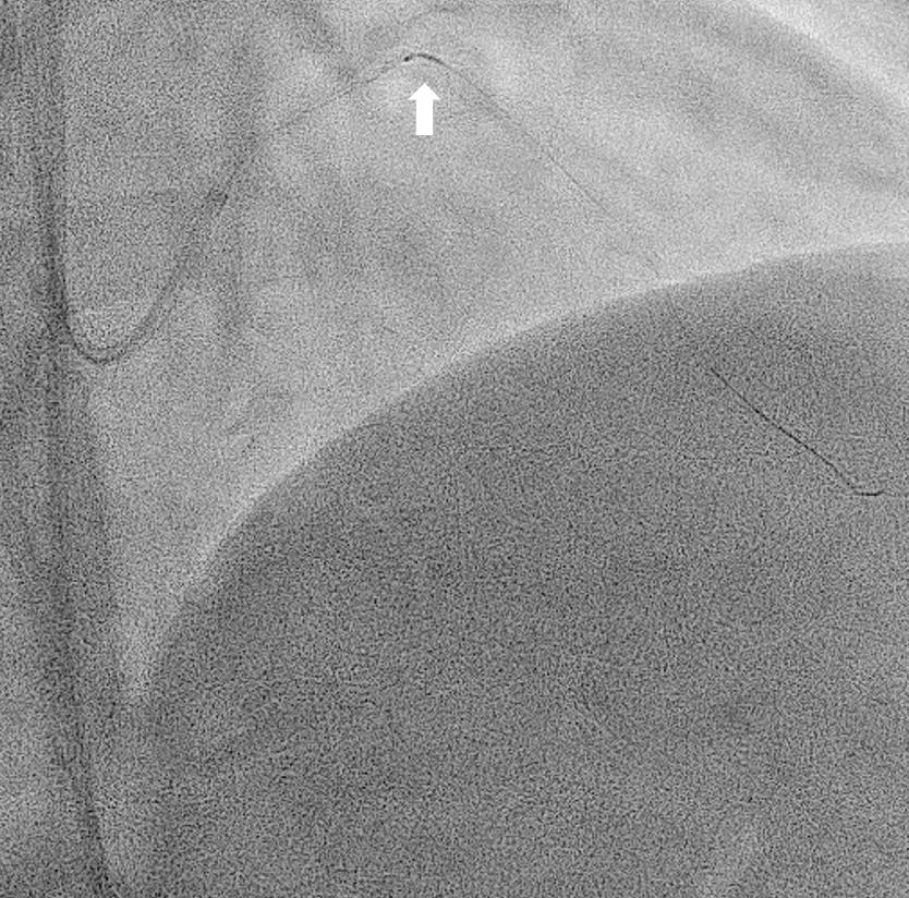
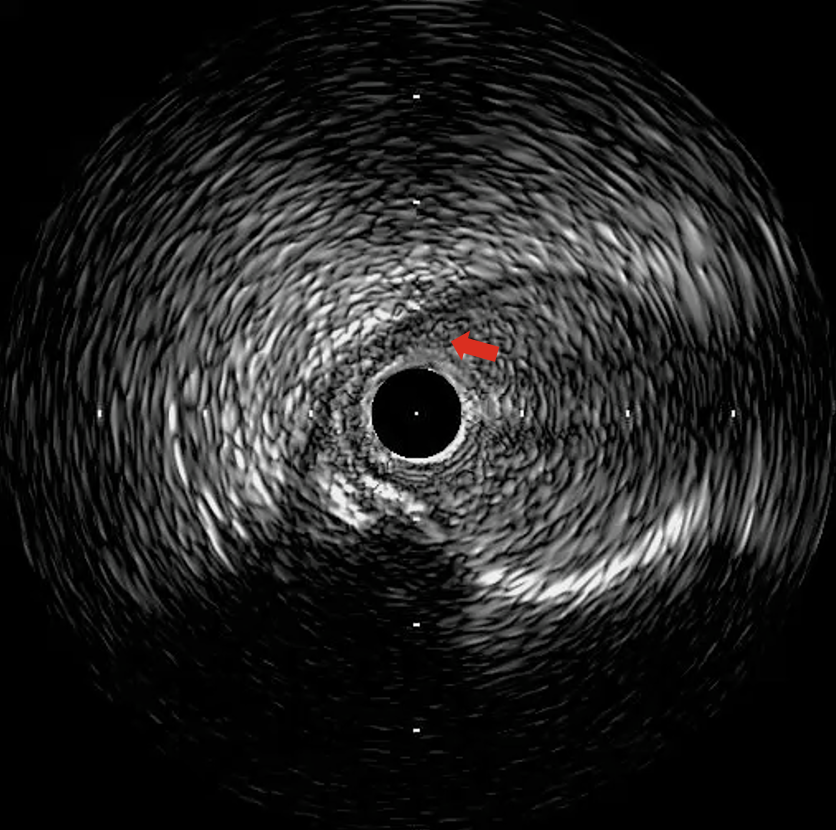
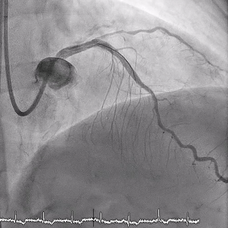
Rotablation across this segment carries risks of vessel injury, perforation, or burr entrapment (Figure 5). To mitigate these risks, we performed “halfway rotablation” with a ROTAPRO Rotational Atherectomy System 1.5-mm burr (Boston Scientific). The burr was advanced slowly without crossing beyond the angle of curvature (Video 1). After this partial ablation, wire bias improved, as confirmed by both fluoroscopy and IVUS (Figures 6-8). Full rotablation beyond the angulated segment could then be completed safely (Video 2). Following further balloon dilatation, a 3.0 × 50-mm drug-eluting stent was successfully implanted. Final angiography (Figure 9) and IVUS (Video 3) demonstrated a satisfactory result.
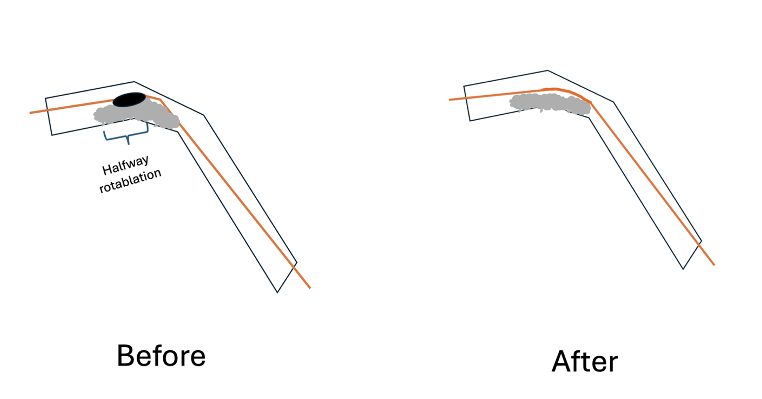
The halfway rotablation technique was originally described by Japanese operators1 for long, diffuse calcified lesions with angulation. In the original approach, rotablation is performed proximal to the angulation, followed by balloon predilatation of the lesion distal to the angle. In the present case, we applied the technique specifically to improve wire bias, thereby enabling safe completion of rotablation across the angulated segment.
Affiliations and Disclosures
Kwok-Ho Yau, MBBS; Justin Ka-Ho Wong, MBBS; John Cheong-Hin Chan, MBBS
1From the Cardiology Team, Department of Medicine, Pamela Youde Nethersole Eastern Hospital, Chai Wan, Hong Kong SAR.
Disclosures: The authors report no financial relationships or conflicts of interest regarding the content herein.
Consent statement: The authors confirm that informed consent was obtained from the patient for the intervention(s) described in the manuscript and for the publication of thereof, including any and all images.
Address for correspondence: Kwok-Ho Yau, MBBS, 3 Lok Man Road, Chai Wan, Hong Kong. Email: jason200200200@hotmail.com
References
1. Sakakura K, Taniguchi Y, Yamamoto K, Wada H, Momomura SI, Fujita H. Halfway rotational atherectomy for calcified lesions: comparison with conventional rotational atherectomy in a propensity-score matched analysis. PLoS One. 2019;14(7):e0219289. doi:10.1371/journal.pone.0219289