Transcatheter Stenting for Severe Native Aortic Coarctation
© 2026 HMP Global. All Rights Reserved.
Any views and opinions expressed are those of the author(s) and/or participants and do not necessarily reflect the views, policy, or position of the Journal of Invasive Cardiology or HMP Global, their employees, and affiliates.
J INVASIVE CARDIOL 2026. doi:10.25270/jic/26.00265. Epub August 31, 2026.
A 20-year-old woman presented with resistant hypertension despite taking 3 antihypertensives. On examination, she had a blood pressure of 180/100 mm Hg and a systolic murmur heard under the left scapula. Additionally, the femoral pulse was feeble, and there was an appreciable brachio-femoral delay; blood pressure in the right upper limb was 180/100 mm Hg and in the left lower limb was 100/70 mm Hg.
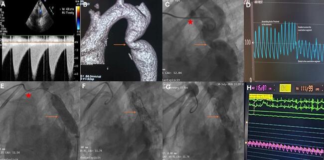
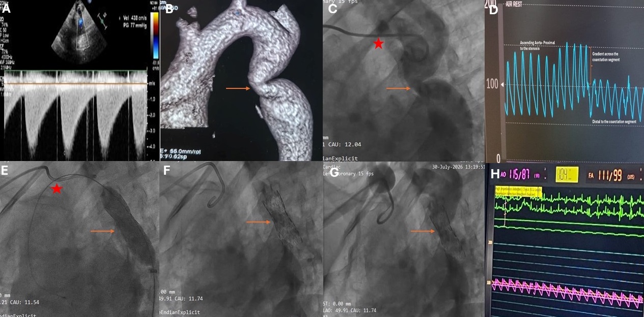
Twelve-lead electrocardiogram showed left ventricular (LV) hypertrophy with a strain pattern and left axis deviation. Two-dimensional transthoracic echocardiography revealed concentric hypertrophy with a normal LV ejection fraction. The supra-sternal view revealed a narrowed descending aorta with a posterior shelf, and the gradient across the aorta was 77 mm Hg (Figure A). Computed tomography (CT) aortogram revealed a coarctation of the aorta which was juxta-ductal in location and away from the origin of the subclavian artery (Figure B).
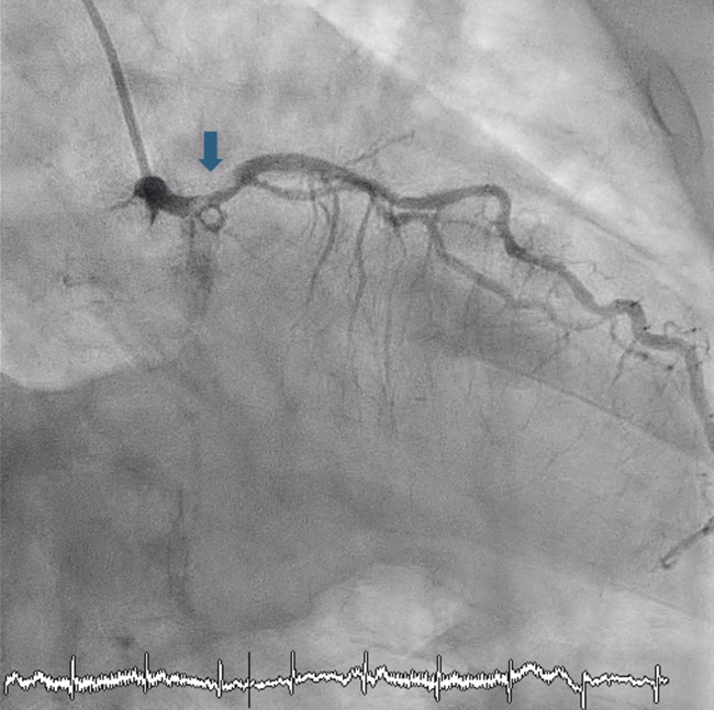
After obtaining informed consent, the patient underwent cardiac catheterization with pullback from the ascending aorta across the coarctation segment, which revealed a gradient of 60 mm Hg. Subsequently, the patient was planned for coarctation stenting. We established 6F right radial access and 6F right femoral arterial and venous access. Using the radial access, a pigtail catheter was inserted into the distal aortic arch, and the coarctation segment was profiled (Figure C, Video 1). Pullback trace from the ascending aorta across the coarctation segment revealed a gradient of 60 mm Hg (Figure D). The narrowest segment of the coarctation measured 8.5 mm, the isthmus diameter was 22 mm, and the descending thoracic aorta was 20 mm at the level of the diaphragm.
Subsequently, the coarctation segment was crossed from the right femoral access using a hydrophilic guidewire wire (Terumo), followed by a multipurpose catheter. The Terumo wire was then exchanged for an Amplatz Extra-Stiff wire (Cook Medical). Following this, the femoral arterial sheath was removed, and the access site was dilated with 8F, 10F, and 12F dilators, and the sheath was upgraded to 12F. The bare-metal stent was advanced through the sheath and deployed at the rated burst pressure of the balloon (Figure E, Video 2). Following deployment, pressures across the coarcted segment were nearly zero, and the stent was well inflated. The stent balloon was then removed (Figure F and G, Video 3).
The procedure was successful, with no residual gradient (Figure H). The patient's blood pressure was 128/60 mm Hg the day after the procedure, and at 3-month follow-up her blood pressure was well controlled without the need for any antihypertensive medications. She is planned for a CT aortogram at 1 month, 6 months, and 1 year.
Coarctation of the aorta is a common cause of secondary hypertension and, if left untreated, can have profound implications; median survival is 35 years. In adults, it may present with uncontrolled hypertension, LV systolic dysfunction, or early-onset coronary artery disease. It is usually diagnosed with echocardiography and confirmed with a CT aortogram. In children under 10 years of age, if percutaneous management is chosen, balloon dilation of the aorta is preferred. In contrast, stenting is the default strategy in adults, with either bare-metal or covered stents.
Affiliations and Disclosures
Devesh Kumar, MD, MRCP, DM; Anwar Hussan Ansari, MD, DM; Archana Malik, MD; Surbhit Bhaduria, MD, DM; Pahul Ahuja, MD, DM; Puneet Gupta, MD, DM; Anunay Gupta, MD, DM; Ranjith Nath, MD, DM
From the Department of Cardiology, Vardhman Mahavir Medical College and Safdarjung Hospital, New Delhi, India.
Disclosures: The authors report no financial relationships or conflicts of interest regarding the content herein.
Consent statement: The authors confirm that informed consent was obtained from the patient for the intervention described in the manuscript and for the publication of thereof, including photographs.
Address for correspondence: Ranjith Nath, MD, DM, Department of Cardiology, Vardhman Mahavir Medical College and Safdarjung Hospital, New Delhi 110029, India. Email: ranjitknath@yahoo.com