A Comparison Between Visual, Robotic, and Intracoronary Imaging Lesion Length Assessment During Robotic-Assisted Percutaneous Coronary Intervention
© 2026 HMP Global. All Rights Reserved.
Any views and opinions expressed are those of the author(s) and/or participants and do not necessarily reflect the views, policy, or position of the Journal of Invasive Cardiology or HMP Global, their employees, and affiliates.
J INVASIVE CARDIOL 2026. doi:10.25270/jic/26.00208. Epub September 29, 2026.
Key Clinical Summary
- Inaccurate coronary lesion length assessment during percutaneous coronary intervention (PCI) may contribute to adverse clinical outcomes.
- Visual estimation routinely underestimates true coronary lesion length.
- Robotic-assisted PCI may provide more accurate and reproducible lesion-length measurements than visual estimation, with stronger correlation and agreement with intracoronary imaging–derived measurements.
Abstract
Objectives. Inaccurate lesion length assessment can lead to adverse clinical outcomes after percutaneous coronary intervention (PCI). Robotic-assisted PCI (R-PCI) may provide more accurate lesion assessment. The authors aimed to compare the accuracy of visual, robotic, and intracoronary imaging-derived lesion length assessment.
Methods. Sixty-three patients (69 lesions) who underwent R-PCI using the CorPath GRX (Siemens Healthineers) system were analyzed. The operator provided a visual estimate of lesion length, which was compared to the measurement using the robotic system. In cases in which intracoronary imaging (ICI) was performed (n=21), the visual and robotic estimates were compared against the ICI measurements, which were considered the gold standard.
Results. The median visual length measurement was 23.0 mm (IQR: 18.0-28.0) and the median robotic visual length measurement was 24.0 mm (IQR: 18.7-33.0 mm). In only 10.1% of lesions did the visual and robotic estimates agree. In 66.7% of the lesions, the visual estimate was shorter than the robotic measurement, whereas the visual estimate was longer than the robotic measurement in 23.2% of lesions. Using a threshold of greater than 5 mm too long/short, 22 lesions (32%) were inaccurately measured. Both visual (r=0.82) and robotic (r=0.97) estimates had good correlation with ICI measurements; however, this difference in correlation was statistically significant (P = .004). Visual assessment consistently underestimated lesion length compared with ICI (mean difference -4.2 ± 6.9 mm).
Conclusions. Visual assessment of lesion length often underestimates true lesion length. R-PCI may provide more accurate and reproducible lesion measurements, with better correlation and agreement with ICI measurements than visual estimation.
Introduction
The concept of geographic miss (GM) was first developed to describe the failure of intracoronary radiation therapy to completely cover a segment of balloon-injured artery.1 Once deployment of stents during percutaneous coronary intervention (PCI) became routine, the term longitudinal geographic miss (LGM) was adapted to describe when the deployed coronary stent did not completely cover the stenotic segment of the diseased coronary artery. Sabate et al demonstrated that edge restenosis rates were 4-times higher in patients with GM compared with those without (40.9% vs 10%, P < .001).1
LGM is associated with worse longer-term clinical outcomes, including increased rates of restenosis, target lesion/vessel revascularization, and myocardial infarction (MI).2 In the STLLR (Stent deployment Techniques on cLinicaL outcomes of patients treated with the cypheR stent) trial, patients with LGM were more likely to require target vessel revascularization (6.1% vs 2.6%, P < .04) or experience MI (2.4% vs 0.8%, P = .04) than patients without LGM.2 Several factors can contribute to increased likelihood of LGM; however, the authors postulated that appropriate stent size selection and positioning were the most important factors in avoiding LGM. As such, techniques and technologies to improve stent selection and deployment are crucial to reduce incidence of LGM.
Robotic-assisted PCI (R-PCI) may provide interventional cardiologists more precise machine-assisted control of the guiding catheter, coronary wires, and intracoronary devices during PCI. Initial experience has shown high technical and clinical success with low complication rates.3,4 Other potential benefits that R-PCI may provide include improved procedural precision, better equipment delivery, and stent deployment.4 It may also allow for more precise and objective lesion assessment, which may lead to more appropriate selection of stent length and, therefore, avoid deployment of unnecessary stents and the associated procedural risks.5
Previous studies have highlighted the possible benefits of R-PCI in terms of more accurate lesion length assessment and reduced LGM. In a 60-patient cases series, Campbell et al showed that only 21 visually estimated lesions (35%) were accurate when compared with a robotic measurement.6 In 5 cases (8.3%), using robotic measurements would have prevented 1 unnecessary stent being deployed. Furthermore, in 19 cases (32%), the visual estimate was shorter than the robotic measurement. Using the visual estimate could potentially result in LGM, requiring another stent to be implanted. Bezerra et al showed that R-PCI was associated with significantly lower rates of LGM compared with manual PCI (M-PCI, 12.2% vs 43.1%, P < .0001).7 However, it was unclear if lesion length assessment with R-PCI was more accurate, as no gold standard reference was used.
The PARTY (Percutaneous coronary intervention using Assisted Robotic TechnologY) trial was the world’s first randomized clinical trial comparing R-PCI to M-PCI. It enrolled 148 patients and demonstrated a numerical reduction in patient radiation exposure with significant reductions to operator radiation. R-PCI was associated with reduced contrast use and less operator strain with low complication rates at 30-day follow-up.8 This current study compared visual, robotic, and intracoronary imaging (ICI) lesion length measurements performed during R-PCI cases enrolled in the PARTY trial.
Methods
The PARTY trial was an unblinded, investigator-initiated, randomized controlled trial comparing R-PCI to M-PCI. The rationale and design of the trial have been previously published.9 In summary, symptomatic patients aged 18 to 85 years who underwent coronary angiography and required PCI were suitable for recruitment. Patients whom the operator deemed had unsuitable anatomy for R-PCI were excluded. R-PCI procedures were completed using the CorPath GRX robot (Siemens Healthineers), the setup and operation of which has been previously described.4 The PARTY trial was approved by the
South-Western Sydney Local Health District Human Research Ethics Committee (2022/STE03770) with prospective registration with the Australian New Zealand Clinical Trials Registry (ANZCTR12623000480684). All patients provided informed written consent prior to their procedure.
Lesion length assessment
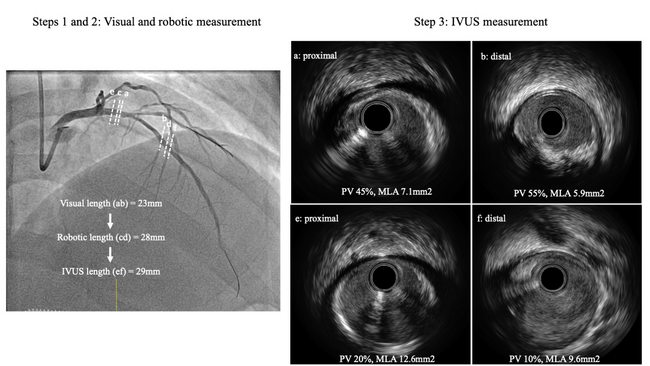
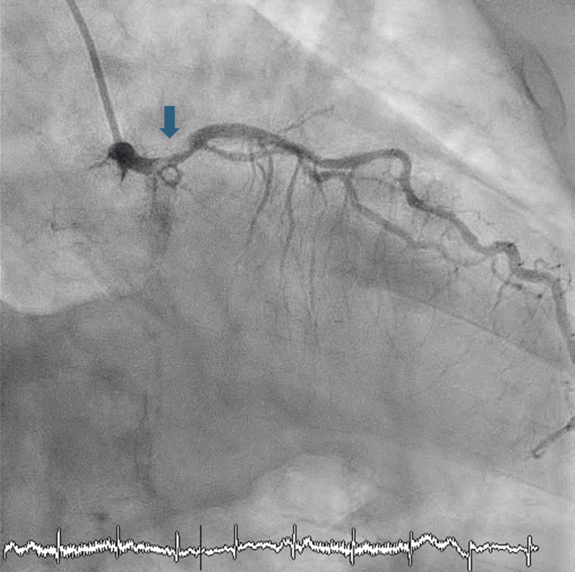
During the R-PCI procedure, the operator provided a visual estimate of lesion length after the lesion was wired and pre-dilated. This was compared to the robotic assessment of lesion length. The operator advanced an intracoronary balloon with the radio-opaque marker past the distal edge of the lesion. The length counter on the cockpit touchscreen was zeroed before the operator retracted the marker on the balloon to the proximal edge of the lesion. The robotic lesion length assessment was displayed on the touchscreen (Figure 1). In cases in which ICI was used, either with intravascular ultrasound (IVUS) or optical coherence tomography (OCT), the visual and robotic assessments were compared to the length measurement by ICI. ICI lesion length was measured from the most normal segment distal to the lesion to the most normal proximal segment, where the plaque volume was less than 50%. Choice of ICI used was at the operator’s preference.
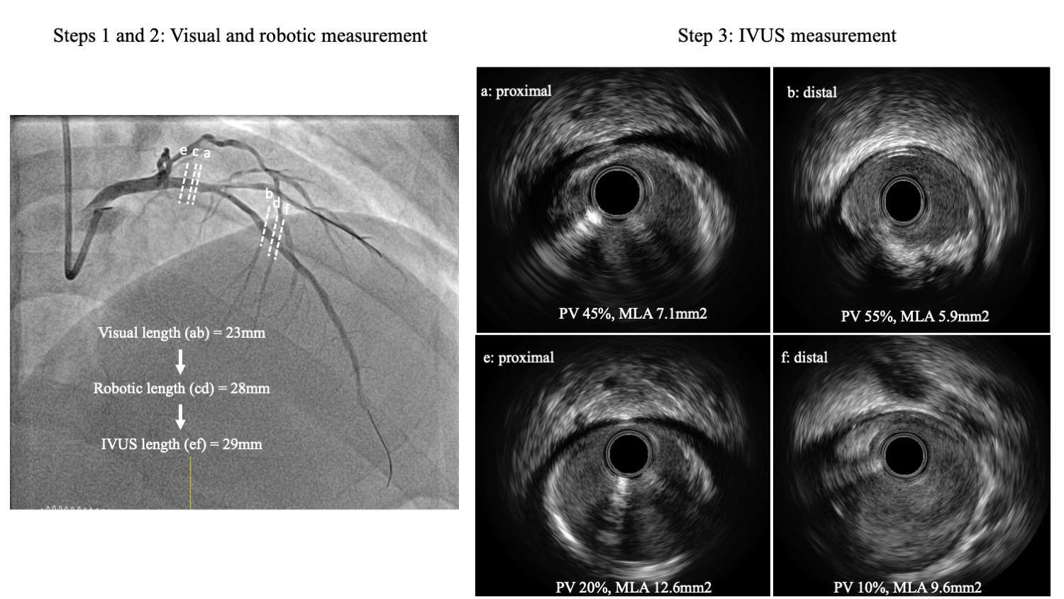
Lesion length comparison
This study compared lesion length estimates performed visually by the operator to those calculated by the robotic system. The visual estimate was considered accurate if the 2 estimates were identical. However, the visual estimates were considered “too short” or “too long” if they were shorter or longer than the robotic estimate, respectively. The visual estimates were also assessed against a secondary threshold of ± 5 mm too long or too short. In cases where ICI was performed, the visual and robotic estimates were compared against the ICI length measurement using the same criteria.
Statistical analyses
Continuous variables were reported as mean ± standard deviation or median with interquartile range (IQR) if not normally distributed. Categorical variables were presented as an absolute number with percentages. Paired t-tests were used to compare the visual and robotic length measurements. For the patients who had ICI, linear regression was used to evaluate correlations between visual and ICI and between robotic and ICI measurements. Bland-Altman plots were generated to assess agreement between visual and ICI and between robotic and ICI measurements. Fisher’s z-test was performed to compared correlation coefficients, with a 2-sided P-value of less than 0.05 considered statistically significant.
Results
Seventy-two patients (with 78 lesions) underwent R-PCI in the PARTY trial. Visual and robotic length measurements were performed for 63 of these patients (69 lesions), which were analyzed in this study. The median visual length measurement was 23.0 mm (IQR: 18.0-28.0) and the median robotic visual length measurement was 24.0 mm (IQR: 18.7-33.0 mm).8 Because of clinical urgency to proceed with PCI, in 9 cases the visual and robotic measurements were not performed.
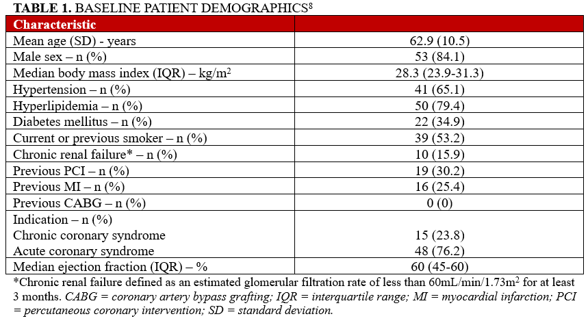
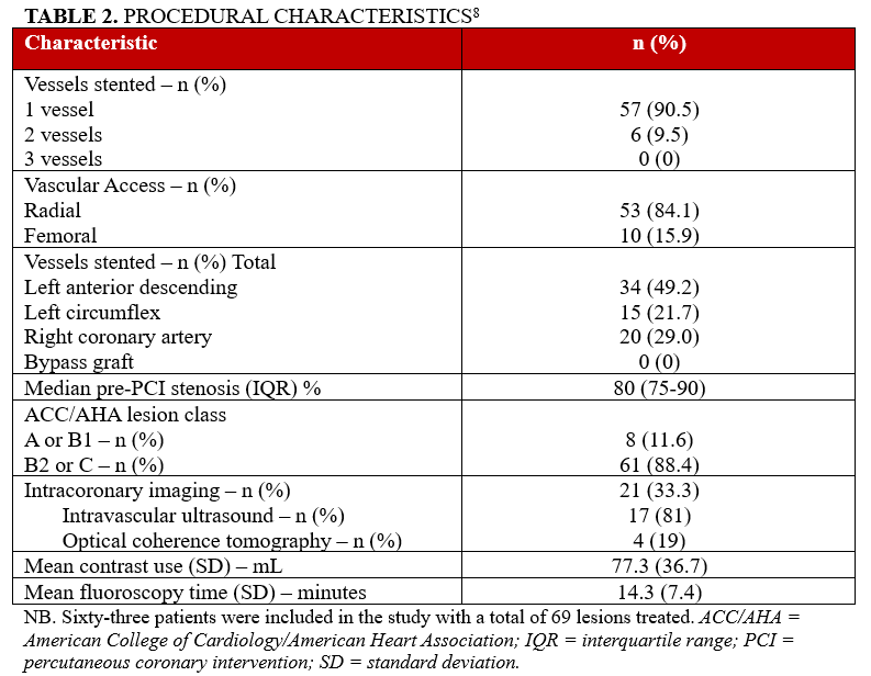
Key baseline patient characteristics are summarized in Table 1. Most patients were male (84% [53]), with the majority presenting with complex lesions (88.4% of lesions [61] were American College of Cardiology/American Heart Association class B2/C) and acute coronary syndrome requiring PCI (76.2% [48]). Most patients underwent single-vessel PCI, with the radial approach used in 84.1% of cases (53). The left anterior descending artery was the most commonly stented artery (Table 2). ICI was used in 33.3% of cases (21), 81% of IVUS (17), and 19% of OCT (4).
Lesion length assessment
The visual assessment was shorter than the robotic measurement in 46 lesions (66.7%) and was longer than robotic assessment in 16 lesions (23.2%). The visual and robotic assessments were identical in 7 lesions (10.1%). Using a threshold of greater than 5 mm too long/short, 47 lesions (68%) were accurately measured visually, while 22 lesions (32%) were inaccurately measured: visual assessment was shorter in 21 lesions (95.4%) and longer in only 1 lesion (4.5%).
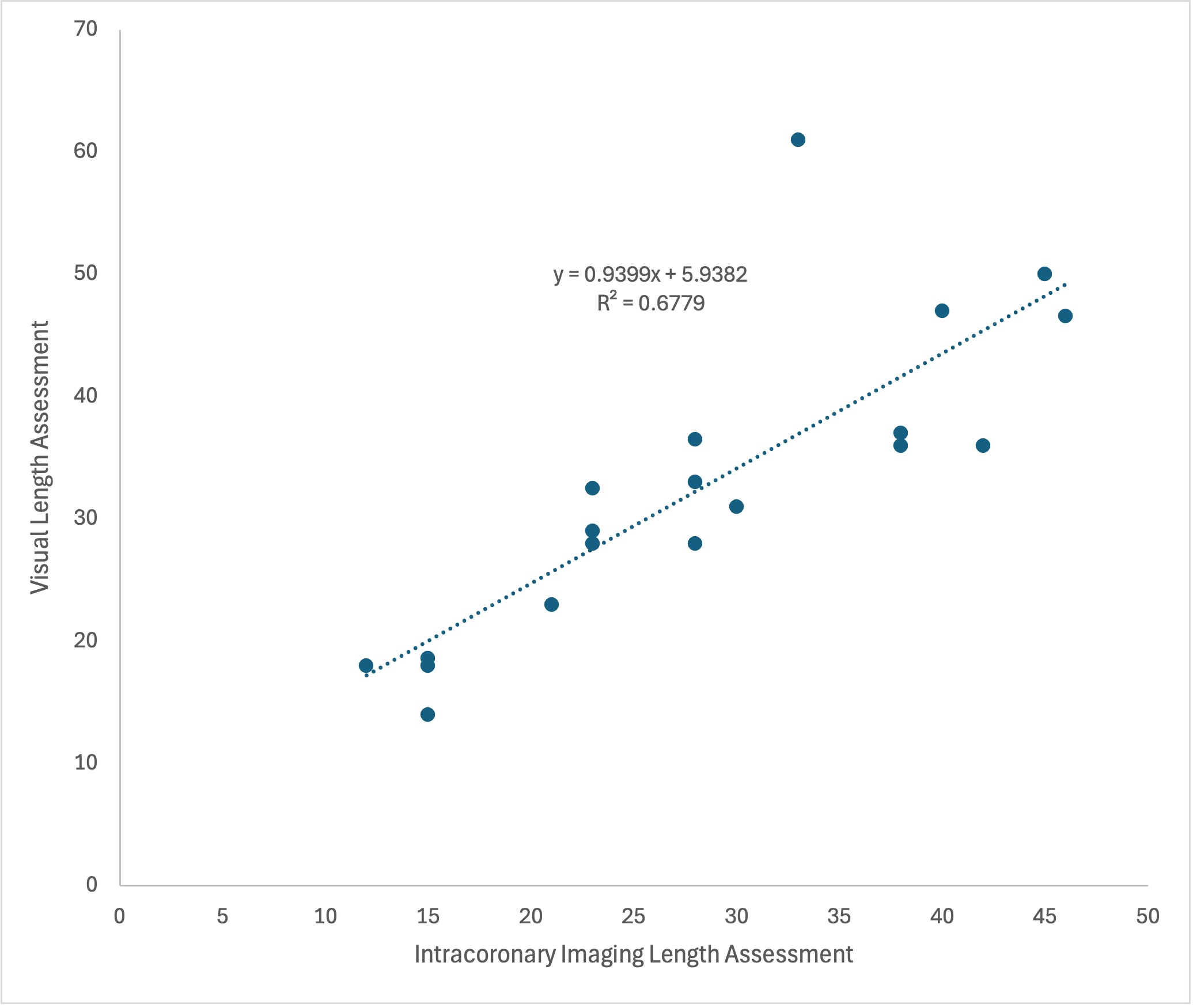
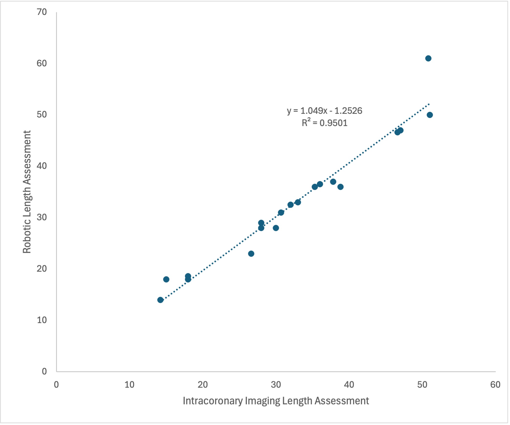
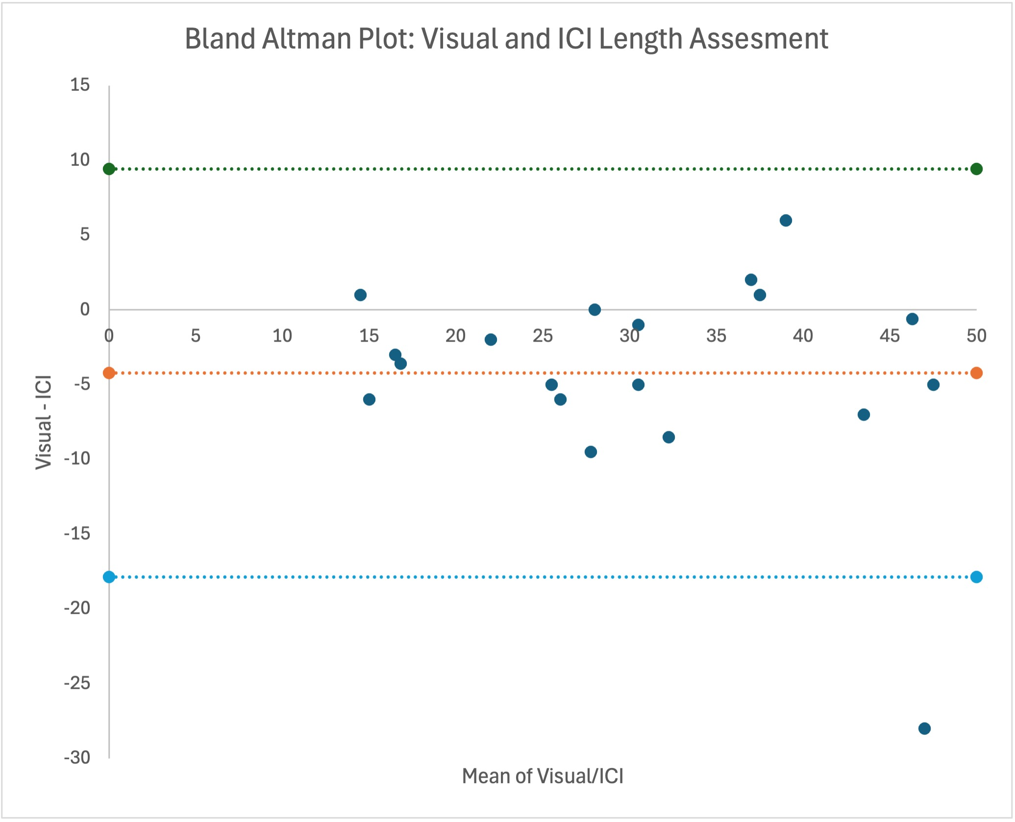
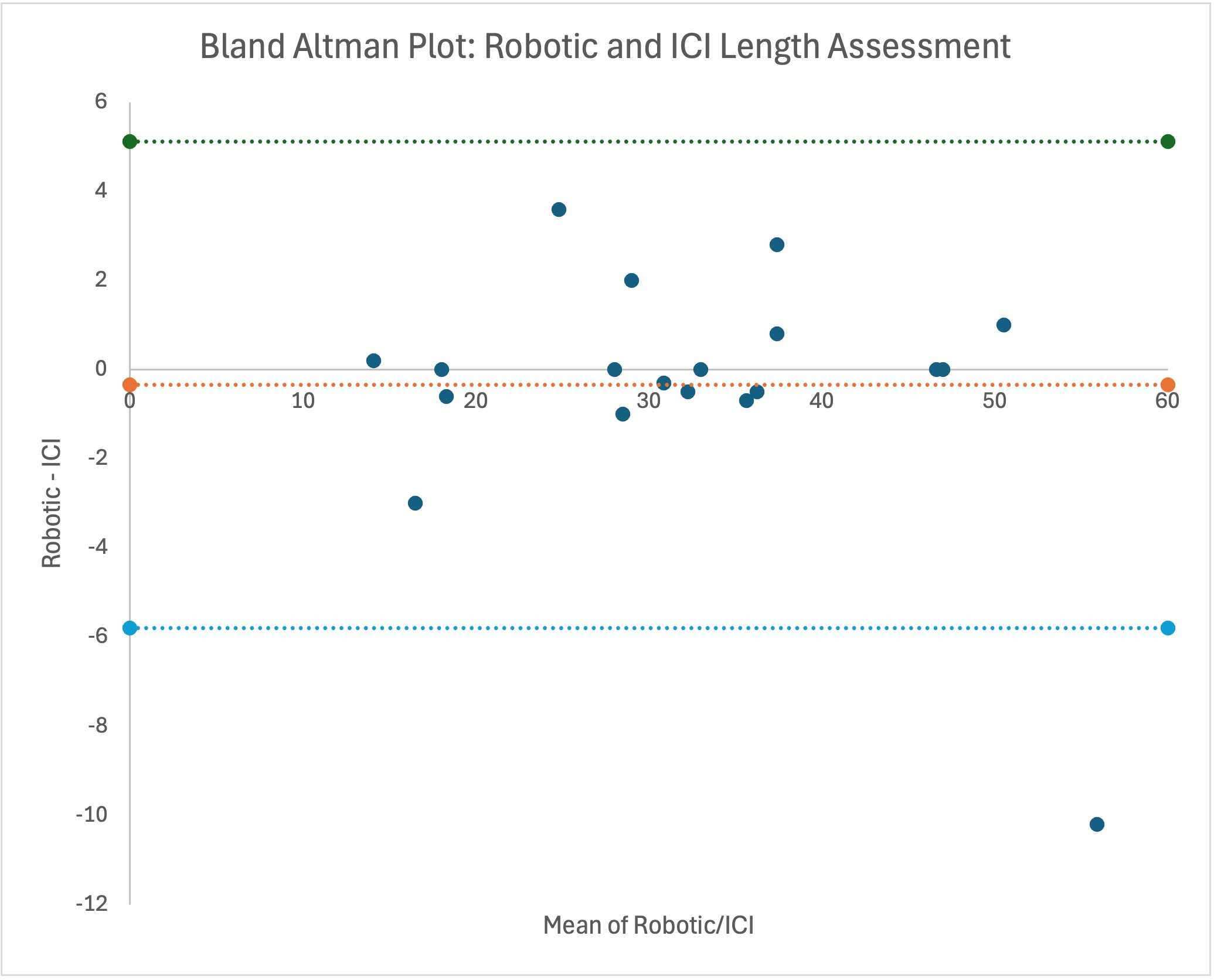
In the cases in which ICI was performed, both the visual and robotic assessments showed good correlation with the ICI lesion length assessment with 1 outlier: r = 0.82 and r = 0.97, respectively (Figure 2A and B). The correlation between robotic and ICI measurements was significantly higher than that between visual and ICI measurements (P = .004). The Bland-Altman analyses showed good agreement between visual/ICI and robotic/ICI assessments (Figures 3A and B). However, the visual estimate systematically underestimated the true lesion length calculated by ICI, with a mean difference of -4.22 ± 6.97 mm. The mean difference between robotic and ICI estimates was -0.34 ± 2.79 mm.
Discussion
This study highlights the inaccuracies inherent to visual estimation of lesion length and, as a result, assessment of appropriate stent length for implantation. In our study, only 10.1% of lesions were accurately measured visually when compared with robotic measurement, with the majority of cases being visually estimated too short (66.7%) and a smaller proportion of lesions estimated too long (23.2%). Allowing a more generous threshold of 5 mm, 32% of lesions were inaccurately measured; all except one were underestimated. We selected a threshold of greater than 5 mm as too long or short based on endovascular radiotherapy delivered post-PCI to prevent restenosis. In those initial studies, the authors found that the injured artery segments that were more than 5 mm away from the radiotherapy seed were at the highest risk of failure at follow-up.1
Both the visual and robotic length assessments showed good correlation with the “gold standard” of ICI10; however, the robotic length assessment showed significantly higher correlation. Furthermore, the visual assessments consistently underestimated the lesion length compared with the ICI measurements (4.22 mm too short). This did not occur when robotic measurements were used, where the mean difference was a clinically insignificant 0.34 mm too short.
Our results differ from Kimura et al, who found that measured lesion length was significantly greater for IVUS compared with robotic assessments (25.7 ± 9.4 mm vs 21.0 ± 8.89 mm, P < .001).11 This difference may potentially be due to the fact that they based their length measurements on parameters such as plaque volume and lumen area, which we did not assess in our study. Visual estimations of lesion length have previously been identified to be unreliable. Leow et al showed that when estimated visually, length was often overestimated, with the degree of overestimation increasing with increasing length.12 This differs from our findings which showed that most visual measurements were underestimated when compared with robotic measurements.
Deploying either too-short or too-long stents can potentially lead to adverse clinical outcomes. Deploying too short a stent can cause LGM and its associated adverse sequalae. If identified, further stents are usually deployed to correct the LGM, prolonging the PCI procedure and increasing total equipment and procedural costs. Deploying too-long stents can also potentially lead to adverse events, as was demonstrated by Kong et al. In their registry analysis of 9217 patients undergoing PCI, patients with long stents (>40 mm) had significantly higher rates of target lesion failure and early stent thrombosis.13 This association persisted after adjustment for factors such as lesion complexity and patient comorbidities, which may necessitate longer stents and contribute to poorer long-term outcomes.
ICI has become an important tool during PCI to improve procedural and longer-term clinical outcomes.14 ICI can assess plaque location and morphology pre-PCI to help guide lesion preparation strategies as well as appropriate selection of stent length and diameter. It can also help check lesion preparation and proper stent deployment, potentially avoiding procedural complications. The growing evidence supporting ICI use during PCI has been reflected in multiple guideline updates, including the 2025 ACC/AHA/ACEP/NAEMSP/SCAI (American College of Cardiology/American Heart Association/American College of Emergency Physicians/National Association of EMS Physicians/Society for Cardiovascular Angiography and Interventions) guidelines for the management of acute coronary syndromes, where ICI has a Class IA recommendation to guide PCI in left main or complex lesions.15 The high correlation between lesion length assessment using R-PCI and ICI suggests that R-PCI could be similarly employed to provide comparable lesion length assessment in cases where ICI is not used.
R-PCI may improve lesion assessment and stent delivery via several mechanisms. The robotic system allows movement and positioning of intracoronary devices in 1-mm increments, which is more precise and reproducible than can be achieved by hand, leading to higher accuracy. Its measurements also account for the 3-dimensional (D) nature of the coronary artery rather than a 2D visual assessment. Furthermore, during R-PCI the operator can sit much closer to the fluoroscopy screen, allowing improved visualization of the PCI procedure.
Limitations
Our study was performed at a single center with a relatively small sample size, particularly the subgroup that underwent ICI. Thus, our results may not be generalizable to other patient populations. Furthermore, they are at risk of being unduly influenced by random chance. The unblinded nature of the study could have potentially introduced bias into the lesion measurements. The fact that the measurements were performed sequentially meant that the knowledge of the first measurement performed (usually visual) could influence the operators’ subsequent measurements. The study was not powered for a head-to-head comparison between visual, robotic, and ICI measurements. We were also not able to assess inter-observer variability in our current study. These factors and limitations could all be investigated in future studies.
There are inherent limitations to using the robotic system to measure lesion length, which still requires interpretation of the coronary angiogram, a 2D representation of a 3D structure. It has been previously shown that visual assessment of coronary artery stenoses or using quantitative coronary angiography are both unreliable in estimating the severity of stenoses.16 ICI, the “gold standard” for lesion length assessment, was used in only one-third of our cases. A larger sample size would allow for more robust comparisons. We did not assess for LGM via core lab analysis of the PCI images, nor did we assess other ICI parameters such as minimal lumen or minimal stent area. We did not specifically perform any clinical follow-up for this analysis. However, overall complication rates for the R-PCI arm of the PARTY trial at 30 days were low.8 Another avenue for future investigation would be performing a cost-effectiveness analysis to see if R-PCI, despite the extra upfront equipment costs, could produce longer-term cost savings by reducing unnecessary stent usage and avoiding adverse cardiovascular outcomes associated with LGM or excessive over-stenting.
Conclusions
Visual lesion assessment can be inaccurate, often underestimating true lesion length. Robotic lesion assessment has improved correlation with ICI, with less under- or overestimation than visual assessments. R-PCI can potentially help reduce the incidence of LGM by providing more accurate lesion assessment, procedural visualization, and equipment and stent delivery. More studies with larger sample sizes are required to further assess these potential benefits and perform larger comparisons with ICI.
Affiliations and Disclosures
James Leung, MBBS1,2; John French, MBChB, PhD1,2; James Xu, MBBS, PhD1,2; Kathryn Wales, MD1; Hashim Kachwalla, MBBS3; Krishna Kaddapu, MBBS, PhD3; Sarah Fares, MPhil1; Viet Dang, MBBS1; Tamer Badie, MBBS3; Christian Mussap, MBBS, PhD1,2; Rohan Rajaratnam, MBBS1,2; Dominic Leung, MBBS, PhD1,2; Sidney Lo, MBBS1; Craig Juergens, MBBS, DMedSc1,2
From the 1Department of Cardiology, Liverpool Hospital, Sydney, New South Wales, Australia; 2South West Sydney Clinical School, University of New South Wales, Australia; 3Department of Cardiology, Campbelltown Hospital, Sydney, New South Wales, Australia.
Disclosures: The authors report no financial relationships or conflicts of interest regarding the content herein.
Funding: The Percutaneous coronary intervention using Assisted Robotic TechnologY (PARTY) trial on which this study is based was supported by philanthropic donation via the Ingham Institute for Applied Medical Research. Dr Leung is supported as a PhD candidate through the University of New South Wales and also by a National Heart Foundation PhD Scholarship (ID 107684).
Address for correspondence: James Leung, MBBS, Department of Cardiology, Liverpool Hospital, Corner of Elizabeth and Goulburn Streets, Liverpool, NSW, 2170, Australia. Email: james.t.leung@unsw.edu.au
References
1. Sabaté M, Costa MA, Kozuma K, et al. Geographic miss: a cause of treatment failure in radio-oncology applied to intracoronary radiation therapy. Circulation. 2000;101(21):2467-2471. doi:10.1161/01.cir.101.21.2467
2. Costa MA, Angiolillo DJ, Tannenbaum M, et al; STLLR Investigators. Impact of stent deployment procedural factors on long-term effectiveness and safety of sirolimus-eluting stents (final results of the multicenter prospective STLLR trial). Am J Cardiol. 2008;101(12):1704-1711. doi:10.1016/j.amjcard.2008.02.053
3. Gupta R, Malik AH, Chan JSK, et al. Robotic assisted versus manual percutaneous coronary intervention: systematic review and meta-analysis. Cardiol Rev. 2024;32(1):24-29. doi:10.1097/CRD.0000000000000445
4. Leung J, French J, Xu J, et al. Robotic assisted percutaneous coronary intervention: initial australian experience. Heart Lung Circ. 2024;33(4):493-499. doi:10.1016/j.hlc.2024.01.019
5. Kapur V, Smilowitz NR, Weisz G. Complex robotic-enhanced percutaneous coronary intervention. Catheter Cardiovasc Interv. 2014;83(6):915-921. doi:10.1002/ccd.25271
6. Campbell PT, Kruse KR, Kroll CR, Patterson JY, Esposito MJ. The impact of precise robotic lesion length measurement on stent length selection: ramifications for stent savings. Cardiovasc Revasc Med. 2015;16(6):348-350. doi:10.1016/j.carrev.2015.06.005
7. Bezerra HG, Mehanna E, W Vetrovec G, A Costa M, Weisz G. Longitudinal feographic miss (LGM) in robotic assisted versus manual percutaneous coronary interventions. J Interv Cardiol. 2015;28(5):449-455. doi:10.1111/joic.12231
8. Leung J, French J, Xu J, et al. A randomised clinical trial investigating robotic-assisted percutaneous coronary intervention: the PARTY trial. AsiaIntervention. 2026;12(2):e136-e145. doi:10.4244/AIJ-D-25-00066
9. Leung J, Xu J, French JK, Kachwalla H, et al. Rationale and design of the randomised, controlled Percutaneous coronary intervention using Assisted Robotic TechnologY (PARTY) trial. Open Heart. 2024;11(2):e002950. doi:10.1136/openhrt-2024-002950
10. Räber L, Mintz GS, Koskinas KC, et al; ESC Scientific Document Group. Clinical use of intracoronary imaging. Part 1: guidance and optimization of coronary interventions. An expert consensus document of the European Association of Percutaneous Cardiovascular Interventions. Eur Heart J. 2018;39(35):3281-3300. doi:10.1093/eurheartj/ehy285
11. Kimura T, Koeda Y, Ishida M, et al. Safety and feasibility of intravascular ultrasound-guided robotic percutaneous coronary intervention. Coron Artery Dis. 2023;34(7):463-469. doi:10.1097/MCA.0000000000001274
12. Leow MQ, Teo WZW, Lim JX, et al. Visual estimation of lengths and angles: how accurate are we? Singapore Med J. 2026;67(Suppl 2):S15-S18. doi:10.4103/singaporemedj.SMJ-2021-123
13. Kong MG, Han JK, Kang JH, et al; Collaborators. Clinical outcomes of long stenting in the drug-eluting stent era: patient-level pooled analysis from the GRAND-DES registry. EuroIntervention. 2021;16(16):1318-1325. doi:10.4244/EIJ-D-19-00296
14. Truesdell AG, Alasnag MA, Kaul P, et al; ACC Interventional Council. Intravascular imaging during percutaneous coronary intervention: JACC state-of-the-art review. J Am Coll Cardiol. 2023;81(6):590-605. doi:10.1016/j.jacc.2022.11.045
15. Rao SV, O'Donoghue ML, Ruel M, et al. 2025 ACC/AHA/ACEP/NAEMSP/SCAI guideline for the management of patients with acute coronary syndromes: a report of the American College of Cardiology/American Heart Association joint committee on clinical practice guidelines. Circulation. 2025;151(13):e771-e862. doi:10.1161/CIR.0000000000001309
16. Fischer JJ, Samady H, McPherson JA, et al. Comparison between visual assessment and quantitative angiography versus fractional flow reserve for native coronary narrowings of moderate severity. Am J Cardiol. 2002;90(3):210-215. doi:10.1016/s0002-9149(02)02456-6