Seeing the Mechanism: A Transesophageal Echocardiographic Demonstration of Dynamic Left Ventricular Outflow Tract Obstruction in Hypertrophic Obstructive Cardiomyopathy
© 2026 HMP Global. All Rights Reserved.
Any views and opinions expressed are those of the author(s) and/or participants and do not necessarily reflect the views, policy, or position of the Journal of Invasive Cardiology or HMP Global, their employees, and affiliates.
J INVASIVE CARDIOL 2026. doi:10.25270/jic/26.00320. Epub October 8, 2026.
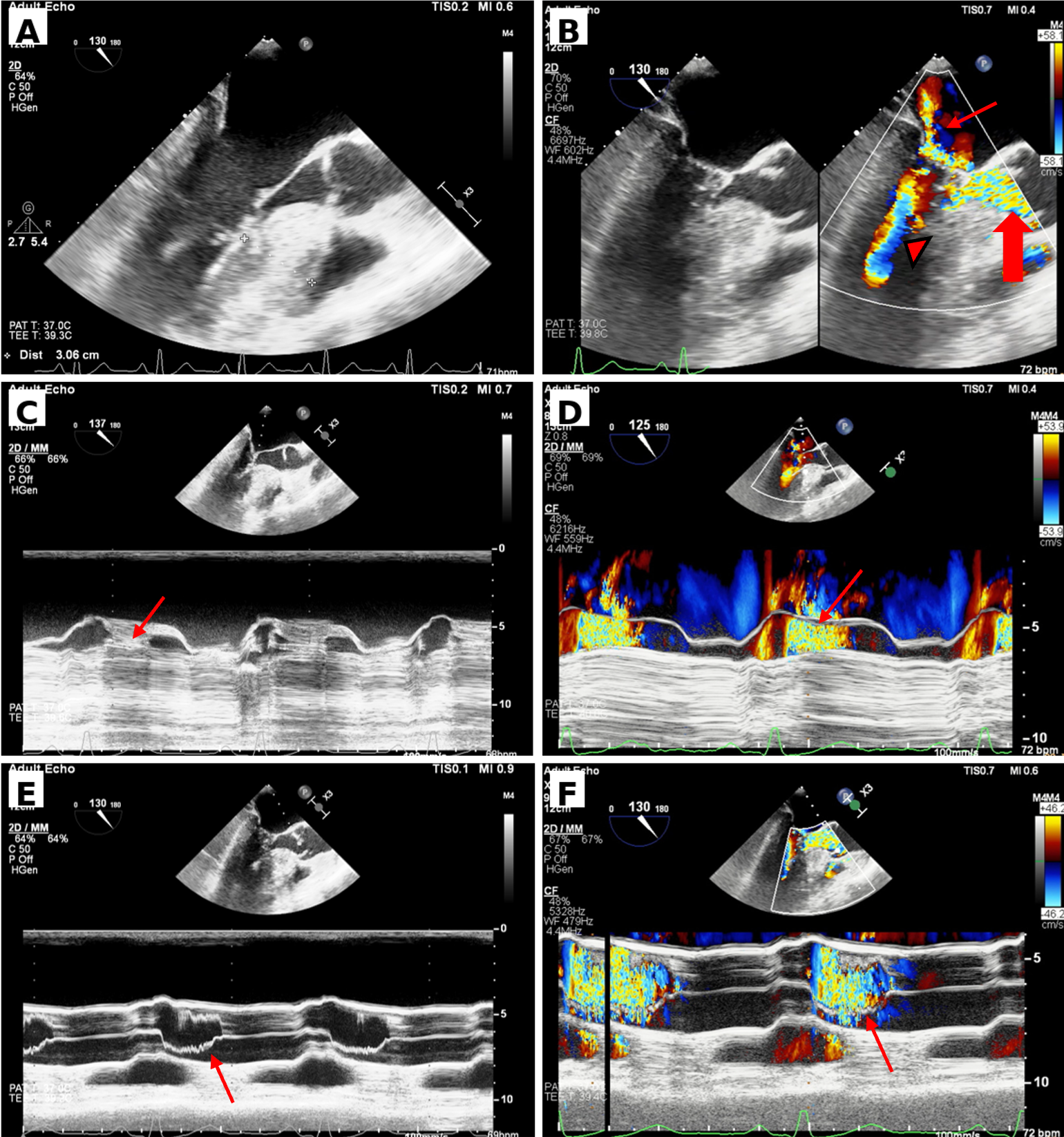
With these 6 transesophageal echocardiographic (TEE) images (Figure and Video), the authors provide a striking visual demonstration of the anatomical and dynamic mechanisms underlying left ventricular outflow tract (LVOT) obstruction in hypertrophic obstructive cardiomyopathy (HOCM). All images were obtained at approximately 130° in the mid-esophageal TEE position.
The sequence begins with marked asymmetric septal hypertrophy and consequent narrowing of the LVOT (Figure A). Figure B demonstrates the pivotal mechanism that is prominent systolic anterior motion (SAM) of the anterior mitral leaflet, producing a posteriorly directed mitral regurgitation jet. Color Doppler simultaneously reveals intense turbulence within the LVOT and mid-left ventricular cavity.
The severity of SAM is particularly evident on M-mode imaging at the level of mitral valve, with grade IV SAM demonstrated in Figure C. Corresponding color M-mode (Figure D) provides a dynamic visualization of the resulting LVOT obstruction, demonstrating high-velocity turbulent systolic flow generated by the SAM-related narrowing.
The downstream hemodynamic consequences are further illustrated at the level of the aortic valve (AV). M-mode imaging at the level of AV in Figure E demonstrates fine systolic fluttering of the AV leaflets, reflecting the marked turbulence generated by high-velocity flow through the narrowed LVOT. Figure F, using color M-mode, directly visualizes this turbulent high-velocity flow traversing the AV.
Together, these images capture the complete pathophysiologic cascade of HOCM—from asymmetric septal hypertrophy and SAM to dynamic LVOT obstruction, secondary mitral regurgitation, and characteristic turbulent flow across the AV—within a single TEE examination.
Affiliations and Disclosures
Keshav Sachdeva, MD, DM1; Hardik Jain, MD1; Tejinder Singh Malhi, MD, DM1; Suraj Kumar, MD, DM1; Jasneet Kaur, MD2; Bhupinder Singh, MD, DM1
From the 1Department of Cardiology, All India Institute of Medical Sciences, Bathinda, India; 2Department of Cardiology, Nassau University Medical Center, New York.
Disclosures: The authors report no financial relationships or conflicts of interest regarding the content herein.
Consent statement: The authors confirm that informed consent was obtained from the patient and family for the intervention described in the manuscript and for the publication of thereof, including photographs.
Address for correspondence: Bhupinder Singh, MD, DM, Department of Cardiology, All India Institute of Medical Sciences, Bathinda 151001, India. Email: dr_bhupinders@yahoo.in