Clinical Videos
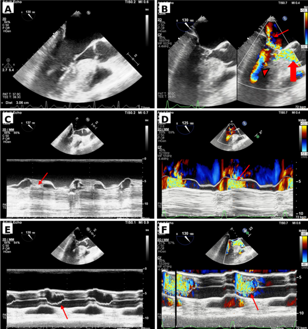
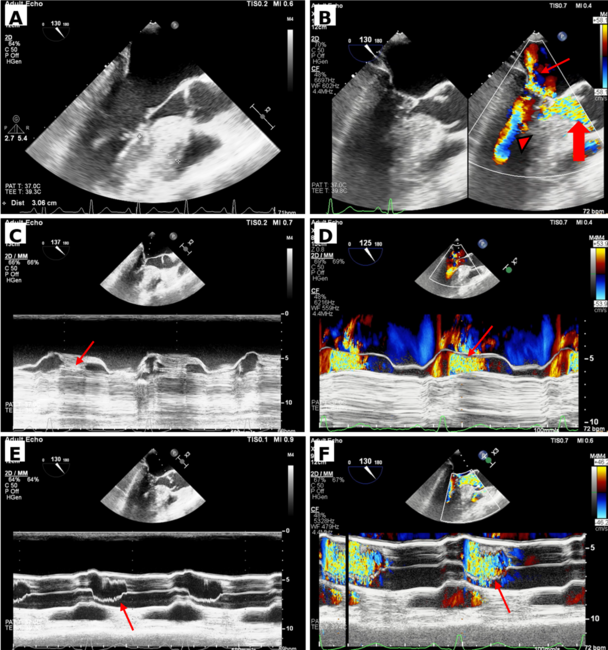
Video Supplement: When the Left Ventricle Dances: Ballerina Foot Appearance in Hypertrophic Cardiomyopathy
05/27/2026
Video Supplement to "When the Left Ventricle Dances: Ballerina Foot Appearance in Hypertrophic Cardiomyopathy" (Clinical Image).
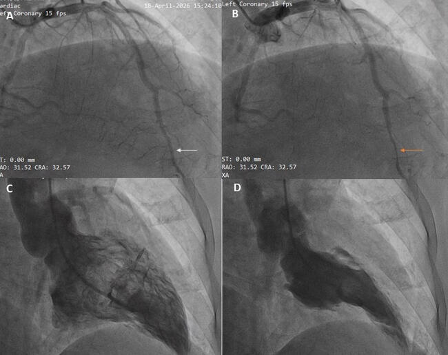
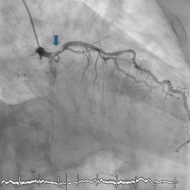
Video 1. Coronary angiogram in the right anterior oblique cranial view with evidence of dynamic compression (milking) seen in the distal left anterior descending artery as a sign of myocardial bridging.
© 2026 HMP Global. All Rights Reserved.
Any views and opinions expressed are those of the author(s) and/or participants and do not necessarily reflect the views, policy, or position of the Journal of Invasive Cardiology or HMP Global, their employees, and affiliates.