




Agreement of Noninvasive Multimodality Imaging Measurements With Balloon Sizing in the Cardiac Catheterization Laboratory Prior to Percutaneous Pulmonary Valve Implantation
© 2026 HMP Global. All Rights Reserved.
Any views and opinions expressed are those of the author(s) and/or participants and do not necessarily reflect the views, policy, or position of the Journal of Invasive Cardiology or HMP Global, their employees, and affiliates.
J INVASIVE CARDIOL 2026. doi:10.25270/jic/26.00166. Epub November 12, 2026.
Key Clinical Summary
- Cardiac magnetic resonance imaging (CMR) and computed tomography (CT) provide complementary, essential information before transcatheter pulmonary valve implantation for right ventricular outflow tract (RVOT) dysfunction in repaired congenital heart disease.
- CMR- and CT-derived landing-zone measurements correlated substantially with invasive balloon sizing performed in the catheterization laboratory.
- Future studies incorporating computational fluid dynamics and 4D multimodality imaging may help tailor transcatheter pulmonary valve selection and assess the risk of coronary artery compression.
Abstract
Objectives. Right ventricular outflow tract (RVOT) dysfunction is common in repaired congenital heart defects resulting in right ventricular (RV) dilation and dysfunction. Emerging transcatheter options enable placement of a competent pulmonary valve, obviating the need for surgery. Multimodality imaging plays a key role in determining the type and timing of intervention prior to percutaneous pulmonary valve implantation (PPVI). The authors sought to assess correlation of noninvasive RVOT measurements with cardiac magnetic resonance imaging (CMR) and cardiac computed tomography (CT) with balloon sizing in the catheterization laboratory prior to PPVI.
Methods. A retrospective review was performed on patients who had undergone PPVI with preprocedural CMR and cardiac CT scan. Three-dimensional multiplanar reconstruction (MPR) of the narrowest RVOT diameter on magnetic resonance angiography (MRA), whole heart sequence (WHS) images, and CT scan images were performed using commercially available software; balloon sizing prior to PPVI was recorded. Concordance correlation coefficients were used to assess agreement with the clinically acceptable limit between the modalities set at +/- 2 mm. Additionally, mean difference was used to quantify the difference in measurements among the 2 modalities.
Results. For maximal waist dimensions, correlation between MRA and WHS images with balloon sizing was 0.70 to 0.79, while correlation between CT scan and balloon sizing was 0.65 to 0.75. For minimal waist dimensions, correlation between MRA and WHS images was 0.72 to 0.82; correlation between CT scan and balloon sizing was 0.64 to 0.73.
Conclusions. Cardiac CT and CMR are complementary modalities that provide essential information for preprocedural planning for PPVI.
Introduction
The problem of right ventricular outflow tract (RVOT) dysfunction is a common denominator for numerous conotruncal anomalies including tetralogy of Fallot (TOF), transposition of great arteries, double outlet right ventricle, congenital pulmonary stenosis, pulmonary atresia with intact ventricular septum, and truncus arteriosus. With emerging successful palliation of these anomalies, residual or recurrent RVOT dysfunction and RV failure with time is an expected sequela. Initial palliative procedures to augment pulmonary blood flow in infancy include stenting of patent ductus arteriosus, modified Blalock-Taussig shunt, or creation of an extracardiac conduit between the RV and the pulmonary artery (RV-PA).1,2 Transannular patch technique of repair in TOF creates free pulmonary regurgitation (PR) with detrimental effects.3 While surgical bioprosthetic valves or RV-PA conduits serve to palliate residual RVOT dysfunction, they have limited durability, necessitating periodic replacement.
Since the implantation of the first transcutaneous pulmonary valve (PV) by Bonhoeffer et al in 2000,4 Percutaneous pulmonary valve implantation (PPVI) has revolutionized management options for older children and adults with repaired congenital heart disease, minimizing the risk for open heart surgeries4 and thereby reducing the number of sternotomies and the need for cardiopulmonary bypass,5 as well as prolonging conduit lifespan.6 However, PPVI can pose numerous technical challenges with regard to assessing postoperative RVOT size and determining the narrowest dimension or landing zone for suitable valve implantation because of its heterogenous anatomy and complex 3-dimensional (3D) configuration, especially in patients with native, expansile RVOTs and no rigid landing zone.5 Extending our capabilities to implant valves in native RVOTs5 with no rigid landing zone—unlike calcified conduits—emphasizes the importance of preprocedural imaging to understand the dynamic nature of the RVOT, the presence of calcifications, the spatial relationship to coronary arteries, proximity to the sternum, and the anatomy of branch pulmonary arteries.6
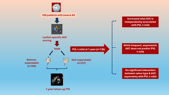
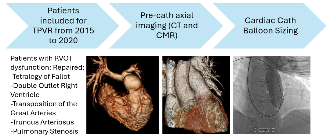
Patients warranting PV implantation often have axial imaging with cardiac magnetic resonance imaging (CMR) and computed tomography (CT) scans prior to anticipated intervention. The purpose of this study was to determine correlation between noninvasive imaging modalities and balloon sizing in the cath lab (gold standard) to assess the landing zone of the RVOT in patients being considered for PPVI (Central illustration).
Methods
Study design
This was a retrospective cross-sectional study of patients with conotruncal abnormalities who had undergone RVOT revision and were being followed at the Cleveland Clinic Children’s Hospital between 2015 and 2020. The study was approved by the Cleveland Clinic Institutional Review Board with a waiver of informed consent because of the retrospective nature of the study.
Inclusion criteria
Patients with repaired conotruncal abnormalities and RVOT dysfunction (stenosis/regurgitation/both) with attempted balloon sizing during cardiac catheterization along with preprocedural axial imaging studies prior to cardiac catheterization were included. Timing of referral for intervention and desired intervention was dictated by the clinician following these patients. Patients with stents, pacemakers, implantable cardioverter defibrillators (ICD), or any other metallic implant that precluded CMR underwent CT scan.
Exclusion criteria
Patients with incomplete data and those with noninvasive imaging studies who did not have the relevant images prior to cardiac catheterization were excluded.
Demographic data and clinical parameters
Baseline demographic data, including age, height, weight, gender, diagnosis, prior surgical procedures, and type of RVOT were obtained by retrospective chart review. Echocardiographic variables of right and left ventricular dilation and function as well as degree of pulmonary stenosis and regurgitation were reviewed. CMR variables including right and left ventricular end diastolic volumes and function, pulmonary regurgitant fraction, and CT data were reviewed in select patients. Catheterization data with balloon size and type of PPVI implanted were reviewed.
Noninvasive imaging studies
CMR
CMR examinations were performed on standard 1.5T and 3.0T magnetic resonance scanners (Phillips Achieva and Phillips Ingenia) and the 1.5T MAGNETOM Avanto scanner (Siemens Healthineers) using electrocardiographic (ECG) gating and prescribed institutional protocols which included non-gated contrast enhanced magnetic resonance angiography (MRA) and free-breathing navigator and ECG-gated 3D steady-state free precession whole heart sequence (WHS).
Contrast-enhanced multidetector computerized tomography (MDCT) protocol
Patients were scanned on a dual-source MDCT scanner (Definition [Siemens Healthineers]) following administration of iodinated contrast agent. A contrast bolus monitoring technique using a region of interest in the central pulmonary artery was used to determine the scan delay time. Once the desired attenuation was reached, scanning was initiated in the craniocaudal direction during a single inspiratory breath-hold from the level of the thoracic inlet to the base of the heart with prospective ECG triggering. Images were reconstructed with reconstructed slice thickness of 1 mm for post processing and analysis.
Cardiac catheterization procedure
Balloon sizing, i.e., inflating low pressure balloons in the RVOT, was performed until the appearance of a waist, which would serve as the landing zone for the valve. The maximum and minimum diameters of the waist obtained during balloon sizing were measured. In patients with no waist, the minimum waist diameter was assumed to be the maximal diameter obtained during balloon sizing. Pre-stenting was performed in some patients to decrease the RVOT size to make it amenable to PPVI. All patients underwent testing for potential coronary compression by simultaneous balloon inflation in the RVOT with aortic root angiogram.
RVOT measurements
Three-dimensional multiplanar reconstruction (MPR) was performed using a commercially available post-processing software, the Aquarius iNtuition viewer (TeraRecon Inc.) - an independent vendor neutral image viewing solution for both CMR and CT studies.
First, the RVOT was assessed by multiplanar reformatting with the centerline extending from the bottom of the RV and ending at the pulmonary artery bifurcation. The RVOT was defined as the area between the crest of the RV and the PV, with the area without any trabeculations below the PV defined as the crest of the RV. The narrowest area, or landing zone, was measured on orthogonal images obtained from double-oblique reconstruction of the RVOT-conduit and pulmonary artery (Figure 1). Measurement of the minimum and maximum diameters of the narrowest waist in plane 1 (superior-inferior, S-I) and plane 2 (right-left, R-L) were performed on both the MRA and WHS, with manual feature tracking and distance to the pulmonary artery bifurcation being used as reliable landmarks.
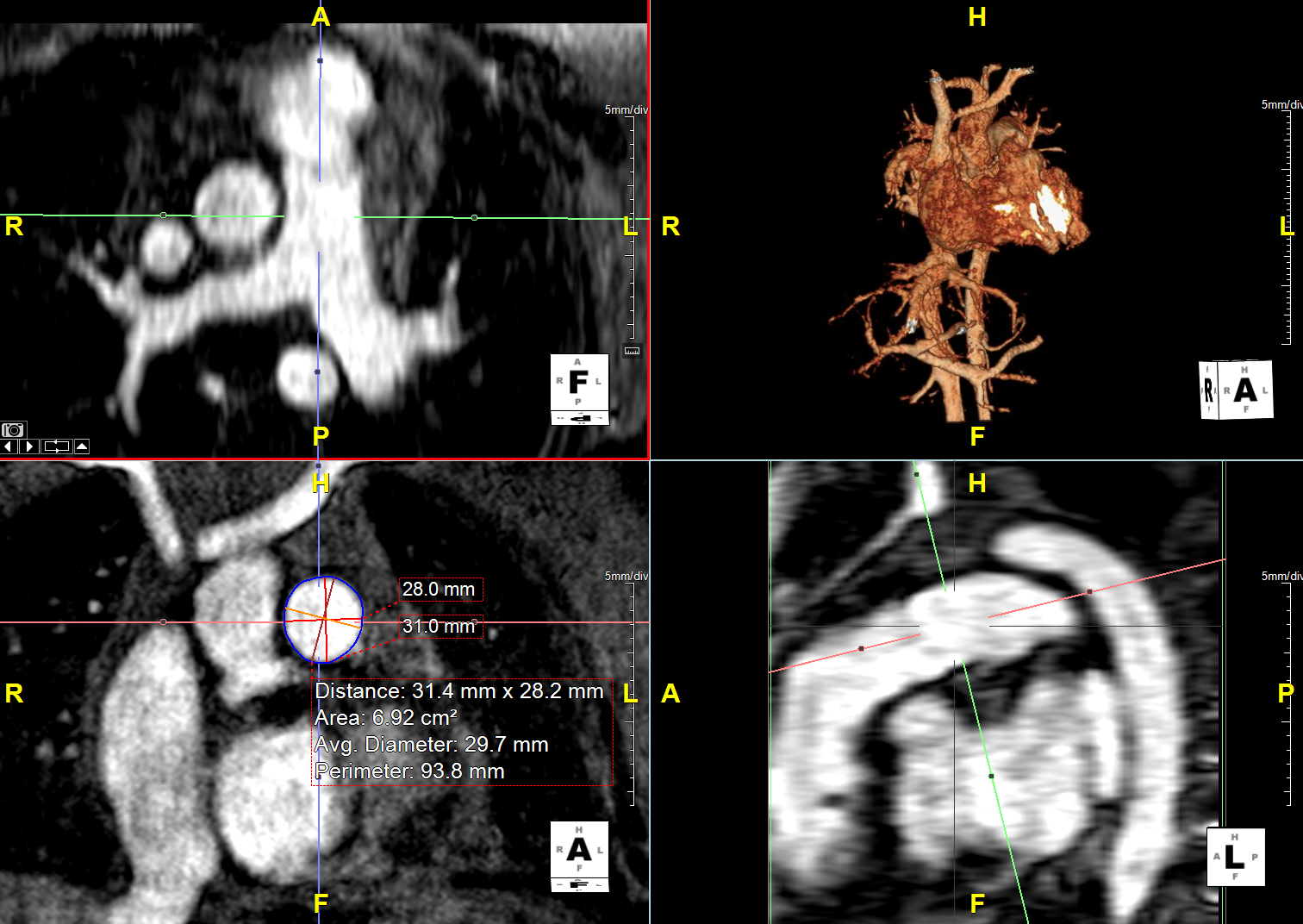
All studies were assessed by a single observer (SR), with subset analysis performed by another observer (RK) to assess interobserver variability. Up to 4 measures of RVOT diameter were collected from the MRA and WHS images, and 2 measures of RVOT diameter were obtained from CT images.
Statistical analysis
Data were presented as median and range for continuous variables and counts and percentages for categorical variables. We used concordance correlation coefficients (CCC) to evaluate the agreement between the RVOT diameter by noninvasive imaging assay (CMR or CT scan) and waist diameter by balloon sizing. The CCC quantifies how closely paired measurements match, accounting for both their correlation and systematic differences in their values. A CCC of 1 indicates perfect agreement, with all paired measurements lying on the line of identity (y = x).7 A diameter difference between imaging and balloon sizing falling between ± 2 mm was preferred. Paired mean differences in diameters with a 95% confidence interval (CI) and proportion within 2 mm were provided as a supplement for concordance correlation. The inter-rater reproducibility on CMR measurements (RVOT diameter, area, perimeter) was assessed on a random subsample by using the intraclass correlation coefficients (ICCs) for the 2-way mixed model. CCCs or ICCs equal to or lower than 0.40 indicate poor to fair agreement, 0.41 to 0.60 moderate, 0.61 to 0.80 good, and greater than 0.80 indicate excellent agreement.8
All analyses were performed on a complete-case basis; subjects with missing data on any variables were only excluded for analyses in which those variables were used. All tests were 2-tailed and performed at an overall significance level of 0.05. SAS 9.4 software (SAS Institute, Inc.) was used for all analyses and plots.
Results
Eighty-three patients who underwent balloon sizing prior to PPVI during cardiac catheterization were identified during the study period; of these patients, 75 met inclusion criteria: 49 with MRA measurements, 53 with WHS measurements, and 21 with CT scan measurements (Figure 2). For patients with MRA, WHS, or CT scan measurements, 87.8%, 88.7%, and 85.7%, respectively, had cardiac catheterization within 12 months of the imaging studies.
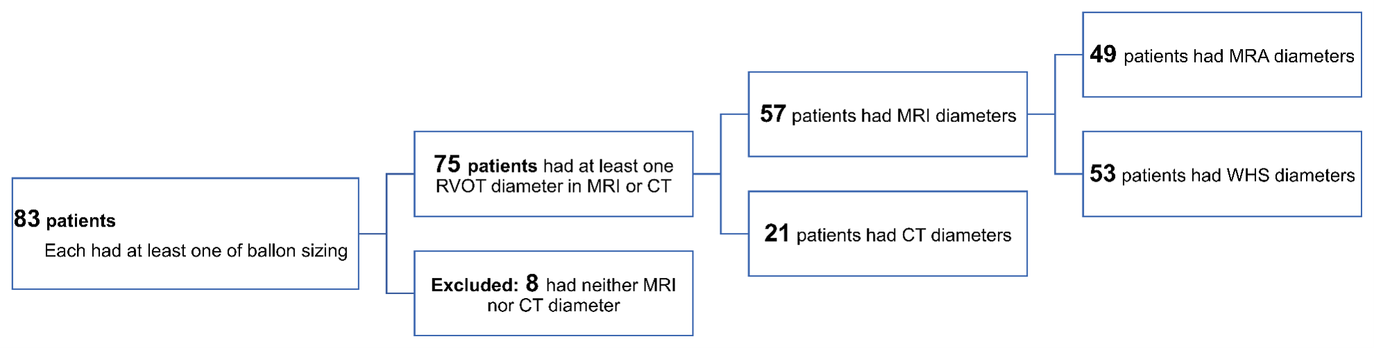
Table 1 outlines the demographic characteristics and echocardiographic assessments as well as the type of transcatheter pulmonary valve (TPV) implanted. In the study sample, patients were 8 to 79 years old and 54.7% were male. The most common underlying diagnosis was TOF (47 patients, 62.7%), followed by PV stenosis (13 patients, 17.3%). Of the 75 patients, 62 (82.7%) underwent PPVI and 13 (17.3%) underwent surgical PV replacement (PVR). In the PPVI group, Melody valve (Medtronic) implantation was performed in 49 patients (79%), and 13 patients (21%) underwent SAPIEN valve (Edwards Lifesciences) implantation. In the entire cohort, based on echocardiographic data, the median maximum gradient was 25 mm Hg (range, 3.0-82.0 mm Hg), 68 patients (90.7%) had moderate or severe PR, 47 (62.7%) exhibited moderate or severe RV dilation, and 14 (18.7%) displayed moderate or severe RV dysfunction. Moderate or severe LV dysfunction was uncommon in the study sample (3 patients, 4%). More than half of the patients (41, 54.7%) had a native RVOT, 24 (32.0%) had RV-PA conduit, and 10 (13.3%) had a bioprosthetic valve. The majority of the patients with RV-PA conduit had pulmonary homografts (17-30 mm in size). The surgically placed bioprosthetic valves ranged in size from 21 to 29 mm.
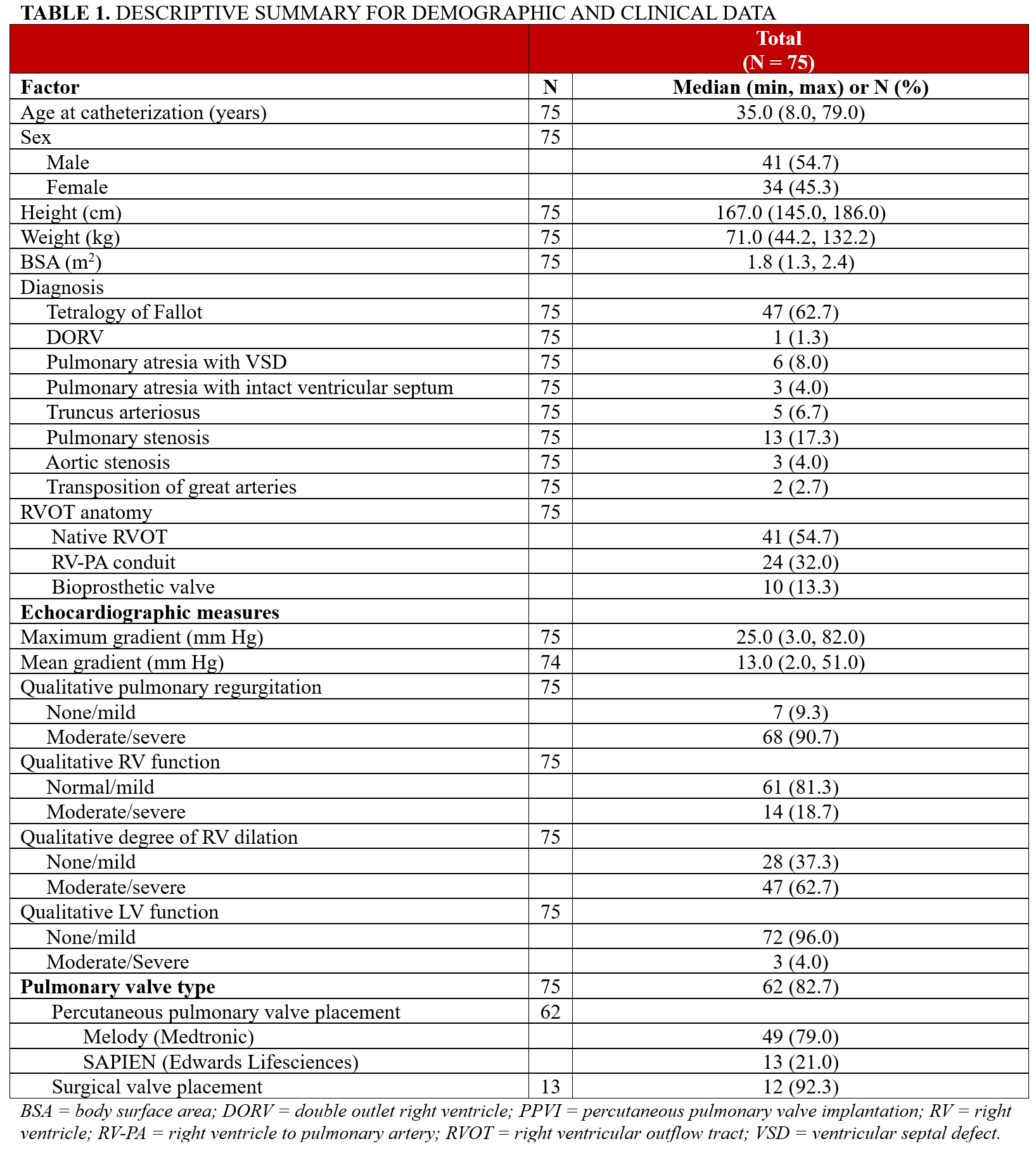
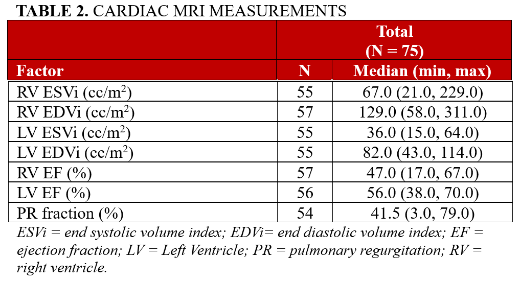
Table 2 outlines the Cardiac MRI measures of ventricular volumes and function. The median PR fraction was 41.5% (range, 3.0%-79.0%). The median RV end diastolic volume index was 129.0 mL/m2 (range, 58.0-311.0 mL/m2). The median RV ejection fraction was 47% (range, 17.0%-67.0%).
Among patients with a pure regurgitant lesion, the median indexed RV end-diastolic volume was 143 cc/m² (range, 87-331 cc/m²).
Table 3 outlines the various measures of RVOT based on cardiac catheterization and axial imaging modalities. Based on cardiac catheterization data, the peak systolic ejection gradient ranged between 0.0 to 58.0 mm Hg with median gradient of 13 mm Hg. The largest balloon used for sizing ranged between 16.0 to 30.0 mm, with a median of 24.0 mm. The median RVOT diameter by MRA was 20.0 (plane 1; S-I) x 21.0 (plane 2; R-L) mm, while by WHS the median RVOT diameter was 19.0 (plane 1; S-I) x 21.0 (plane 2; R-L) mm. By CT scan, the median RVOT diameter was 24.0 (plane 1; S-I) x 23.0 (plane 2; R-L) mm. Median RVOT perimeter and areas were larger on the CT scan.
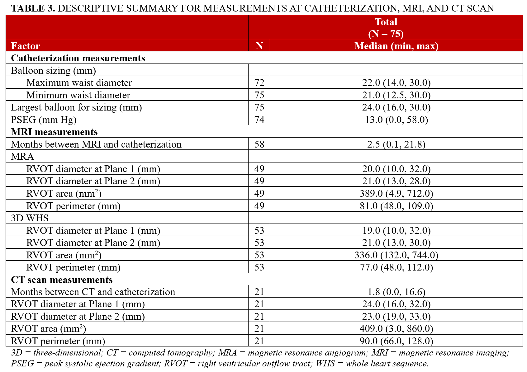
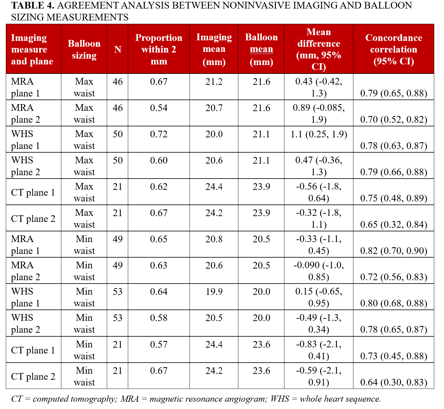
Table 4 outlines the agreement analysis between noninvasive imaging measurements and balloon sizing measurements. The measurements of RVOT diameters from CMR or CT scan did not significantly differ from the maximum or minimum waist diameters on balloon sizing (95% CIs covering 0); an exception was the WHS plane 1 dimension, with a difference smaller than 2-mm limits (1.1 mm, 95% CI, 0.25-1.9). CMR WHS and balloon sizing diameters for each set of planes showed good agreement or above (CCC = 0.78, 0.79, 0.80, 0.78, respectively; 95% CIs falling above 0.60); MRA and balloon sizing diameters exhibited moderate agreement or above (CCC = 0.79, 0.70, 0.82, 0.72, respectively; 95% CIs falling above 0.50). The dimension of 1 plane in CT scan showed moderate agreement or above with balloon sizing diameters (CCC = 0.75, 0.73, respectively; 95% CIs falling above 0.40). There were no obvious heterogeneous trends in subgroup agreement analysis (Figures 3 and 4).


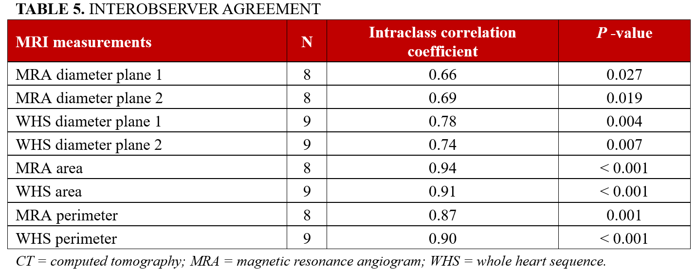
A random sample of patients (10%) was chosen to assess inter-rater reproducibility on MRA and WHS diameters. The agreement on diameters was reasonably high and significant (MRA plane 1: CCC= 0.66, P = .027; MRA plane 2: CCC = 0.69, P = .019; WHS plane 1: 0.78, P = .004, WHS plane 2: CCC=0.74, P = .007) (Table 5).
Discussion
Our study re-emphasizes the value of various noninvasive imaging modalities prior to anticipated PPVI. The question of whether CMR or CT can predict the suitability of the RVOT for transcatheter PVR (TPVR) is very important, and we designed this study to at least partially answer this question. We found that both CMR and CT parameters showed good-to-moderate correlation or above when compared with invasive balloon sizing for assessment of the appropriate site of PPVI placement. The concordance correlation between noninvasive measurements of the landing zone and the gold standard balloon sizing ranged between 0.65 to 0.79 for the maximum waist and between 0.64 to 0.82 for the minimum waist, achieving between substantial and almost perfect correlations. The inter-rater reproducibility yielded reasonably high-significant agreement, indicating its replicability amongst multiple users.
In general, before this study, if we found that the landing zone diameter was too large on preprocedural axial imaging, we might have excluded that patient from attempting percutaneous valve implantation and sent them straight to surgery. Therefore, we sought to identify the imaging modality that best predicts this outcome, thereby avoiding unnecessary cardiac catheterization, selecting appropriate candidates for intervention, and referring unsuitable candidates directly for surgery. As we based the valve size on balloon sizing rather than on 3D rotational angiography or biplane angiography alone, we have designed this study to compare balloon sizing with preprocedural imaging.
However, the heterogeneity of the RVOT in terms of size and distensibility makes prediction of successful PPVI a distinct challenge. Multiplanar 3D image reconstruction is now considered the mandatory gold standard for safe patient selection.9 With our growing adult congenital heart disease population, PPVI is a rapidly expanding alternative for those with multiple prior sternotomies who have had dysfunctional conduits or bioprosthetic valves.10 Unlike transcatheter aortic valve replacement procedures where the landing zone is relatively rigid, PPVI has considerable variability because the valve can be deployed anywhere along the RVOT that has the narrowest dimension and is of suitable length. Given this heterogeneity, even with known diameter of the existing conduits or bioprosthetic valves and considering hemodynamic changes over time, we still perform balloon sizing to see the true balloon waist to evaluate the landing zone and prepare the lesion for valve implantation. In addition, especially with conduits, this would allow us to evaluate the compliance of the RVOT. Furthermore, coronary compression testing is performed. For bioprosthetic valves, after balloon sizing with a compliant balloon, we typically intentionally fracture the valve ring with a noncompliant balloon if indicated. Though we incorporated existing conduit/bioprosthetic valve size in the decision making, the ultimate valve size and type were largely dependent on the balloon sizing.
WHS enable assessment of the pulmonary, systemic, and venous anatomy with a single acquisition, but MRA depends on the timing of contrast media with more than one acquisition needed to assess the entire vasculature. While previous studies have shown that 3D steady-state free precession (SSFP) WHS images obtained in systole have a larger diameter and significant differences between ECG-gated WHS and non-gated MRA sequences,6 our study found substantial correlation using both the sequences with balloon sizing despite WHS images being obtained in mid diastole.
The prototype lesion of long-term RV dysfunction has been TOF and, as expected, was the most common lesion in our series. The resultant RVOT abnormalities can lead to progressive RV dilation and dysfunction, serving as an arrhythmogenic substrate, but can also cause associated left ventricular dysfunction and clinical symptoms such as exercise intolerance and decrease in functional status.11 Optimum timing of PVR prevents irreversible electromechanical cardiomyopathy in repaired TOF.12 Patients with repaired TOF often receive serial assessments with CMR to determine appropriate timing of intervention. CMR has the added value of accurately determining the RV function and RV mass/volume ratios that serve as important pre-PVR markers associated with poor clinical outcomes, as described in the INDICATOR cohort.13 Additionally, CMR has the advantage of no radiation exposure with the added assessment of valve function, and now assumes a more important role in preprocedural planning.
Echocardiographic assessment for a potential landing zone is limited because it is a 2D dataset and does not provide an accurate estimation, as the landing zone could be out of the plane during the cardiac cycle while the images are obtained. As expected, there was extremely poor correlation between this single-plane 2D measurement of RVOT dimension and balloon sizing; therefore, we did not include it in this study. However, echocardiography has the unique advantage of assessing pulmonary stenosis as well as ventricular size and function and is often the most commonly used diagnostic imaging modality to follow these patients.
In our study, correlation coefficients obtained on CMR with balloon sizing were comparable to CT scan with balloon sizing when considering the entire cohort, with both having good to superior correlation. However, the smaller sample size of patients undergoing CT scan may have contributed to these results. Higher spatial resolution and thinner slices obtained with CT scan make it an attractive option. With decreased artifacts, thin collimation, and newer 4D CT, detailed visualization of smaller structures is possible, as is assessment of the dynamicity of the RVOT/PA morphoigies.14,15 Though 4D CT provides dynamic imaging of the RVOT during the entire cardiac cycle, its limitations include exposure to high radiation dose in young patients, the need for potentially nephrotoxic contrast medium, and limited assessment of the PV or RV-to-PA conduit function.
Assessment of coronary arteries is vital prior to PPVI.16 CT scan offers the advantage of visualizing coronary anatomy in detail, with Tezza et al17 describing a proposed algorithm for identifying patients at high and intermediate risk of coronary compression if PPVI was undertaken. But factors such as asymmetric RVOT expansion, scar tissue, and conduit calcification can affect the overall risk, thus making invasive testing with balloon inflation the continued standard of care. Additionally, patients with complex congenital heart disease with varying coronary artery anatomy may have compression only during attempted RVOT expansion, making balloon sizing crucial in these scenarios. The 2019 study by Ebel et al showed that high-resolution SSFP sequences acquired during systole allow for assessment of the relation between the landing zone to the left coronary vessel and sternum.6 However, there are no guidelines about the minimum distance that would prevent coronary artery occlusion or the optimal distance between the stent and the sternum, warranting further studies in the future. Of the entire cohort in our study, 4 patients did not undergo PPVI because of coronary artery compression found during balloon sizing and were instead referred for surgical PVR.
Native RVOTs are often more expansile and distended. Importantly, the absence of calcification makes them distensible, which predisposes to dislocation and embolization of stents. In such circumstances, balloon interrogation with large and long balloons at low inflation pressure helps to prevent this complication as well as delineate a clear waist for valve deployment.5 In these patients, when no balloon waist was seen, RV angiogram was performed during balloon inflation; however, these procedures were done before self-expandable valve platform. With the balloon-expandable valves available during the study period, valves sized 1- to 2-mm larger than the balloon waist was chosen to ensure that the valve would be stable.
For larger RVOT dimensions, the newer SAPIEN 3 valve and the Alterra Adaptive Prestent system (Edwards Lifesciences) are PPVI options; alternatively, a hybrid technique may be employed using a limited sternotomy.18-20 The increasing availability of larger TPVs and hybrid approaches are now obviating the need for repeat sternotomies; as a result, the dimensions mentioned above are not a critical cutoff. Our study did not take into consideration alternative valve options for native RVOTs and larger distensible RVOTs, such as the Harmony valve (Medtronic), Alterra Adaptive Prestent system, or the newly emerging Venus P valve (Venus Medtech) which has received investigational device exemption from the US Food and Drug Administration with trials expected to start soon in the United States.18 Nine patients in our study received surgical PVR because of native RVOT morphology, with consequent higher RV end diastolic volumes, higher PR fraction, and larger RVOT dimensions.
Elastic properties of the pulmonary arterial wall can affect the landing zone. Rapid prototype models have gained popularity; however, because of their rigidity, they are unable to mirror the distensibility of the arterial wall as seen on balloon inflation. Since they do not undergo deformation, they may not exactly replicate an in vivo scenario.21 We did not perform prototypes of the RVOT prior to PPVI in our study.
Malone et al22 demonstrated that CMR-derived measurements and effective diameters with 3D SSFP/WHS sequence and CT measurements of diameter-derived circumference and perimeters on MRI correlated well with invasive balloon sizing and the ultimate implant size. However, the area and perimeter measurements in our cohort did not show good agreement/correlation with balloon sizing.
Future directions
Further studies are warranted to assess optimal distance between coronary arteries and the pulmonary valve/RV-PA conduit by non-invasive imaging modalities prior to PPVI. Integration of 4D flow CMR and computational models can allow for virtual simulation of landing zone and avoid dangerous coronary artery compression.
Limitations
Our study was retrospective in nature and has many limitations. During the time period our study took place, most of the transcatheter valves implanted were Melody valves, and there is now a wider range and size of TPVs available. There is no standard practice for performance of balloon sizing for TPVR across institutions. Rotational angiography was not performed, only biplane angiography was performed prior to TPVR. MRA is an imaging technique without ECG gating, which was the standard protocol, and coronary artery assessment could not be performed on the MRA. ECG-gated 3D SSFP-WHS sequences were performed only in mid-diastole per institutional protocol, which could potentially underestimate the maximum systolic dimensions of the narrowest diameter. Finally, only a small number of patients with CT were included in the study, leading to slightly lower correlation.
Conclusions
Both CT and CMR can be utilized for assessment of the landing zone prior to PPVI. Maximum diameters on CMR and CT showed substantial correlation with invasive balloon sizing. Although there is reasonable correlation with axial imaging and catheterization, testing for coronary compression during balloon sizing is of paramount importance, and computational fluid dynamics and modeling of the RVOT with attempted valve implantation can help simulate this condition to overcome some of the limitations.
Affiliations and Disclosures
Sruti Rao, MD1; Patcharapong Suntharos, MD2; Wei Liu, MS3; Lourdes Prieto, MD4; Sarah Worley, MS3; Paul Schoenhagen, MD5; Rukmini Komarlu, MD2
From the 1Department of Pediatric Cardiology, Helen Devos Children’s Hospital, Grand Rapids, Michigan; 2Department of Pediatric Cardiology, Cleveland Clinic Children’s Hospital, Cleveland, Ohio; 3Department of Quantitative Health Sciences, Cleveland Clinic, Cleveland, Ohio; 4Department of Pediatric Cardiology, Nicklaus Children’s Hospital, Miami, Florida; 5Department of Cardiovascular Imaging, Heart and Vascular Institute, Cleveland Clinic, Cleveland, Ohio.
Disclosures: The authors report no financial relationships or conflicts of interest regarding the content herein.
Address for correspondence: Rukmini Komarlu, MD, Associate Professor of Pediatrics, Lerner College of Medicine of Case Western Reserve University, Department of Pediatric Cardiology, Children's Institute Department of Heart, Vascular & Thoracic, Division of Cardiology and Cardiovascular Medicine, 9500 Euclid Avenue, Cleveland, OH 44195, USA. Email: komarlr@ccf.org
References
1. Bradley SM, Erdem CC, Hsia TY, Atz AM, Bandisode V, Ringewald JM. Right ventricle-to-pulmonary artery shunt: alternative palliation in infants with inadequate pulmonary blood flow prior to two-ventricle repair. Ann Thorac Surg. 2008;86(1):183-188; discussion 188. doi:10.1016/j.athoracsur.2008.03.047
2. Gladman G, McCrindle BW, Williams WG, Freedom RM, Benson LN. The modified Blalock-Taussig shunt: clinical impact and morbidity in Fallot's tetralogy in the current era. J Thorac Cardiovasc Surg. 1997;114(1):25-30. doi:10.1016/S0022-5223(97)70113-2
3. Cocomello L, Meloni M, Rapetto F, et al. Long-term comparison between pulmonary homograft versus bioprosthesis for pulmonary valve replacement in tetralogy of Fallot. J Am Heart Assoc. 2019;8(24):e013654. doi:10.1161/JAHA.119.013654
4. Bonhoeffer P, Boudjemline Y, Saliba Z, et al. Percutaneous replacement of pulmonary valve in a right-ventricle to pulmonary-artery prosthetic conduit with valve dysfunction. Lancet. 2000;356(9239):1403-1405. doi:10.1016/S0140-6736(00)02844-0
5. Georgiev S, Tanase D, Ewert P, et al. Percutaneous pulmonary valve implantation in patients with dysfunction of a "native" right ventricular outflow tract - mid-term results. Int J Cardiol. 2018;258:31-35. doi:10.1016/j.ijcard.2017.11.091
6. Ebel S, Gottschling S, Buzan MTA, et al. 3D-assessment of RVOT dimensions prior percutaneous pulmonary valve implantation: comparison of contrast-enhanced magnetic resonance angiography versus 3D steady-state free precession sequence. Int J Cardiovasc Imaging. 2019;35(8):1453-1463. doi:10.1007/s10554-019-01578-w
7. Lin LI. A concordance correlation coefficient to evaluate reproducibility. Biometrics. 1989;45(1):255-268.
8. Kouris NT, Kostopoulos VS, Psarrou GA, Kostakou PM, Tzavara C, Olympios CD. Left ventricular ejection fraction and Global Longitudinal Strain variability between methodology and experience. Echocardiography. 2021;38(4):582-589. doi:10.1111/echo.15025
9. Chung R, Taylor AM. Imaging for preintervention planning: transcatheter pulmonary valve therapy. Circ Cardiovasc Imaging. 2014;7(1):182-189. doi:10.1161/CIRCIMAGING.113.000826
10. Phillips AB, Nevin P, Shah A, Olshove V, Garg R, Zahn EM. Development of a novel hybrid strategy for transcatheter pulmonary valve placement in patients following transannular patch repair of tetralogy of fallot. Catheter Cardiovasc Interv. 2016;87(3):403-410. doi:10.1002/ccd.26315
11. Khambadkone S, Coats L, Taylor A, et al. Percutaneous pulmonary valve implantation in humans: results in 59 consecutive patients. Circulation. 2005;112(8):1189-1197. doi:10.1161/CIRCULATIONAHA.104.523266
12. Geva T, Gauvreau K, Powell AJ, et al. Randomized trial of pulmonary valve replacement with and without right ventricular remodeling surgery. Circulation. 2010;122(11 Suppl):S201-S208. doi:10.1161/CIRCULATIONAHA.110.951178
13. Geva T, Mulder B, Gauvreau K, et al. Preoperative predictors of death and sustained ventricular tachycardia after pulmonary valve replacement in patients with repaired tetralogy of Fallot enrolled in the INDICATOR cohort. Circulation. 2018;138(19):2106-2115. doi:10.1161/CIRCULATIONAHA.118.034740
14. Saremi F, Gera A, Ho SY, Hijazi ZM, Sánchez-Quintana D. CT and MR imaging of the pulmonary valve. Radiographics. 2014;34(1):51-71. doi:10.1148/rg.341135026
15. Schievano S, Capelli C, Young C, et al. Four-dimensional computed tomography: a method of assessing right ventricular outflow tract and pulmonary artery deformations throughout the cardiac cycle. Eur Radiol. 2011;21(1):36-45. doi:10.1007/s00330-010-1913-5
16. Chung R, Taylor AM. Imaging for preintervention planning: transcatheter pulmonary valve therapy. Circ Cardiovasc Imaging. 2014;7(1):182-189. doi:10.1161/CIRCIMAGING.113.000826
17. Tezza M, Witsenburg M, Nieman K, van de Woestijne PC, Budde RPJ. Cardiac CT to assess the risk of coronary compression in patients evaluated for percutaneous pulmonary valve implantation. Eur J Radiol. 2019;110:88-96. doi:10.1016/j.ejrad.2018.11.018
18. Murphy SP, Sultana S, Zern EK, et al. Multimodality imaging evaluation of diseases of the pulmonic valve and right ventricular outflow tract for the adult cardiologist. Circ Cardiovasc Imaging. 2025;18(2):e017126. doi:10.1161/CIRCIMAGING.124.017126
19. Ghobrial J, Levi DS, Aboulhosn J. Native right ventricular outflow tract transcatheter pulmonary valve replacement without pre-stenting. JACC Cardiovasc Interv. 2018;11(6):e41-e44. doi:10.1016/j.jcin.2017.11.014
20. Morgan GJ. Pulmonary regurgitation- is the future percutaneous or surgical? Front Pediatr. 2018;6:184. doi:10.3389/fped.2018.00184
21. Schievano S, Migliavacca F, Coats L, et al. Percutaneous pulmonary valve implantation based on rapid prototyping of right ventricular outflow tract and pulmonary trunk from MR data. Radiology. 2007;242(2):490-497. doi:10.1148/radiol.2422051994
22. Malone L, Fonseca B, Fagan T, et al. Preprocedural risk assessment prior to PPVI with CMR and cardiac CT. Pediatr Cardiol. 2017;38(4):746-753. doi:10.1007/s00246-017-1574-0