




Recurrent Prosthetic Mitral Valve Dysfunction: A Multimodality Imaging and Surgical Challenge
© 2026 HMP Global. All Rights Reserved.
Any views and opinions expressed are those of the author(s) and/or participants and do not necessarily reflect the views, policy, or position of the Journal of Invasive Cardiology or HMP Global, their employees, and affiliates.
J INVASIVE CARDIOL 2026. doi:10.25270/jic/26.00343. Epub October 22, 2026.
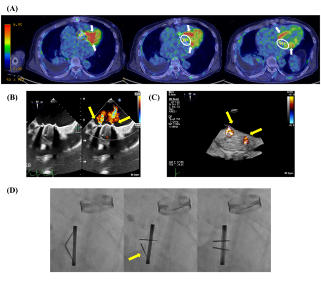
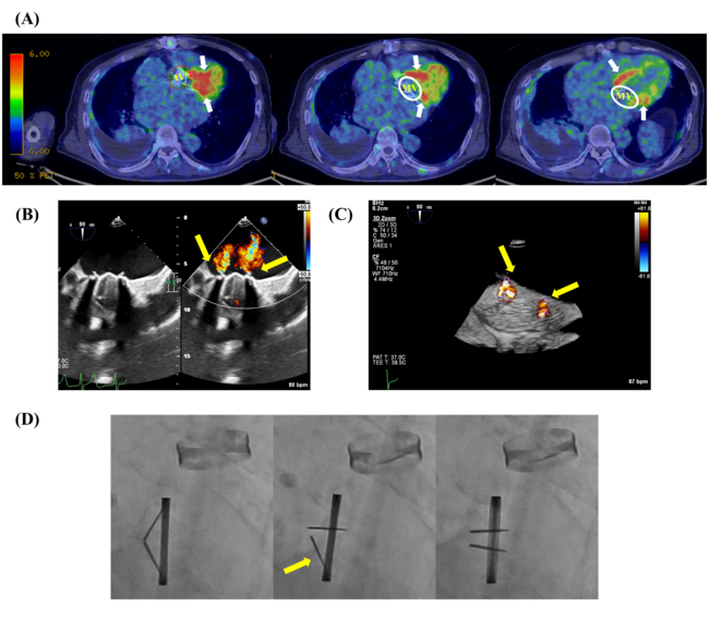
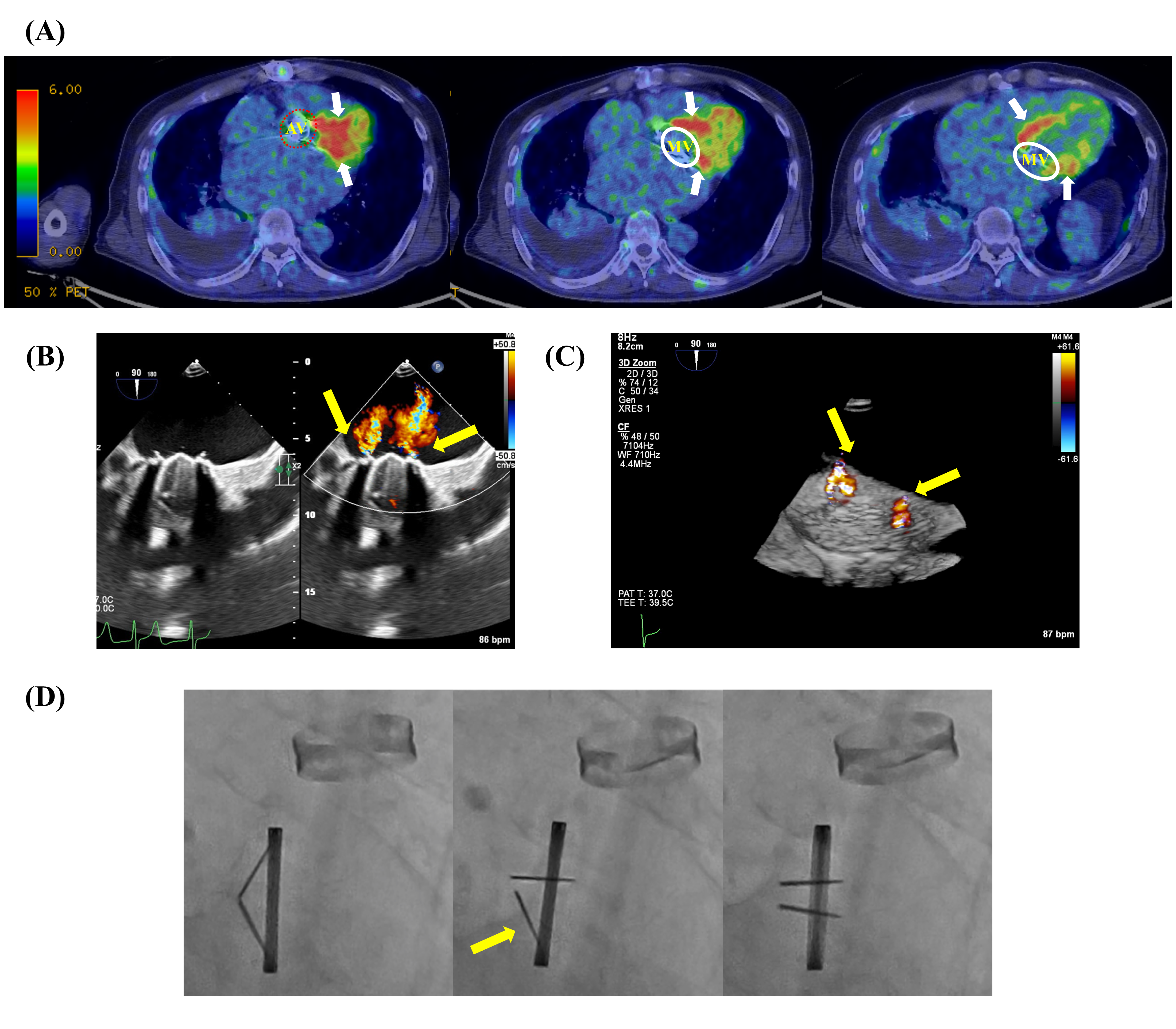
A 70-year-old man had undergone mechanical mitral and aortic valve replacement in 2005 and redo mechanical mitral valve replacement in 2018 for prosthetic mitral valve dysfunction. In 2023, during hospitalization for lung abscess and pleuritis, 18F-fluorodeoxyglucose positron emission tomography/computed tomography (FDG-PET/CT) showed focal uptake around the prosthetic mitral annulus, whereas no abnormal uptake was observed around the aortic prosthesis (Figure A). His blood cultures were negative.
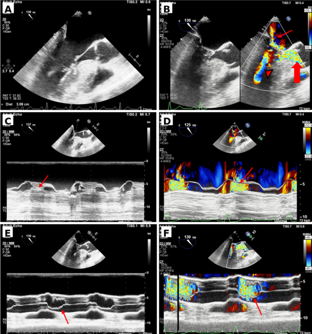
In 2025, the patient presented with progressive dyspnea and hemolytic anemia. Transesophageal echocardiography (TEE) demonstrated eccentric paravalvular regurgitation adjacent to the prosthetic sewing ring (Figure B, Video 1). Three-dimensional TEE further localized multiple paravalvular regurgitant jets arising outside the sewing ring (Figure B and C). Fluoroscopy demonstrated intermittent delayed opening of the anterior mechanical leaflet without persistent fixed obstruction (Figure D, Video 2).
The combination of paravalvular regurgitation, hemolysis, and prior focal annular FDG uptake raised concern for structural failure of the mitral annular interface, although the etiology of the FDG uptake remained uncertain and occult low-grade infection could not be excluded.
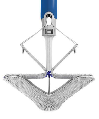
At the third mitral valve replacement, the prosthesis was partially detached at the posterior annulus. The annular tissue was friable, and the sutures were removed easily; no pannus, gross vegetation, or abscess was identified. A 25-mm Epic porcine bioprosthesis (Abbott) was implanted with pericardial skirt reinforcement.
This case demonstrates the complementary value of multimodality imaging: echocardiography defined and localized paravalvular regurgitation, fluoroscopy assessed leaflet motion, and prior FDG-PET/CT demonstrated focal annular metabolic activity of uncertain etiology. Operative inspection ultimately confirmed posterior annular dehiscence and guided reinforced surgical reconstruction.
Affiliations and Disclosures
Daiki Kasahara, MD1; Rie Aoyama, MD, PhD1; Kana Horie, MD1; Hironao Sudo, MD1; Yo Iwata, MD, PhD1; Shinichi Okino, MD1; Shigeru Fukuzawa, MD, PhD1; Ryosuke Marushima, MD2; Masaaki Omori, MD2; Manabu Sakurai, MD, PhD2
From the Departments of 1Cardiology and 2Cardiovascular Surgery, Heart and Vascular Institute, Funabashi Municipal Medical Center, Chiba, Japan.
Acknowledgments: The authors used ChatGPT (OpenAI) and DeepL to assist with English-language editing. All output was reviewed by the authors, who take responsibility for the final content.
Disclosures: The authors have no relationships with industry or other relevant entities to disclose.
Consent statement: Written informed consent for publication of this case, including images and videos, was obtained from the patient.
Address for correspondence: Rie Aoyama, MD, PhD, Department of Cardiology, Heart and Vascular Institute, Funabashi Municipal Medical Center, 1-21-1 Kanasugi, Funabashi, Chiba 273-8588, Japan. Email: r-aoyama@nms.ac.jp